. 2017 Mar 23;2(6):e90521.
doi: 10.1172/jci.insight.90521.

Priming is key to effective incorporation of image-guided thermal ablation into immunotherapy protocols

Affiliations

Priming is key to effective incorporation of image-guided thermal ablation into immunotherapy protocols

Matthew T Silvestrini et al. JCI Insight. .

Abstract

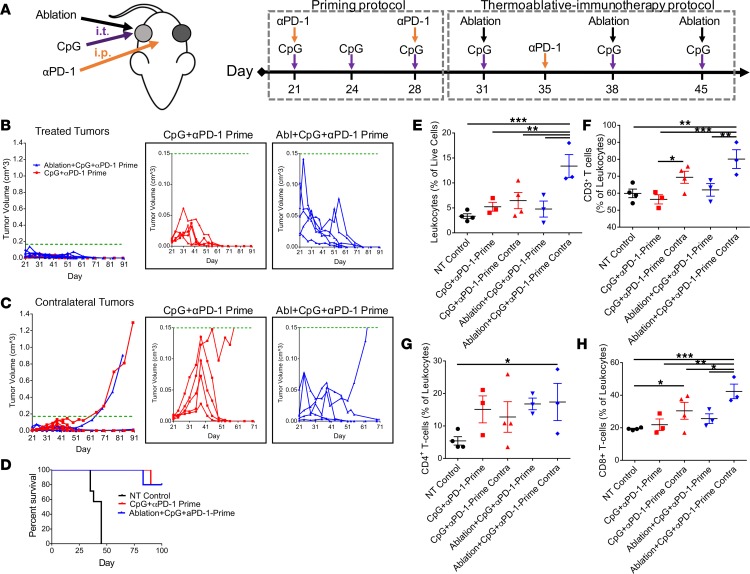
Focal therapies play an important role in the treatment of cancers where palliation is desired, local control is needed, or surgical resection is not feasible. Pairing immunotherapy with such focal treatments is particularly attractive; however, there is emerging evidence that focal therapy can have a positive or negative impact on the efficacy of immunotherapy. Thermal ablation is an appealing modality to pair with such protocols, as tumors can be rapidly debulked (cell death occurring within minutes to hours), tumor antigens can be released locally, and treatment can be conducted and repeated without the concerns of radiation-based therapies. In a syngeneic model of epithelial cancer, we found that 7 days of immunotherapy (TLR9 agonist and checkpoint blockade), prior to thermal ablation, reduced macrophages and myeloid-derived suppressor cells and enhanced IFN-γ-producing CD8+ T cells, the M1 macrophage fraction, and PD-L1 expression on CD45+ cells. Continued treatment with immunotherapy alone or with immunotherapy combined with ablation (primed ablation) then resulted in a complete response in 80% of treated mice at day 90, and primed ablation expanded CD8+ T cells as compared with all control groups. When the tumor burden was increased by implantation of 3 orthotopic tumors, successive primed ablation of 2 discrete lesions resulted in survival of 60% of treated mice as compared with 25% of mice treated with immunotherapy alone. Alternatively, when immunotherapy was begun immediately after thermal ablation, the abscopal effect was diminished and none of the mice within the cohort exhibited a complete response. In summary, we found that immunotherapy begun before ablation can be curative and can enhance efficacy in the presence of a high tumor burden. Two mechanisms have potential to impact the efficacy of immunotherapy when begun immediately after thermal ablation: mechanical changes in the tumor microenvironment and inflammatory-mediated changes in immune phenotype.


Conflict of interest statement

Conflict of interest: The authors have declared that no conflict of interest exists.

Figures

Figure 1
Figure 1. MRgFUS ablation promotes local antitumor immune response.
(A) Regimen of thermal ablation in mice orthotopically transplanted with neu deletion line (NDL) tumor biopsies in the fourth and ninth mammary fat pad. (B and C) Magnetic resonance–guided focused ultrasound (MRgFUS) ablation protocol and temperature mapping for thermal ablation in vivo (white color indicates minimum threshold of 70°C). (B) T1w images of localized region to be ablated (red dotted circle). (C) MR thermometry image following ablation protocol (red arrow indicates heated region). (DF) NDL tumor growth following MRgFUS ablation (n = 7) compared with no-treatment (NT) control NDL tumor growth (n = 34 total, 8 representative growth curves shown for this individual study). Mice per group examined in 3 separate experiments. (D) Thermally ablated tumors exhibited a transient suppression in growth compared with (E) contralateral and (F) NT control tumors. (GI) The entire fourth and ninth mammary fat pad (tumor and embedded lymph node) was harvested at day 28, and immune cells were quantified by flow cytometry (n = 4 per group). Effect of therapy on (G) IFN-γ CD4+ T cells, (H) CD8+ T cells, and (I) Tregs in the treated and contralateral tumors. For box-and-whiskers plots, the whiskers represent the minimum and maximum values, the box boundaries represent the 25th and 75th percentiles, and the middle line is the median value. (J) High mobility group box 1 (HMGB1) protein release in vitro quantified via ELISA. Treatment with doxorubicin (Dox) at 37°C represents a positive control. Each dot represents a sample. Data are mean ± SEM of 3 measurements. (KM) H&E staining performed at day 31 confirmed that, compared with (K) control tumors (n = 3), viable tumor was reduced in (L) treated tumors (n = 3), where residual viable tumor tissue (red arrows) existed outside the path of the ultrasound beam. Thermal ablation did not reduce viable (M) contralateral tumor tissue (n = 3). *P < 0.05, **P < 0.01, ****P < 0.0001, †P < 0.05 compared with 37°C group, ‡P < 0.01 compared with 37°C + Dox group. Scale bar: 3 mm. Statistics for GI were determined by ANOVA followed by Fisher’s LSD without multiple comparisons correction, and those for J were determined by ANOVA followed by Tukey multiple comparison correction.
Figure 2
Figure 2. Thermal ablation alters the tumor microenvironment and intratumoral transport kinetics of small molecules and proteins.
(A–F) Tumor permeability of proteins after thermal ablation was assessed in vivo by tracking Copper-64–labeled BSA (64Cu-BSA) with positron emission tomography at (A) 0.5, (B) 6, (C) 18, (D) 24, and (E) 48 hours. A single tumor in a bilateral NDL tumor–bearing mouse was ablated (white arrow). Also visible are contralateral tumors (red arrow). Tracer kinetics and biodistribution were plotted for the (F) maximum intratumoral accumulation of 64Cu-BSA versus time, revealing a temporal peak of the spatial mean and maximum occurring at 6 hours after injection (n = 4 per cohort, data are mean ± SEM). (G and H) Increased vascularization of the tumor-draining lymph node 0.5 hours after tumor ablation upon H&E histological staining (n = 3). Scale bar: 300 μm. (G) Draining lymph node following thermal ablation with green box at location of interest. (H) View of enhanced vascularization within the lymph node indicated by green arrows. (IN) Tumor permeability to small molecules after thermal ablation was assessed in vivo with contrast enhanced T1-weighted magnetic resonance imaging (CET1wMRI) (n = 3). (I) Gadoteridol was administered immediately before (J) ablation (red arrow). (K) Gadoteridol accumulated in the ablated region 3 hours after injection and ablation. (L) Contrast was readministered at 3 hours; additional accumulation was not detected. (M) At 20 hours after injection and ablation, intratumoral gadoteridol accumulation had cleared. (N) After 20 hours, gadoteridol readministration resulted in accumulation in the surrounding tumor rim (blue arrows) but not within in the ablated tissue. (OQ) Mechanisms for enhanced accumulation 48 hours after thermal ablation with H&E (n = 8). (O) Representative tumor following ablation with green and red box at locations of interest. Scale bar: 3 mm. (P) Image from red box of heat-fixed tissue with shrunken but intact nuclei and preservation of tissue architecture (black arrow) surrounded by discohesive and nonviable tumor tissue with some ghosted nuclei and local edema (yellow arrow). Scale bar: 60 μm. (Q) Image from green box of inflammation observed, where leukocytes are densely located in the periphery of the tumor (yellow arrows). Scale bar: 60 μm.
Figure 3
Figure 3. Coincident thermoablative immunotherapy protocol (TA-immunotherapy) diminishes the abscopal effects of immunotherapy.
(A) Regimen of coincident TA-immunotherapy, CpG (100 μg per injection, intratumoral [i.t.]) and anti–PD-1 (αPD-1) (200 μg per injection, i.p.) in mice orthotopically transplanted with NDL tumor biopsies in the fourth and ninth mammary fat pad. (B and C) Tumor growth was followed until an animal from the group was euthanized (tumor diameter >1.5 cm). Treatment cohorts were NT control (n = 8), CpG + αPD-1 (n = 6) and ablation + CpG + αPD-1 (Abl + CpG + αPD-1, n = 7). Tumor growth data result from survival study (n = 21) but are representative of trends in 7 studies with varied end points (n = 90 total). (B) Immunotherapy alone and coincident TA-immunotherapy induced suppression of local tumor growth. (C) Contralateral tumor growth suppression was greater for immunotherapy alone. Data plotted as mean ± SEM. (D) By day 180, survival outcomes for mice treated with immunotherapy alone (CpG + αPD-1, n = 6) were improved compared with those treated coincident with ablation (ablation + CpG + αPD-1, n = 7) or control treatments (NT Control [n = 8], αPD-1 [n = 3], CpG [n = 7], ablation [n = 7], and ablation + CpG [n = 4]). (EH) The fourth and ninth mammary fat pad (tumor and the embedded node) was harvested at day 28, and immunocytes were quantified via flow cytometry (n = 4 per group). Number of (E and F) leukocytes and (G and H) IFN-γ CD8+ T cells in treated (E and G) and contralateral (F and H) tumors. For box-and-whiskers plots, the whiskers represent the minimum and maximum values, the box boundaries represent the 25th and 75th percentiles, and the middle line is the median value. (I) IHC on day 50 verified that both CpG + αPD-1 (n = 3) and ablation + CpG + αPD-1 (n = 3) increased infiltrating CD8+ T cells (brown stain) in contralateral tumors compared with NT controls. C was analyzed using an unpaired t test assuming unequal variance comparing mean tumor volume of CpG + αPD-1 and ablation + CpG + αPD-1 at each day. EH were analyzed by ANOVA followed by Fisher’s LSD test without multiple comparisons correction. Scale bars: 150 μm.*P < 0.05, **P < 0.01, ***P < 0.001.
Figure 4
Figure 4. Comparison of coincident TA-immunotherapy (ablation + CpG + αPD-1) with immunotherapy alone (CpG + αPD-1).
CpG+αPD-1 reduces macrophages and myeloid-derived suppressor cells (MDSCs) in contralateral tumors and increases PD-L1 expression in tumor-infiltrating leukocytes. Coincident thermoablative immunotherapy (TA-immunotherapy) increases macrophages in treated tumors. (AE) Animals were treated as described in Figure 3A with thermal ablation, CpG (100 μg per injection), and αPD-1 (200 μg per injection). Treatment cohorts were NT control (n = 8), CpG + αPD-1 (n = 8), and ablation + CpG + αPD-1 (n = 8). (A and B) The frequency of (A) macrophages and (B) MDSCs in the treated and contralateral tumors following treatment. The fourth and ninth mammary fat pads (tumor and the embedded node) were harvested at day 35 and immunocytes were quantified via flow cytometry. (CE) Expression of PD-1 and PD-L1 in treated and contralateral tumors. Tumors were harvested at day 28, and immunocytes were quantified via flow cytometry. (C) The amount of PD-1 expressed on CD4+ T cells, the (D) expression of PD-L1 on tumor/stromal cells (CD45), and (E) the expression of PD-L1 on CD45+ leukocytes in treated and contralateral tumors. For box-and-whiskers plots, the whiskers represent the minimum and maximum values, the box boundaries represent the 25th and 75th percentiles, and the middle line is the median value. Statistics in AE were determined by ANOVA followed by Fisher’s LSD test without multiple comparisons correction. *P < 0.05, **P < 0.01, ***P < 0.001, ‡P < 0.05 compared with all groups.
Figure 5
Figure 5. Priming the immune system is required for efficacious incorporation of thermal ablation into an immunotherapeutic protocol.
(A) Regimen of primed thermoablative immunotherapy (TA-immunotherapy). Immunotherapy was administered prior to thermal ablation in priming protocol. Following priming, mice received a combination of thermal dosing and immunotherapy in TA-immunotherapy protocol. Treatments included CpG + αPD-1-Prime (n = 5), ablation + CpG + αPD-1-Prime (Abl + CpG + αPD-1-Prime, n = 5), and NT control (n = 4). CpG was injected intratumorally (i.t., 100 μg), and αPD-1 was injected i.p. (200 μg). Tumor growth data result from survival study (n = 14), but trends are representative of 4 separate experiments with varied end points (total n = 50). (B and C) CpG + αPD-1-Prime and ablation + CpG + αPD-1-Prime achieved an 80% complete response by day 90 (n = 5). (B) 100% of all treated tumors and (C) 80% (4 of 5) of all contralateral tumors from both groups were eradicated after treatment; enhanced views (tumor volume plotted below dotted green line) of the treated (B) and contralateral (C) tumor growth from both treatment groups. (D) Survival outcomes for mice treated with the priming protocol (CpG + αPD-1-Prime, ablation + CpG + αPD-1-Prime, and NT Control, n = 4 per group). (EH) On day 35, tumors were harvested, and leukocytes and T cells were quantified via flow cytometry (data are plotted as mean ± SEM, n = 4 per group). Effect of therapy on (E) CD45+ leukocytes, (F) CD3+ T cells, (G) CD4+ T cells, and (H) CD8+ T cells. Significance was determined via 1-way ANOVA followed by a Fisher’s LSD test without multiple comparisons correction. *P < 0.05, **P < 0.01, ***P < 0.001.
Figure 6
Figure 6. Direct comparison of results from primed thermoablative immunotherapy (TA-immunotherapy), coincident TA-immunotherapy, and immunotherapy alone demonstrates enhanced response.
(A) Priming prior to thermal ablation (ablation + CpG + αPD-1-Prime; n = 5) suppressed contralateral tumor growth compared with other treatments that incorporated ablation, including NT control (n = 8), ablation (n = 7), ablation + CpG (n = 4), and ablation + CpG + αPD-1 (n = 7). For bilateral tumors, the growth of primed TA-immunotherapy was similar to primed immunotherapy alone (n = 5). Data are plotted as mean ± SEM. (BF) Infiltrating CD8+ T cells (brown stain) in contralateral tumors were increased by (B) primed TA-immunotherapy as compared with (C) ablation, (D) ablation + CpG, (E) coincident TA-immunotherapy, and (F) primed immunotherapy (n = 3 per group) upon immunohistochemical staining of CD8. Scale bars: 150 μm.
Figure 7
Figure 7. Sequential multisite thermoablative immunotherapy (TA-immunotherapy) enhances response.
(AC) Treatment with single-site TA-immunotherapy (ablation + CpG + αPD-1-Prime) results in heterogeneous lesions by day 38 (n = 4). (A) Histological section of ablation + CpG + αPD-1-Prime–treated tumor with boxes at regions of interest. Scale bar: 4 mm. (B) Enlarged view of red box, with local inflammation (infiltration of leukocytes), hemorrhage (black arrows), and necrotic tumor cells containing ghosted nuclei (red arrow). Scale bar: 200 μm. (C) Enlarged view of green box with heat fixation (red arrow), tumor necrosis, and collagen remodeling (black arrow). Scale bar: 200 μm. (DH) Mice bearing 3 NDL tumors were treated with priming and multisite ablation. Tumor growth was followed until an animal from the group was euthanized (tumor diameter >1.5 cm). Treatment cohorts were NT control (n = 4), CpG + αPD-1-Prime (n = 4), and ablation + CpG + αPD-1-Prime (n = 5), where data are plotted as mean ± SEM. Mice per group examined in 2 separate experiments. (D) Mice were orthotopically transplanted with NDL tumor biopsies in the ninth (i), fourth (ii), and second mammary fat pad (iii). (E) Regimen of multisite administration of ablation + CpG + αPD-1-Prime. CpG was injected intratumorally (i.t., 100 μg), and αPD-1 was injected i.p. (200 μg). (F) Effect of multisite administration on tumor growth in nontreated (iii) and (G) left-treated (i) tumors. (H) Survival outcomes for mice treated with the multisite priming protocol CpG + αPD-1-Prime (n = 4), ablation + CpG + αPD-1-Prime (n = 5), and NT Control (n = 4).

References

    1. Topalian SL, Drake CG, Pardoll DM. Immune checkpoint blockade: a common denominator approach to cancer therapy. Cancer Cell. 2015;27(4):450–461. doi: 10.1016/j.ccell.2015.03.001. - DOI - PMC - PubMed
    1. Nguyen LT, Ohashi PS. Clinical blockade of PD1 and LAG3--potential mechanisms of action. Nat Rev Immunol. 2015;15(1):45–56. - PubMed
    1. Postow MA, Callahan MK, Wolchok JD. Immune Checkpoint Blockade in Cancer Therapy. J Clin Oncol. 2015;33(17):1974–1982. doi: 10.1200/JCO.2014.59.4358. - DOI - PMC - PubMed
    1. Nowak AK, Robinson BW, Lake RA. Synergy between chemotherapy and immunotherapy in the treatment of established murine solid tumors. Cancer Res. 2003;63(15):4490–4496. - PubMed
    1. Baxevanis CN, Perez SA, Papamichail M. Combinatorial treatments including vaccines, chemotherapy and monoclonal antibodies for cancer therapy. Cancer Immunol Immunother. 2009;58(3):317–324. doi: 10.1007/s00262-008-0576-4. - DOI - PMC - PubMed

Publication types

MeSH terms

Substances