

Role of FDG-PET scan in staging of pulmonary epithelioid hemangioendothelioma
- PMID: 28352786
- PMCID: PMC5329812
- DOI: 10.1515/med-2016-0025
Role of FDG-PET scan in staging of pulmonary epithelioid hemangioendothelioma
Abstract
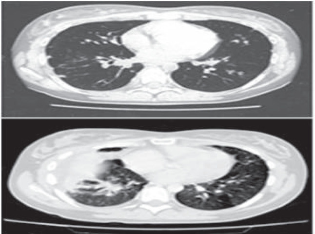
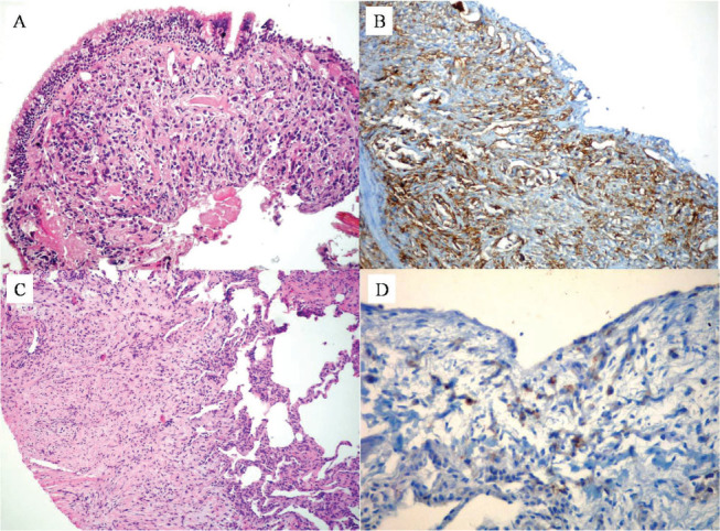
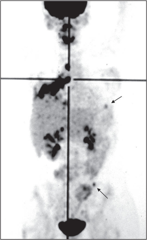
In this report we describe a case of pulmonary epithelioid hemangioendothelioma (PEH) in a young woman. The neoplasm manifested with dry cough, chest pain, finger clubbing, and multiple bilateral pulmonary nodules on chest x-ray and computed tomographic (CT) scan. She underwent thoracoscopy, and the histological features of the lung biopsies were initially interpreted as consistent with a not-well-defined interstitial lung disease. Our patient was clinically and radiologically stable over a period of four years, after which the disease progressed to involve not only the lung but also mediastinal lymph nodes, liver and bone. Fiberoptic bronchoscopy showed subtotal occlusion of the right middle and lower lobe bronchi. The histologic examination of bronchial biopsies revealed a poorly differentiated neoplasm immunohistochemically positive for vimentin and vascular markers CD31, CD34 and Factor VIII. A diagnosis of malignant hemangioendothelioma was made. Positron emission tomography (PET) is more sensitive than CT scan and bone scintigraphy in detecting PEH metastases. Furthermore, 18-fluorodeoxyglucose (FDG) uptake seems to be related to the grade of malignancy of PEH lesions. Therefore, we suggest that FDG-PET should be included in the staging system and follow-up of PEH.
Keywords: CT-scan; FDG-PET; lung cancer staging; pulmonary epithelioid hemangioendothelioma.
Figures
References
-
- Baldi C, Ieni A, Cozzolino I, Cerbone V, Memoli D, Zeppa P. Ultrasound-guided fine needle aspiration cytology of a primary lymph node leiomyoma: a flexible procedure for a complex case. Acta Cytol. 2014;58:303–308. - PubMed
-
- Cozzolino I, Vigliar E, Todaro P, Peluso AL, Picardi M, Sosa Fernandez LV, Mignogna MD, Tuccari G, Selleri C, Zeppa P. Fine needle aspiration cytology of lymphoproliferative lesions of the oral cavity. Cytopathology. 2014;25:241–249. - PubMed
-
- Zeppa P, Barra E, Napolitano V, Cozzolino I, Troncone G, Picardi M, De Renzo A, Mainenti PP, Vetrani A, Palombini L. Impact of endoscopic ultrasound-guided fine needle aspiration (EUS-FNA) in lymph nodal and mediastinal lesions: a multicenter experience. Diagn Cytopathol. 2011;39:723–729. - PubMed
-
- Cozzolino I, Nappa S, Picardi M, De Renzo A, Troncone G, Palombini L, Zeppa P. Clonal B-cell population in a reactive lymph node in acquired immunodeficiency syndrome. Diagn Cytopathol. 2009;37:910–914. - PubMed
LinkOut - more resources
Full Text Sources
Other Literature Sources