

Uncommon primary hydatid cyst occupying the adrenal gland space, treated with laparoscopic surgical approach in an old patient
- PMID: 28352829
- PMCID: PMC5329862
- DOI: 10.1515/med-2016-0075
Uncommon primary hydatid cyst occupying the adrenal gland space, treated with laparoscopic surgical approach in an old patient
Abstract
Hydatid disease (HD) is caused by Echinococcus Granulosus (EG), which is a larva endemic in many undeveloped areas. The most common target is the liver (59%-75%). The retroperitoneal space is considered as a rare localization. We report an uncommon case of HD located in the adrenal gland space.
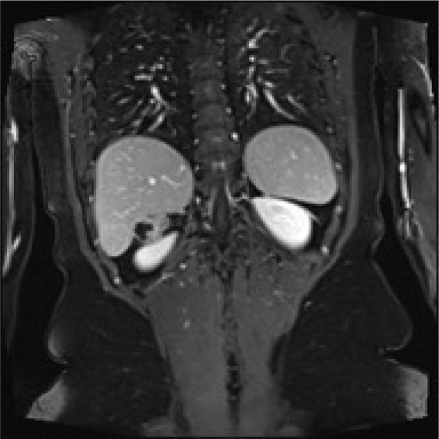
Presentation of case: This is a 78-year-old Moroccan woman, with right flank pain for eight months previously. She denied contact with dogs or sheep. Her physical examination was normal. There was no pathological alteration of laboratory exams. CT scan measuring 5 cm without clear signs for a sure diagnosis found a round lesion in the right adrenal gland. An abdominal MRI showed a round mass of 34 x 27 mm with fluid component without a clear plane of dissection from kidney and liver. A laparoscopic procedure was performed to obtain a histological diagnosis. We reached a conclusive diagnosis of Hydatid cyst of right adrenal gland space. Hydatid cysts often develop in the liver. The location in the adrenal bed is rare without clinical signs related to alteration of the gland's secretion. Hydatid cyst identification in the adrenal gland space is based on ultrasonography, CT or MRI scans. The differential diagnosis includes various benign and malignant lesions. Laparoscopic procedure is the best approach available to obtain a histological diagnosis and a curative treatment. The best treatment for HD is the pericystectomy. Laparoscopic surgery can guarantee a radical resection of these lesions when it performed by an expert surgeon.
Keywords: Adrenal Gland; Hydatid disease; Laparoscopic Adrenalectomy; Laparoscopic Pericystectomy; MRI Cystic lesions.
Conflict of interest statement
Conflict of interest: The authors declare no conflict of interest.
Figures


References
-
- Wani Rauf Ahmad. et al. Primary extrahepatic abdominal hydatidosis. International Journal of Surgery. 2005;3:125–127. - PubMed
-
- Di Cataldo A.. et al. Hydatid disease in a very unusual location: the adrenal gland. A case report. Chirurgia italiana. 2002;55:275–278. - PubMed
-
- de Werra C.. et al. Hydatid disease of the liver: thirty years of surgical experience. CHIRURGIA ITALIANA-MILANO THEN ROMA. 2007;59:611. - PubMed
LinkOut - more resources
Full Text Sources
Other Literature Sources
Research Materials