. 2017 Jun 29;53(53):7234-7237.
doi: 10.1039/c7cc01480e.

Chemoproteomics-enabled covalent ligand screen reveals a cysteine hotspot in reticulon 4 that impairs ER morphology and cancer pathogenicity

Affiliations

Chemoproteomics-enabled covalent ligand screen reveals a cysteine hotspot in reticulon 4 that impairs ER morphology and cancer pathogenicity

L A Bateman et al. Chem Commun (Camb). .

Abstract

Chemical genetics has arisen as a powerful approach for identifying novel anti-cancer agents. However, a major bottleneck of this approach is identifying the targets of lead compounds that arise from screens. Here, we coupled the synthesis and screening of fragment-based cysteine-reactive covalent ligands with activity-based protein profiling (ABPP) chemoproteomic approaches to identify compounds that impair colorectal cancer pathogenicity and map the druggable hotspots targeted by these hits. Through this coupled approach, we discovered a cysteine-reactive acrylamide DKM 3-30 that significantly impaired colorectal cancer cell pathogenicity through targeting C1101 on reticulon 4 (RTN4). While little is known about the role of RTN4 in colorectal cancer, this protein has been established as a critical mediator of endoplasmic reticulum tubular network formation. We show here that covalent modification of C1101 on RTN4 by DKM 3-30 or genetic knockdown of RTN4 impairs endoplasmic reticulum and nuclear envelope morphology as well as colorectal cancer pathogenicity. We thus put forth RTN4 as a potential novel colorectal cancer therapeutic target and reveal a unique druggable hotspot within RTN4 that can be targeted by covalent ligands to impair colorectal cancer pathogenicity. Our results underscore the utility of coupling the screening of fragment-based covalent ligands with isoTOP-ABPP platforms for mining the proteome for novel druggable nodes that can be targeted for cancer therapy.


Figures
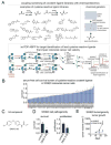
Figure 1
Figure 1. Coupling Screening of Cysteine-Reactive Covalent Ligands with isoTOP-ABPP to Identify Anti-Cancer Compounds and Druggable Hotspots for Colorectal Cancer
(A) We screened a library of cysteine-reactive fragment-based covalent ligands in colorectal cancer cells to identify compounds that impair colorectal cancer pathogenicity and used isoTOP-ABPP to identify the druggable hotspots targeted by hits. (B) We screened a cysteine-reactive fragment library consisting of acrylamides and chloroacetamides in SW620 colorectal cancer cells (50 μM) to identify any leads that significantly impaired SW620 serum-free cell survival. (C, D) Shown is the structure of the lead covalent ligand DKM 3-30 (C) that significantly (p<0.05) impaired SW620 cell survival and proliferation (D). (E) SW620 tumor xenograft growth in immune-deficient SCID mice. Mice were subcutaneously injected with SW620 cells to initiate the tumor xenograft study and treatments of mice were initiated with vehicle or DKM 3-30 (50 mg/kg ip, once per day) ten days initiation of the xenograft study. Data in (B, D, E) are presented as mean ±sem, n=3-8/group. Significance expressed as *p<0.05 compared to vehicle-treated controls.
Figure 2
Figure 2. DKM 3-30 Targets C1101 on RTN4
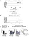
(A) IsoTOP-ABPP analysis of DKM 3-30 in SW620 colorectal cancer cells. SW620 proteomes were pre-treated with DMSO or DKM 3-30 (50 μM) prior to labeling proteomes with IAyne and isoTOP-ABPP analysis. Shown are mean light to heavy ratios for those probe-modified peptides identified in at least 2 out of 3 biological replicates. Also shown is gel-based ABPP validation of DKM 3-30 to RTN4. Pure human RTN4 protein was preincubated with DMSO or DKM for 30 min followed by IAyne labelling. Rhodamine-azide was conjugated by CuAAC and probe-labeled RTN4 was visualized by SDS/PAGE and in-gel fluorescence. (B) IsoTOP-ABPP analysis of cysteine-reactivity in pooled primary human colorectal tumors. Nine primary human colorectal tumors were pooled together and labeled with 100 or 10 μM of IAyne followed by subsequent isoTOP-ABPP analysis. Shown are ratios of heavy (100 μM) to light (10 μM) peptides. (C, D) Serum-free cell survival and proliferation (48 h) and tumor xenograft growth in SCID mice from transient siRNA or stable shRNA knockdown of RTN4 in SW620 cells. Expression was determined by qPCR. All data shown represents n=3-6/group. Data in (C, D) are presented as mean ± sem. Significance is expressed as *p<0.05 compared to vehicle-treated or si or shControls. Raw data for (A, B) can be found in Table S2.
Figure 3
Figure 3. DKM 3-30 disrupts the ER tubular network
(A) A schematic illustration depicts the proposed topology of Rtn4 and the position of C1101 modified by DKM 3-30 (red arrow). A homology model of human Rtn4 illustrates the membrane-associated portion (blue), the cytosolically accessible portion (green), and the position of C1101 (red). (B) U2OS cells expressing GFP-tagged Sec61β an ER marker, were treated with DKM 3-30 (50 μM) for 16 hr and the ER (green) and nucleus (blue) of fixed cells visualized by fluorescence microscopy. Scale bar = 10 μm. (C, D) U2OS cells expressing GFP-tagged Sec61β were treated with vehicle (DMSO) (C) or DKM 3-30 (50 μM) (D) and ER morphology visualized by time-lapse fluorescence microscopy. Time (min) is indicated on each panel. Bottom panels indicate boxed region. (E) U2OS cells were transiently transfected with control or RTN4 siRNA and expression determined by qPCR. Data are presented as mean ±sem, n=3. Significance is expressed as *p<0.05. (F) U2OS cells expressing GFP-tagged Sec61β were transfected with siRNAs as in panel (D) and the ER (green) and nucleus (blue) of fixed cells visualized by fluorescence microscopy. Scale bar = 10 μm.
Figure 4
Figure 4. DKM 3-30 disrupts nuclear envelope morphology during mitosis
(A-C) U2OS cells expressing GFP-tagged Sec61β were treated with vehicle (DMSO) or DKM 3-30 (50 μM) and ER morphology of mitotic cells visualized by time-lapse fluorescence microscopy. Time (min) is indicated on each panel. Panels (A, B) provide examples of mitotic cells. Enlarged images following mitosis show the nuclear envelope. White arrowheads indicate a GFP-Sec61β structure bisecting the nucleus of a cell incubated with DKM 3-30. (C) shows alterations in the nuclear envelope structure, followed by cell death at the 800 min time point. Bottom panels indicate boxed region.
Figure 5
Figure 5. DKM 3-30 and analogs
(A) Structures of DKM 3-30 and analogs. (B) Gel-based ABPP analysis showing competition side-by-side competition studies of DKM 3-30, YP 1-46, and AMR 1-125 against IA-rhodamine labelling of pure human RTN4. Shown are the 50 % inhibitory concentration (IC50) values for each compound. (C) Serum-free cell survival of U2OS (48 h) or SW620 (24 h) cells treated with DMSO vehicle or each compound (50 μM). Data in (C) are presented as mean ± sem. Significance is expressed as *p<0.001 compared to vehicle-treated controls.

References

    1. Gangadhar NM, Stockwell BR. Curr Opin Chem Biol. 2007;11:83–87. - PMC - PubMed
    1. Smukste I, Stockwell BR. Annu Rev Genomics Hum Genet. 2005;6:261–286. - PubMed
    1. Weerapana E, Wang C, Simon GM, Richter F, Khare S, Dillon MBD, Bachovchin DA, Mowen K, Baker D, Cravatt BF. Nature. 2010;468:790–795. - PMC - PubMed
    1. Backus KM, Correia BE, Lum KM, Forli S, Horning BD, González-Páez GE, Chatterjee S, Lanning BR, Teijaro JR, Olson AJ, Wolan DW, Cravatt BF. Nature. 2016;534:570–574. - PMC - PubMed
    1. Wang C, Weerapana E, Blewett MM, Cravatt BF. Nat Methods. 2014;11:79–85. - PMC - PubMed

MeSH terms