. 2017 May 23;11(5):4387-4394.
doi: 10.1021/acsnano.6b07760. Epub 2017 Mar 29.

Label-Free and Real-Time Detection of Protein Ubiquitination with a Biological Nanopore

Affiliations

Label-Free and Real-Time Detection of Protein Ubiquitination with a Biological Nanopore

Carsten Wloka et al. ACS Nano. .

Abstract

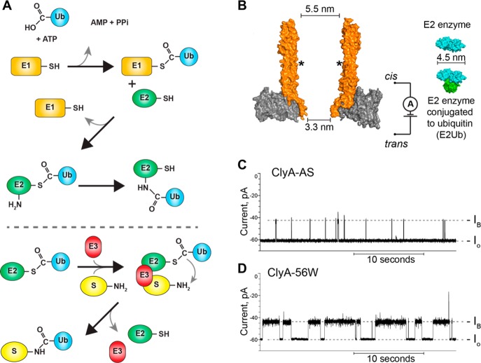
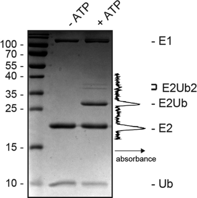
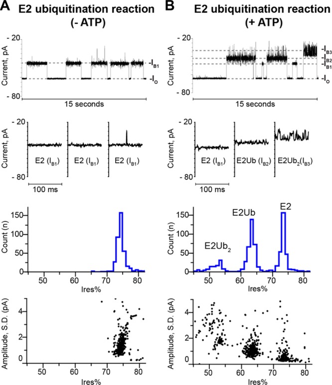
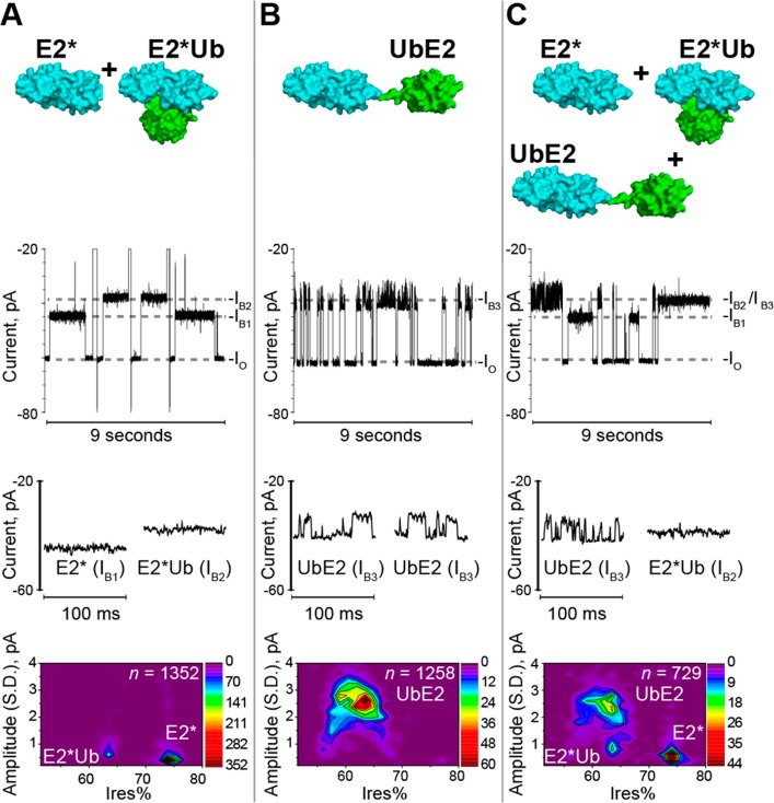
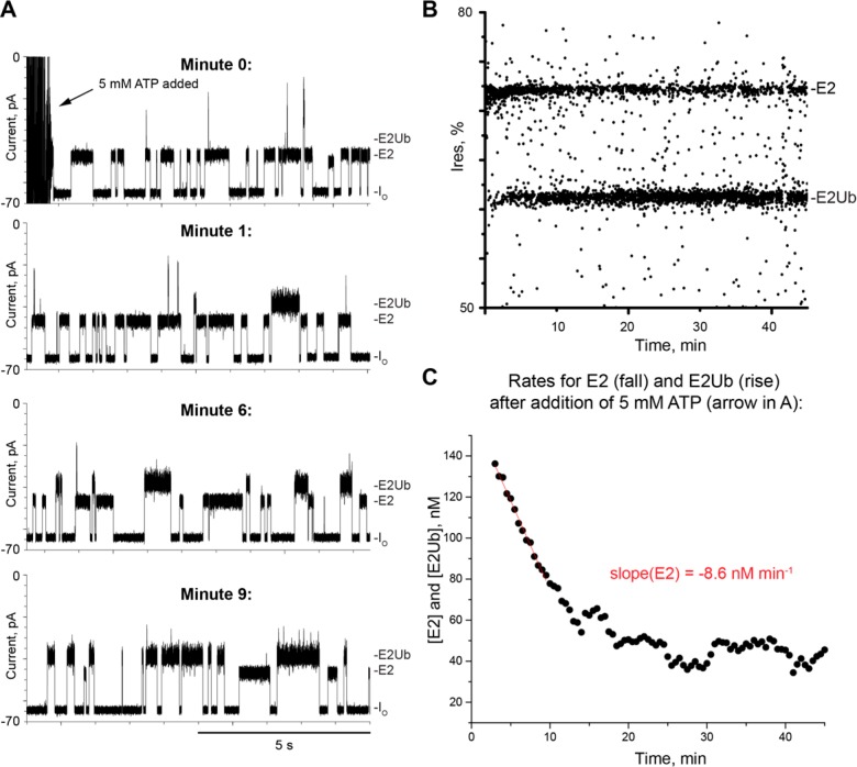
The covalent addition of ubiquitin to target proteins is a key post-translational modification that is linked to a myriad of biological processes. Here, we report a fast, single-molecule, and label-free method to probe the ubiquitination of proteins employing an engineered Cytolysin A (ClyA) nanopore. We show that ionic currents can be used to recognize mono- and polyubiquitinated forms of native proteins under physiological conditions. Using defined conjugates, we also show that isomeric monoubiquitinated proteins can be discriminated. The nanopore approach allows following the ubiquitination reaction in real time, which will accelerate the understanding of fundamental mechanisms linked to protein ubiquitination.

Keywords: nanopore; nanotechnology; protein modifications; single-molecule kinetics; ubiquitin.


Conflict of interest statement

The authors declare no competing financial interest.

Figures

Figure 1
Figure 1
(A) Scheme of the ubiquitin cascade. Ubiquitin activating enzyme (E1) activates Ub through hydrolysis of ATP. Next, the E1 transfers Ub to the active site cysteine of a ubiquitin-conjugating enzyme (E2). E2 can transfer Ub further to one of its lysine residues or, with the help of a ubiquitin ligase (E3), ubiquitinate substrates (S). The part of the cascade above the dashed line we exploited to create E2 and its ubiquitinated form. (B) Left: ClyA nanopore (orange) from Salmonella typhi(47) lodged inside a lipid bilayer (gray) composed of 1,2-dioleoyl-sn-glycero-3-phosphocholine. Asterisks mark the approximate position of the glutamine to tryptophan substitution (Q56W). On the right are surface representations of the E2 Ub-conjugating enzyme Ubc4 (E2, cyan, PDB: 1QCQ) of Saccharomyces cerevisiae shown with and without Ub (green, PDB: 3CMM). The E2Ub conjugate models were constructed with PyMOL. The larger opening of ClyA is facing the cis compartment, whereas trans denotes the location of the “working” electrode. (C) Representative trace obtained with ClyA-AS after addition of the E2 enzyme (50 nM) added to the cis side of the nanopore. (D) E2 (50 nM) blockades elicited to ClyA-56W. IO denotes the unobstructed open pore current; IB is the blocked pore current as E2 dwells inside ClyA. Data in (C) and (D) were collected in 150 mM NaCl, 50 mM TrisHCl, pH 7.5 at −35 mV potential applied to the trans electrode and recorded using a 2 kHz low-pass Bessel filter with a 10 kHz sampling rate. Traces were postacquisition digitally filtered with a Gaussian 500 Hz low-pass filter.
Figure 2
Figure 2
Coomassie-stained 15% SDS-PAGE gel of an E2 ubiquitination reaction with and without ATP. Protein marker (kDa) is shown on the left; the middle lane shows a ubiquitination reaction lacking ATP and on the right is a ubiquitination reaction shown with 5 mM ATP present. Both reactions were incubated for 2.5 h at 35 °C with shaking in buffer containing 75 mM NaCl, 25 mM TrisHCl, pH 8, 5 mM MgCl2, recombinant Ub (12 μM), activating enzyme (E1, 0.4 μM), and E2 (6 μM). Depicted on the right of the gel is the intensity profile of the in vitro reaction ranging from beneath E2 to above E2Ub2 with arbitrary absorbance units as determined with ImageJ (NIH).
Figure 3
Figure 3
Detection of protein ubiquitination with ClyA nanopores. (A) From top to bottom: Representative trace (IO = open pore and IB1 = blockade level, both at −35 mV), zoom-ins, histogram, and residual current percent (Ires%) versus the standard deviation (SD) of individual current blockades elicited by E2 proteins. The blockades appeared after adding a 1:100 dilution (final) of the in vitro ubiquitination reaction described in Figure 2 (not containing ATP) to the cis side of the nanopore. (B) Same as in (A), but the ubiquitination reaction contained ATP. Note that Ub and E1 do not elicit blockades (Figure S1). Buffer used in all electrophysiological experiments: 150 mM NaCl, 50 mM TrisHCl, pH 7.5. Data were collected at −35 mV and recorded using a 2 kHz low-pass Bessel filter with a 10 kHz sampling rate. Traces were postacquisition digitally filtered with a Gaussian 500 Hz low-pass filter.
Figure 4
Figure 4
Discrimination of isomeric E2Ub conjugates. (A) From top to bottom: Surface representation of the E2 mutant (C116K, E2*) and E2* conjugated to Ub at its active site (E2*Ub, constructed with PyMOL) with below given a continuous representative trace of the in vitro ubiquitination reaction of E2* analyzed with ClyA nanopores (IO = open pore, IB1 = blockade level elicited by E2, IB2 = E2*Ub); zoom-ins for blockades elicited by E2 and E2Ub; contour plot of all obtained blockades of an experiment. The same in vitro ubiquitination reaction was run on a Coomassie gel (lane 4, Figure S1A); a 1:50 dilution, or 120 nM E2*/E2*Ub, was used in experiments with the nanopore). (B) From top to bottom: Surface representation of N-terminally fused E2Ub protein (UbE2), constructed with PyMOL, and below a representative trace of 200 nM purified UbE2 analyzed with ClyA nanopores (IB3 = blockade level elicited by UbE2); zoom-ins provided for two blockades of UbE2; and blockades of an experiment depicted as a contour plot. (C) From top to bottom: Surface representation of E2*, E2*Ub, and UbE2; the proteins analyzed here in a mixture. Below are the same plots as in A and B shown but with 100 nM UbE2 and 1:50 dilution of an in vitro reaction (thus about 120 nM of E2* or E2*Ub) added to the cis side. Buffer used in all experiments: 150 mM NaCl, 50 mM TrisHCl, pH 7.5. Data were recorded using a 2 kHz low-pass Bessel filter with a 10 kHz sampling rate. All traces shown were postacquisition digitally filtered with a 500 Hz Gaussian low-pass filter.
Figure 5
Figure 5
Real-time observation of protein-ubiquitination with the ClyA nanopore. (A) Representative traces from a real-time ubiquitination reaction further analyzed in (B) and (C). E1 (26.6 nM), E2 (160 nM), Ub (320 nM), and MgCl2 (5 mM) are present in the cis buffer (150 mM NaCl, 50 mM TrisHCl, pH 7.5, ∼23 °C). Blockades corresponding to E2Ub appear within 1 min and become the dominant type of events within 10 min. (B) All blockades individually represented as dots versus time. (C) Determination of the rate of E2 disappearance. The concentration of E2 was measured from the frequencies of current blockades as described in the Methods section. Linear fits (Origin, OriginLab, thin line) were used to determine the initial rate of the reaction. Data were collected at −35 mV using a 2 kHz low-pass Bessel filter with a 10 kHz sampling rate.

Similar articles

Cited by

References

    1. Hershko A.; Heller H.; Elias S.; Ciechanover A. Components of Ubiquitin-Protein Ligase System. Resolution, Affinity Purification, and Role in Protein Breakdown. J. Biol. Chem. 1983, 258, 8206–8214. - PubMed
    1. Komander D.; Rape M. The Ubiquitin Code. Annu. Rev. Biochem. 2012, 81, 203–229. 10.1146/annurev-biochem-060310-170328. - DOI - PubMed
    1. Kandimalla R. J.; Anand R.; Veeramanikandan R.; Wani W. Y.; Prabhakar S.; Grover V. K.; Bharadwaj N.; Jain K.; Gill K. D. CSF Ubiquitin as a Specific Biomarker in Alzheimer’s Disease. Curr. Alzheimer Res. 2014, 11, 340–348. 10.2174/1567205011666140331161027. - DOI - PubMed
    1. Popovic D.; Vucic D.; Dikic I. Ubiquitination in Disease Pathogenesis and Treatment. Nat. Med. 2014, 20, 1242–1253. 10.1038/nm.3739. - DOI - PubMed
    1. Meier F.; Abeywardana T.; Dhall A.; Marotta N. P.; Varkey J.; Langen R.; Chatterjee C.; Pratt M. R. Semisynthetic, Site-Specific Ubiquitin Modification of Alpha-Synuclein Reveals Differential Effects on Aggregation. J. Am. Chem. Soc. 2012, 134, 5468–5471. 10.1021/ja300094r. - DOI - PMC - PubMed

Publication types