

Peliosis hepatis: 2 case reports of a rare liver disorder and its differential diagnosis
- PMID: 28353584
- PMCID: PMC5380268
- DOI: 10.1097/MD.0000000000006471
Peliosis hepatis: 2 case reports of a rare liver disorder and its differential diagnosis
Abstract
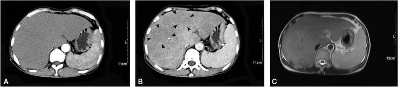
Rationale: Peliosis hepatis (PH) is a rare tumor-like liver lesion composed of multiple blood-filled cavities within the liver parenchyma. It is hard to differentiate PH from other liver lesions by imaging, such as carcinoma, metastases, or abscess.
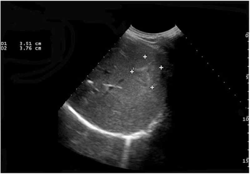
Patient concerns: Here, we reported 2 cases that presented with liver lesions under ultrasound and computed tomography (CT) scanning, without any history of liver diseases or drug usage traced back.
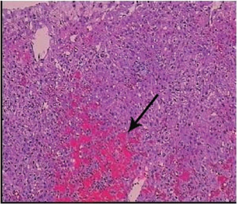
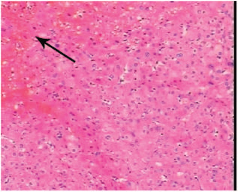
Diagnoses: Liver biopsy and laparoscopy were processed, and the lesions were eventually diagnosed as PH by histopathology, which microscopically presented with multiple sinusoidal dilatations with blood-filled cystic spaces.
Interventions: After the liver biopsy or laparoscopy, the patients were discharged and followed up in the clinic.
Outcomes: Both patients were followed up for at least 1 year with good recovery.
Lessons: PH should always be recognized in the differentiation of liver lesions, particularly indistinctive lesion(s) without any history of liver-related diseases.
Conflict of interest statement
The authors have declared that no conflict of interest exists.
Figures
References
-
- Terlizzi JP, Azizi R, Chow MD, et al. Peliosis hepatis in a child with myotubular myopathy: successful treatment using hepatic artery embolization. J Pediatr Surg 2013;48:e9–12. - PubMed
-
- Iannaccone R, Federle MP, Brancatelli G, et al. Peliosis hepatis: spectrum of imaging findings. AJR Am J Roentgenol 2006;187:W43–52. - PubMed
Publication types
MeSH terms
LinkOut - more resources
Full Text Sources
Other Literature Sources
