. 2017 Mar 29;9(4):338.
doi: 10.3390/nu9040338.

Vitamin D Insufficiency Exacerbates Adipose Tissue Macrophage Infiltration and Decreases AMPK/SIRT1 Activity in Obese Rats

Affiliations

Vitamin D Insufficiency Exacerbates Adipose Tissue Macrophage Infiltration and Decreases AMPK/SIRT1 Activity in Obese Rats

Eugene Chang et al. Nutrients. .

Abstract

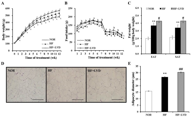
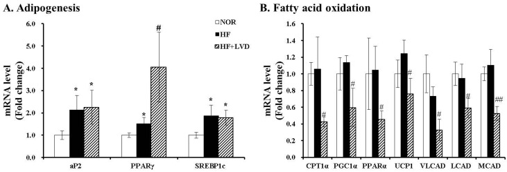
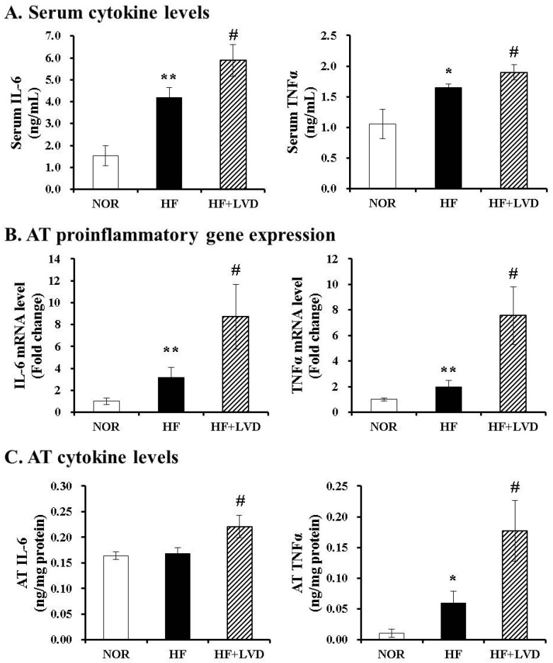
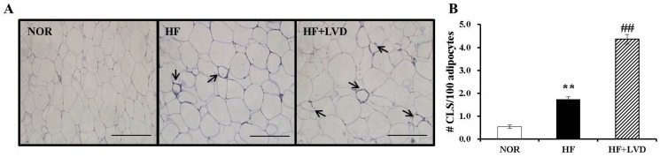
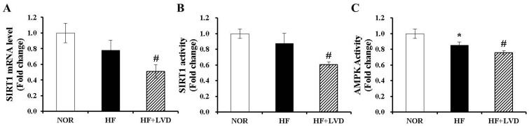
Obesity is recognized as a state of chronic low-grade systemic inflammation due to adipose tissue macrophage infiltration and production of proinflammatory adipokines. Decreased vitamin D status is associated with obesity. The specific aim of the present study is to investigate the effects of vitamin D on obesity-induced adipose tissue inflammation. Male Sprague-Dawley rats were randomized and fed a normal diet (NOR, 1000 IU vitamin D/kg diet), a 45% high-fat diet (HF, 1000 IU vitamin D/kg diet), or a 45% high-fat diet containing 25 IU vitamin D/kg diet (HF+LVD) for 12 weeks. The vitamin D-insufficient diet (HF+LVD) led to vitamin D inadequacy as determined by serum 25(OH)D level, 68.56 ± 7.97 nmol/L. The HF+LVD group exacerbated HF-increased adipocyte size, adipogenic gene expression of PPARγ, adipose tissue macrophage recruitment, and proinflammatory cytokine IL-6 and TNFα levels in epididymal white adipose tissue. In addition, vitamin D insufficiency significantly decreased mRNA levels of β-oxidation-related genes such as CPT1α, PGC1α, PPARα, VLCAD, LCAD, MCAD, and UCP1. Moreover, significant decrements of SIRT1 and AMPK activity were noted in obese rats fed with a vitamin D-insufficient diet. The observed deleterious effects of vitamin D insufficiency on adipose tissue expansion, immune cell infiltration and inflammatory status suggest vitamin D plays a beneficial role in adipocyte metabolic metabolism and obesity progression. SIRT1 and AMPK activity may play a role in the mechanism of vitamin D action.

Keywords: adenosine monophosphate-activated protein kinase (AMPK); adipose tissue macrophage infiltration; obesity; sirtulin 1 (SIRT1); vitamin D.


Conflict of interest statement

The authors declare no conflict of interest.

Figures

Figure 1
Figure 1
Effects of vitamin D-insufficient diet on body weight gain and fat weight in obese rats. Body weight gain (A), food intake (B) and weight of epididymal adipose tissue (EAT) and subcutaneous adipose tissue (SAT) (C). Representative hematoxylin and eosin stained EAT section (scale bar, 50 μm; magnification, 40×) (D). Average adipocyte diameter of adipocytes within adipose tissue (E) and data is expressed as the mean ± SEM. Bars with different letters (a, b, c) show significant difference (p < 0.05). * p < 0.05; ** p < 0.01 compared to NOR. # p < 0.05; ## p < 0.01 compared to HF. NOR, 10% fat diet with 1000 IU vitamin D (n = 9); HF, 45% fat diet with 1000 IU vitamin D (n = 7); HF+LVD, 45% fat diet containing 25 IU vitamin D (n = 7).
Figure 2
Figure 2
Influence of vitamin D insufficiency on gene expression involved in adipogenesis (A) and fatty acid oxidation (B). mRNA levels were determined by RT-PCR and normalized for all samples to β-actin. The results are expressed as the fold change compared to NOR. The value of each bar represents the mean ± SEM. * p < 0.05; ** p < 0.01 compared to NOR. # p < 0.05; ## p < 0.01 compared to HF. NOR, 10% fat diet with 1000 IU vitamin D (n = 9); HF, 45% fat diet with 1000 IU vitamin D (n = 7); HF+LVD, 45% fat diet containing 25 IU vitamin D (n = 7).
Figure 3
Figure 3
Vitamin D insufficiency increases local and systemic proinflammatory cytokine level. Serum levels of IL-6 and TNFα were expressed as ng/mL (A); Gene expression was measured, normalized for all samples to β-actin and expressed as the fold change compared to NOR (B); Measurements of IL-6 and TNFα in adipose tissue (C) were normalized to their respective protein concentrations and expressed as ng/mg protein. Values are expressed as the mean ± SEM. * p < 0.05; ** p < 0.01 compared to NOR. # p < 0.05 compared to HF. NOR, 10% fat diet with 1000 IU vitamin D (n = 9); HF, 45% fat diet with 1000 IU vitamin D (n = 7); HF+LVD, 45% fat diet containing 25 IU vitamin D (n = 7).
Figure 4
Figure 4
Vitamin D insufficiency increases macrophage infiltration in adipose tissue. (A) Macrophage marker, F4/80 immunohistochemistry of epididymal adipose tissue (scale bar, 50 μm; magnification, 40×); the black arrows indicate a CLS; (B) Number of CLS was quantified from multiple histologic sections and expressed as the mean ± SEM. ** p < 0.01 compared to NOR. ## p < 0.01 compared to HF. NOR, 10% fat diet with 1000 IU vitamin D (n = 9); HF, 45% fat diet with 1000 IU vitamin D (n = 7); HF+LVD, 45% fat diet containing 25 IU vitamin D (n = 7).
Figure 5
Figure 5
Vitamin D insufficiency decreases mRNA expression and activity of SIRT1 and AMPK activity in adipose tissue. SIRT1 mRNA levels were measured by quantitative RT-PCR and normalized to β-actin (A); SIRT1 activity was analyzed by a fluorometric SIRT1 activity assay kit (B); AMPK activity was measured using an AMPK kinase kit, normalized to their relative protein contents and expressed as the fold change compared to NOR (C). The value of each bar represents the mean ± SEM. * p < 0.05 compared to NOR. # p < 0.05 compared to HF. NOR, 10% fat diet with 1000 IU vitamin D (n = 9); HF, 45% fat diet with 1000 IU vitamin D (n = 7); HF+LVD, 45% fat diet containing 25 IU vitamin D (n = 7).

Similar articles

Cited by

References

    1. Spalding K.L., Arner E., Westermark P.O., Bernard S., Buchholz B.A., Bergmann O., Blomqvist L., Hoffstedt J., Naslund E., Britton T., et al. Dynamics of fat cell turnover in humans. Nature. 2008;453:783–787. doi: 10.1038/nature06902. - DOI - PubMed
    1. Arner P., Spalding K.L. Fat cell turnover in humans. Biochem. Biophys. Res. Commun. 2010;396:101–104. doi: 10.1016/j.bbrc.2010.02.165. - DOI - PubMed
    1. Weisberg S.P., McCann D., Desai M., Rosenbaum M., Leibel R.L., Ferrante A.W., Jr. Obesity is associated with macrophage accumulation in adipose tissue. J. Clin. Investig. 2003;112:1796–1808. doi: 10.1172/JCI200319246. - DOI - PMC - PubMed
    1. Xu H., Barnes G.T., Yang Q., Tan G., Yang D., Chou C.J., Sole J., Nichols A., Ross J.S., Tartaglia L.A., et al. Chronic inflammation in fat plays a crucial role in the development of obesity-related insulin resistance. J. Clin. Investig. 2003;112:1821–1830. doi: 10.1172/JCI200319451. - DOI - PMC - PubMed
    1. Strissel K.J., Stancheva Z., Miyoshi H., Perfield J.W., II, DeFuria J., Jick Z., Greenberg A.S., Obin M.S. Adipocyte death, adipose tissue remodeling, and obesity complications. Diabetes. 2007;56:2910–2918. doi: 10.2337/db07-0767. - DOI - PubMed

MeSH terms