. 2017 Sep;54(5):855-869.
doi: 10.1177/0300985817698210. Epub 2017 Mar 29.

Pathology of Aging in NOD scid gamma Female Mice

Affiliations

Pathology of Aging in NOD scid gamma Female Mice

Sara F Santagostino et al. Vet Pathol. 2017 Sep.

Abstract

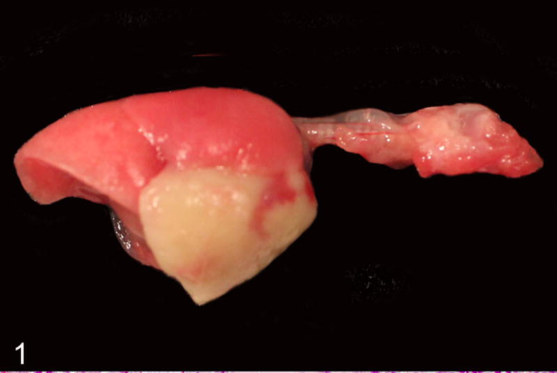
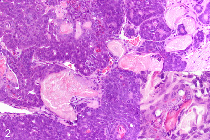
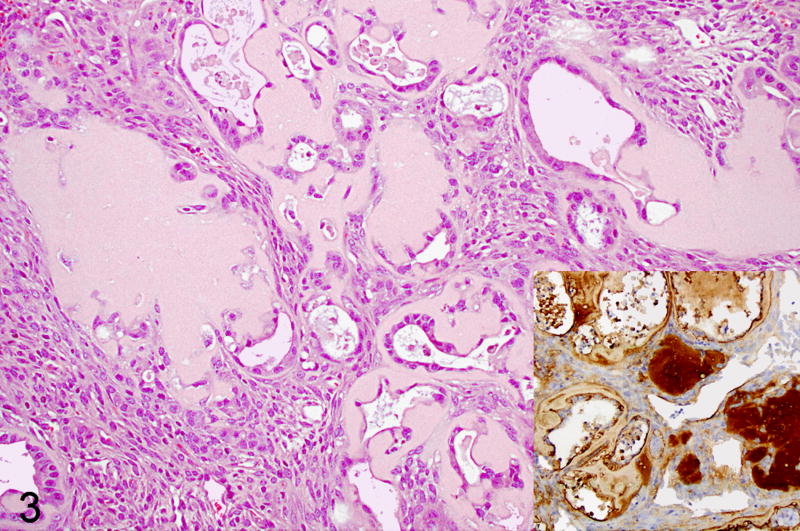
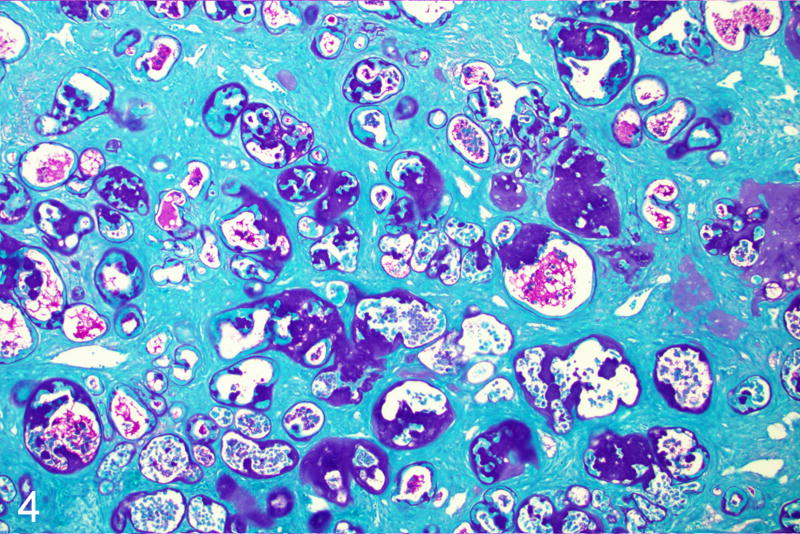
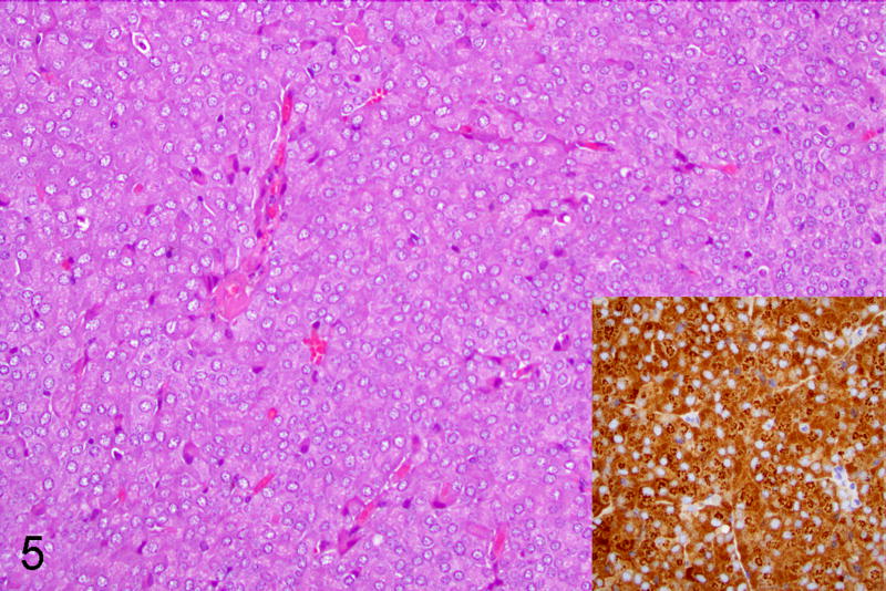
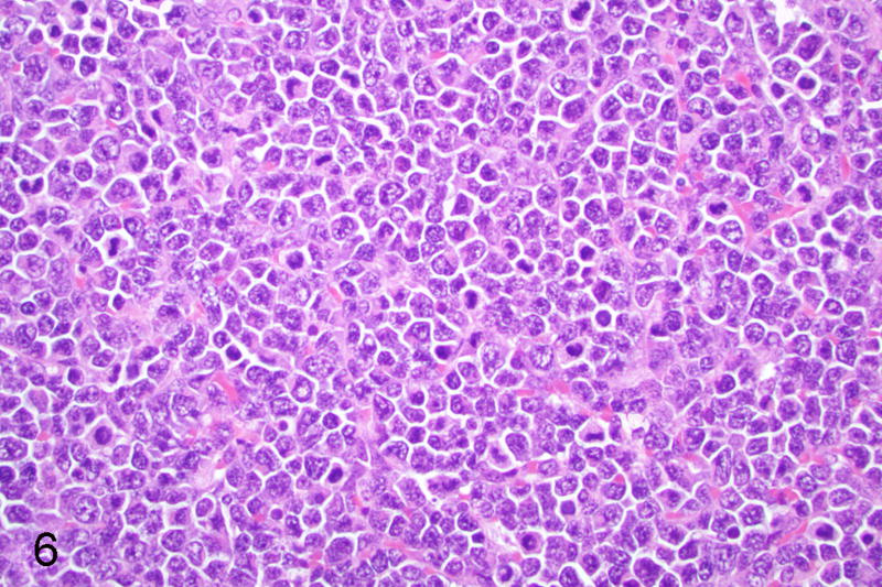
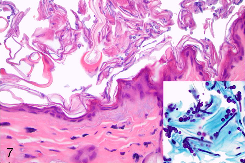
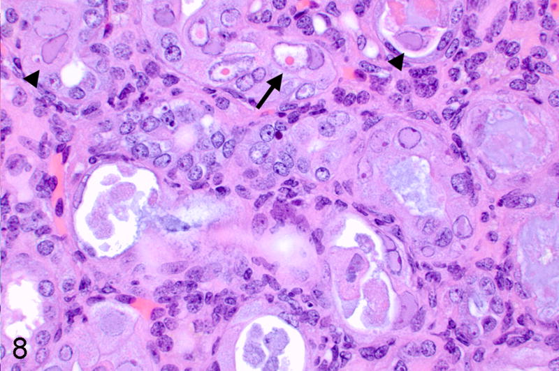
In the past decade, NOD.Cg- Prkdcscid Il2rgtm1Wjl/SzJ (NSG, NOD scid gamma) mice have become a model of choice in several areas of biomedical research; however, comprehensive data on their spontaneous age-related pathology are not currently available in the literature. The prevalence of spontaneous morbidity affecting aged NSG female breeders enrolled in a parasitology study was documented with classification of neoplastic and non-neoplastic (inflammatory, metabolic, degenerative) lesions. Malignant mammary neoplasms were most commonly diagnosed, often accompanied by pulmonary metastases, while a low frequency of lymphoma and histiocytic sarcoma was documented. The major inflammatory conditions were suppurative pleuropneumonia and bronchopneumonia with abscess formation, from which Pasteurella pneumotropica was commonly isolated, followed by otitis media. Both inflammatory and degenerative lesions of the genital tract were identified, along with neoplasms such as endometrial yolk sac carcinomas and granulosa cell tumors. Novel conditions identified included renal tubular degeneration and necrosis associated with 2 concurrent types of intranuclear inclusions, focal or multifocal hyperostosis of the skull, and neuroendocrine tumors of the mesometrium. The majority of degenerative lesions that affected the genital tract, endocrine, and skeletal systems did not represent the actual underlying cause of death but rather were considered incidental findings. This study indicates that both inflammatory and neoplastic conditions contribute to morbidity and mortality in experimentally manipulated aged female NSG mice.

Keywords: aging; background lesions; female; interleukin-2 receptor gamma chain knockout; mouse; non-obese diabetic; pathology; severe combined immunodeficiency.


Conflict of interest statement

Declaration of Conflicting Interests

The authors declared no potential conflicts of interest with respect to the research, authorship, and/or publication of this article.

Figures

Figure 1
Figure 1
Abscesses, lung, mouse. The right cranial lung lobe is obscured by multiple coalescing, variably sized, pale tan to yellow abscesses caused by Pasteurella pneumotropica infection.
Figure 2
Figure 2
Adenosquamous carcinoma, mammary gland, mouse. The neoplasm is composed of microacinar epithelial structures intermingled with cavities lined by stratified squamous epithelium producing keratin. Inset: Scattered keratohyalin and trichohyalin granules are also present. Hematoxylin and eosin (HE).
Figure 3
Figure 3
Parietal yolk sac carcinoma, uterus, mouse. In the endometrium, acini lined by flattened epithelial cells are associated with an abundant extracellular eosinophilic hyaline substance. HE. Inset: This material is immunohistochemically positive for laminin.
Figure 4
Figure 4
Parietal yolk sac carcinoma, uterus, mouse. The hyaline substance is PAS-positive. Periodic acid-Shiff reaction (PAS).
Figure 5
Figure 5
Neuroendocrine carcinoma, mesometrium, mouse. The tumor is arranged in densely cellular packets of cells separated by delicate fibrovascular septa. HE. Inset: The neoplastic cells display strong cytoplasmic expression of chromogranin A, compatible with neuroendocrine differentiation. Immunohistochemistry for chromogranin A.
Figure 6
Figure 6
Precursor T-cell lymphoblastic lymphoma, mesenteric lymph node, mouse. The nodal parenchyma is obscured by dense sheets of intermediate-sized neoplastic lymphoid cells with scant cytoplasm, round nuclei with finely granular chromatin, and several mitotic Figures. HE.
Figure 7
Figure 7
Orthokeratotic hyperkeratosis with intralesional Candida spp., haired skin, mouse. Diffuse superficial accumulation of keratin flakes intermingled with yeasts, septate hyphae and occasional pseudohyphae, compatible with Candida spp. HE. Inset: The aforementioned fungal forms stain positive with periodic acid-Shiff reaction (PAS).
Figure 8
Figure 8
Intranuclear inclusion bodies of unknown etiology, kidney, mouse. Multiple tubular epithelial cells exhibit karyomegaly with large, 15- to 20-µm diameter, amphophilic intranuclear inclusions and margination of chromatin (arrowheads). Scattered small, 3- to 5-µm diameter, round bright eosinophilic intranuclear inclusions are also present (arrow). HE.
Figure 9
Figure 9
Intranuclear inclusion bodies of unknown etiology, renal tubular epithelium, mouse. The nuclei of karyomegalic cells consists of abundant electron-lucent floccular material. The remaining chromatin is peripheralized, and the nuclear membrane is intact. Inset: Rare nuclei display enlarged nucleoli that are expanded by a similar material (arrow). Scale bar: 5 µm. Transmission electron microscopy (TEM).
Figure 10
Figure 10
Proliferative hyperostosis, calvarium, mouse. There is diffuse formation of woven and lamellar bone along the calvarium, with intracranial extension and compression of the brain (arrow). HE. Inset: Despite the compression, the underlying cerebrum appears normal. HE.
Figure 11
Figure 11
Disc protrusion with leukomyelomalacia, spinal cord, mouse. A focal protrusion of the annulus fibrosus is evident at the level of the thoracic segment. HE. Inset: There is evidence of myelin sheath dilation (Wallerian degeneration) of the underlying spinal cord segment. HE.
Figure 12
Figure 12
Multifocal renal atrophy and interstitial fibrosis, kidney, mouse. The cortex and medulla display multifocal interstitial fibrosis, with tubular atrophy and loss, and cystic dilation of the urinary space. HE.

Comment in

Similar articles

Cited by

References

    1. Albassam MA, Courtney CL. Non-neoplastic and neoplastic lesions of the bone. In: Mohr U, Dungworth DL, Capen CC, Carlton WW, Sundberg JP, Ward JM, editors. Pathobiology of the Aging Mouse. Vol. 2. Washington, DC: ILSI Press; 1996. pp. 425–437.
    1. Barthold SW, Griffey SM, Percy DH. Mouse. In: Barthold SW, Griffey SM, Percy DH, editors. Pathology of laboratory rodents and rabbits. 4. Ames, IA: Wiley Blackwell; 2016. pp. 1–118.
    1. Bastidas RJ, Heitman J. Trimorphic stepping stones pave the way to fungal virulence. Proc Natl Acad Sci. USA. 2009;106(2):351–352. - PMC - PubMed
    1. Baze WB, Steinbach TJ, Fleetwood ML, Blanchard TW, Barnhart KF, McArthur MJ. Karyomegaly and intranuclear inclusions in the renal tubules of sentinel ICR Mice (Mus musculus) Comp Med. 2006;56(5):435–438. - PubMed
    1. Boivin GP, Washington K, Yang K, et al. Pathology of mouse models of intestinal cancer: consensus report and recommendations. Gastroenterology. 2003;124(3):762–777. - PubMed

Publication types

LinkOut - more resources