

Value of whole-body magnetic resonance imaging for screening multifocal osteonecrosis in patients with polymyositis/dermatomyositis
- PMID: 28355130
- PMCID: PMC5605105
- DOI: 10.1259/bjr.20160780
Value of whole-body magnetic resonance imaging for screening multifocal osteonecrosis in patients with polymyositis/dermatomyositis
Abstract
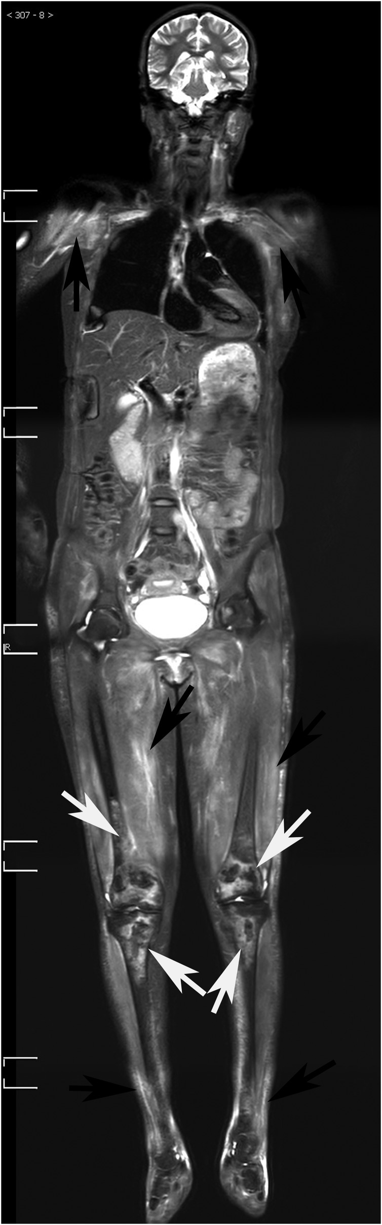
Objective: To assess the value of coronal short-tau inversion recovery whole-body MRI (STIR-WBMRI) for screening osteonecrosis in patients with polymyositis (PM)/dermatomyositis (DM).
Methods: The imaging and medical records of 129 patients with PM/DM who met the Bohan and Peter diagnostic criteria were retrospectively analyzed. STIR-WBMRI was performed in all patients. 18 patients had follow-up STIR-WBMRI. 12 patients underwent regional knee and/or hip MRI while 25 patients underwent radiography of the lower extremities.
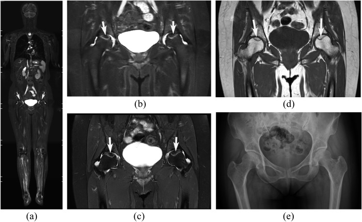
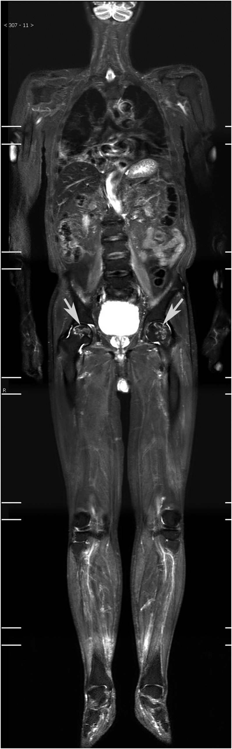
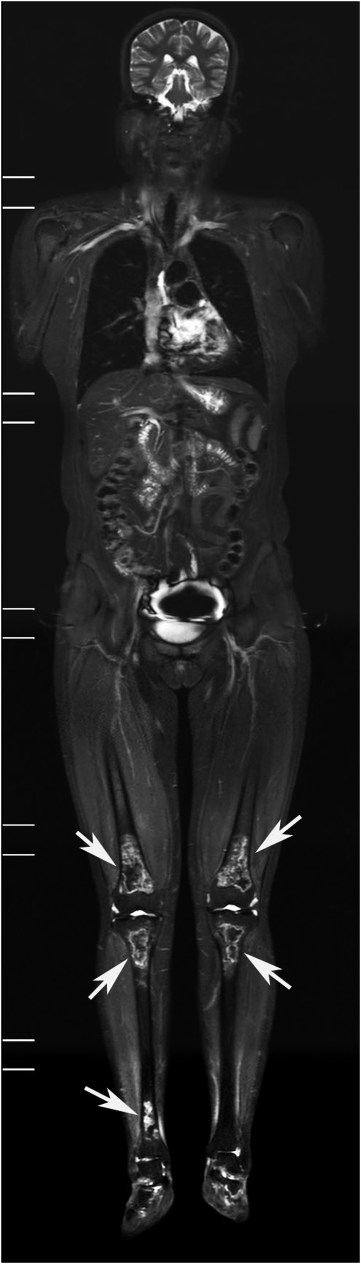
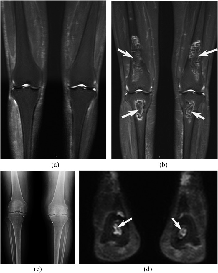
Results: STIR-WBMRI detected osteonecrosis in 15 (11.6%) patients. 38 joints were affected (mean, 2.5 per patient; range, 1-5 joints). Of the 38 joints affected by osteonecrosis, 33 had no clinical symptoms. Among the 12 patients who underwent regional MRI, STIR-WBMRI detected all 10 osteonecrotic sites seen on the regional MRI. The location, shape and size of the osteonecrotic lesions revealed on regional MRI were in accordance with those displayed on STIR-WBMRI. Of the 15 patients with osteonecrosis, 6 performed routine radiography of the affected joints and revealed no osteonecrotic lesions. Follow-up WBMRI detected new osteonecrosis in two patients whose first WBMRI revealed that there was no osteonecrosis in any skeleton.
Conclusion: In addition to displaying muscle inflammation, STIR-WBMRI can efficiently detect early multifocal osteonecrosis in the whole bodies of patients with PM/DM. Advances in knowledge: In patients with PM/DM, WBMRI which takes 12-15 min can display muscular involvement and detect early multisite osteonecrosis in the whole body at the same time. Osteonecrotic lesions revealed by WBMRI are in accordance with those displayed on regional WBMRI.
Figures
Comment in
-
Whole-body MRI in the early detection of multifocal osteonecrosis.Br J Radiol. 2017 Aug;90(1077):20170240. doi: 10.1259/bjr.20170280. Epub 2017 Jul 14. Br J Radiol. 2017. PMID: 28707528 Free PMC article. No abstract available.
References
-
- Dalakas MC, Hohlfeld R. Polymyositis and dermatomyositis. Lancet 2003; 362: 971–82. doi: https://doi.org/10.1016/s0140-6736(03)14368-1 - DOI - PubMed
-
- Del Grande F, Carrino JA, Del Grande M, Mammen AL, Christopher Stine L. Magnetic resonance imaging of inflammatory myopathies. Top Magn Reson Imaging 2011; 22: 39–43. doi: https://doi.org/10.1097/rmr.0b013e31825b2c35 - DOI - PubMed
-
- Tomasová Studynková J, Charvát F, Jarosová K, Vencovsky J. The role of MRI in the assessment of polymyositis and dermatomyositis. Rheumatology (Oxford) 2007; 46: 1174–9. doi: https://doi.org/10.1093/rheumatology/kem088 - DOI - PubMed
-
- Connor A, Stebbings S, Anne Hung N, Hammond-Tooke G, Meikle G, Highton J. STIR MRI to direct muscle biopsy in suspected idiopathic inflammatory myopathy. J Clin Rheumatol 2007; 13: 341–5. doi: https://doi.org/10.1097/rhu.0b013e31815dca0a - DOI - PubMed
-
- Cantwell C, Ryan M, O'Connell M, Cunningham P, Brennan D, Costigan D, et al. A comparison of inflammatory myopathies at whole-body turbo STIR MRI. Clin Radiol 2005; 60: 261–7. doi: https://doi.org/10.1016/j.crad.2004.06.027 - DOI - PubMed
MeSH terms
LinkOut - more resources
Full Text Sources
Other Literature Sources
Medical
Miscellaneous
