Observational Study
. 2017 Mar 29;12(3):e0174581.
doi: 10.1371/journal.pone.0174581. eCollection 2017.

SEARCH 8Es: A novel point of care ultrasound protocol for patients with chest pain, dyspnea or symptomatic hypotension in the emergency department

Affiliations
Observational Study

SEARCH 8Es: A novel point of care ultrasound protocol for patients with chest pain, dyspnea or symptomatic hypotension in the emergency department

Jung Hwan Ahn et al. PLoS One. .

Abstract

Objective: This study was conducted to evaluate a problem-oriented focused torso bedside ultrasound protocol termed "Sonographic Evaluation of Aetiology for Respiratory difficulty, Chest pain, and/or Hypotension" (SEARCH 8Es) for its ability to narrow differential diagnoses and increase physicians' diagnostic confidence, and its diagnostic accuracy, for patients presenting with dyspnea, chest pain, or symptomatic hypotension.

Methods: This single-center prospective observational study was conducted over 12 months in an emergency department and included 308 patients (184 men and 124 women; mean age, 67.7 ± 19.1 years) with emergent cardiopulmonary symptoms. The paired t-test was used to compare the number of differential diagnoses and physician's level of confidence before and after SEARCH 8Es. The overall accuracy of the SEARCH 8Es protocol in differentiating 13 diagnostic entities was evaluated based on concordance (kappa coefficient) with the diagnosis made by the inpatient specialists. Sensitivity, specificity, positive predictive value, and negative predictive value were calculated.

Results: SEARCH 8Es narrows the number of differential diagnoses (2.5 ± 1.5 vs. 1.4 ± 0.7; p < 0.001) and improves physicians' diagnostic confidence (2.8 ± 0.8 vs. 4.3 ± 0.9; p < 0.001) significantly. The overall kappa coefficient value was 0.870 (p < 0.001), with the overall sensitivity, specificity, positive predictive value, and negative predictive value at 90.9%, 99.0%, 89.7%, and 99.0%, respectively.

Conclusion: The SEARCH 8Es protocol helps emergency physicians to narrow the differential diagnoses, increase diagnostic confidence and provide accurate assessment of patients with dyspnea, chest pain, or symptomatic hypotension.


Conflict of interest statement

Competing Interests: The authors have declared that no competing interests exist.

Figures

Fig 1
Fig 1. The algorithm for SEARCH 8Es.
Profiles: A, lung sliding and A lines in both the lungs; A’, A profile without lung sliding and lung point; B, anterior-predominant bilateral B lines with lung sliding; B’, B profile without lung sliding; A/B, anterior-predominant B lines in one lung and predominant A lines in the other; C, anterior alveolar consolidation(s). Abbreviations: AAA, abdominal aortic aneurysm; ACS, acute coronary syndrome; AD, aortic dissection; ARDS, acute respiratory distress syndrome; BLUE, bedside lung ultrasound in emergency; COPD, chronic obstructive pulmonary disease; EF, ejection fraction; HF, heart failure; ILD, interstitial lung disease; IVC, inferior vena cava; LV, left ventricle; PLAPS, posterolateral alveolar/or pleural syndrome; RV, right ventricle; SEARCH, sonographic evaluation of aetiology for respiratory difficulty, chest pain, and/or hypotension; WMA, wall motion abnormality.
Fig 2
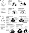
Fig 2. Schematic drawing of transducer, the image planes and key points in the SEARCH 8Es protocol.
The performance sequence of SEARCH 8Es was divided into two steps. (A) The first step of ‘SEARCH’ with the convex probe to search for the pneumothorax, interstitial syndrome (pulmonary edema, ARDS, interstitial disease), pneumonia, pleural effusion, peritoneal effusion, abdominal aortic aneurysm, aortic dissection, or a clue of hypovolemia. The convex probe is located longitudinally on the anterior chest and posterolateral chest to examine whether there are any findings that are suggestive of empty thorax, edematous or wet lung and free fluid above and below the diaphragm. In cases of intraperitoneal fluid, the probe is placed in the same manner as the conventional FAST. The convex probe is used to evaluate the lungs, the abdomen and to look at the IVC. The A profile is defined the conditions appeared lung sliding with A line in both lungs. The A’ profile is an A profile without lung sliding and lung point. The B profile is defined to anterior-predominant bilateral B lines associated with lung sliding. The B’ profile is a B profile without lung sliding. The A/B profile is defined to anterior predominant B lines on one lung and predominant A lines on the other. The C profile is defined to anterior alveolar consolidation(s). (B) The 2nd step of ‘SEARCH’ with the cardiac probe to search for pericardial effusion with or without tamponade physiology (diastolic right ventricle collapse), pulmonary embolism (right ventricle enlargement, paradoxical interventricular septal movement), left ventricle systolic dysfunction, acute myocardial infarction (left and right ventricular regional wall motion abnormality) and it’s mechanical complications (papillary muscular rupture, left ventricle wall rupture), and a clue of a hypovolemic or distributive shock (hypokinetic small-sized left ventricle or hyperkinetic normal-sized left ventricle). In case of chest pain, parasternal short axis view and suprasternal notch view was performed due to the evaluation of regional wall motion and thoracic aorta dissection or aneurysm. If the apical 4 chamber view is difficult due to poor window, a subxiphoid view is used. AAA, abdominal aortic aneurysm; AD, aortic dissection; ARDS, acute respiratory distress syndrome; BLUE, bedside lung ultrasound in emergency; EF, ejection fraction; E-FAST, extended focused assessment with sonography for trauma; FAST, focused assessment with sonography for trauma; IVC, inferior vena cava; LA, left atrium; LUQ, left upper quadrant; LV, left ventricle; PLAPS, posterolateral alveolar and/or pleural syndrome; RA, right atrium; RUQ, right upper quadrant; RV, right ventricle; SEARCH, sonographic evaluation of aetiology for respiratory difficulty, chest pain, and/or hypotension.

Similar articles

Cited by

References

    1. Lichtenstein DA, Meziere GA. Relevance of lung ultrasound in the diagnosis of acute respiratory failure: the BLUE protocol. Chest. 2008;134:117–125. Epub 2008/04/12. 10.1378/chest.07-2800 - DOI - PMC - PubMed
    1. Hernandez C, Shuler K, Hannan H, Sonyika C, Likourezos A, Marshall J. C.A.U.S.E.: cardiac arrest ultra-sound exam—a better approach to managing patients in primary non-arrhythmogenic cardiac arrest. Resuscitation. 2008;76:198–206. Epub 2007/09/08. 10.1016/j.resuscitation.2007.06.033 - DOI - PubMed
    1. Jones AE, Tayal VS, Sullivan DM, Kline JA. Randomized, controlled trial of immediate versus delayed goal-directed ultrasound to identify the cause of nontraumatic hypotension in emergency department patients. Crit Care Med. 2004;32:1703–1708. - PubMed
    1. Jones AE, Stiell IG, Nesbitt LP, Spaite DW, Hasan N, Watts BA, et al. Nontraumatic out-of-hospital hypotension predicts inhospital mortality. Ann Emerg Med. 2004;43:106–113. 10.1016/S0196064403009284 - DOI - PubMed
    1. Amsterdam EA, Kirk JD, Bluemke DA, Diercks D, Farkouh ME, Garvey JL, et al. Testing of low-risk patients presenting to the emergency department with chest pain: a scientific statement from the American Heart Association. Circulation. 2010;122:1756–1776. PubMed Central PMCID: PMC3044644. 10.1161/CIR.0b013e3181ec61df - DOI - PMC - PubMed

Publication types