The value of Gd-EOB-DTPA-enhanced MR imaging in characterizing cirrhotic nodules with atypical enhancement on Gd-DTPA-enhanced MR images
- PMID: 28355258
- PMCID: PMC5371364
- DOI: 10.1371/journal.pone.0174594
The value of Gd-EOB-DTPA-enhanced MR imaging in characterizing cirrhotic nodules with atypical enhancement on Gd-DTPA-enhanced MR images
Abstract
Purpose: To evaluate the utility of Gd-EOB-DTPA-enhanced magnetic resonance imaging (MRI) in characterizing atypically enhanced cirrhotic nodules detected on conventional Gd-DTPA-enhanced MR images.
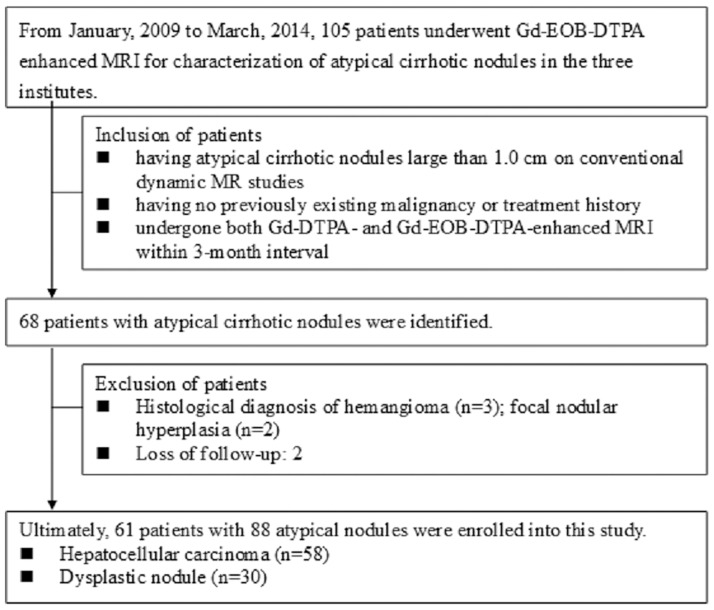
Materials and methods: We enrolled 61 consecutive patients with 88 atypical nodules seen on conventional Gd-DTPA-enhanced MR images who underwent Gd-EOB-DTPA-enhanced MRI within a 3-month period. Using a reference standard, we determined that 58 of the nodules were hepatocellular carcinoma (HCC) and 30 were dysplastic nodules (DNs). Tumor size, signal intensity on precontrast T1-weighted images (T1WI), T2-weighted images (T2WI) and diffusion-weighted images (DWI), and the enhancement patterns seen on dynamic phase and hepatocyte phase images were determined.
Results: There were significant differences between DNs and HCC in hyperintensity on T2WI, hypointensity on T1WI, hypervascularity on arterial phase images, typical HCC enhancement patterns on dynamic MR images, hypointensity on hepatocyte phase images, and hyperintensity on DWI. The sensitivity and specificity were 79.3% and 83.3% for T2WI, 50.0% and 80.0% for T1WI, 82.8% and 76.7% for DWI, 17.2% and 100% for dynamic MR imaging, 93.1% and 83.3% for hepatocyte phase imaging, and 46.8% and 100% when arterial hypervascularity was combined with hypointensity on hepatocyte-phase imaging.
Conclusion: Gd-EOB-DTPA-enhanced hepatocyte phase imaging is recommended for patients at high risk for HCC who present with atypical lesions on conventional Gd-DTPA-enhanced MR images.
Conflict of interest statement
Figures


Similar articles
-
The utility of gadoxetic acid-enhanced MR imaging to characterize atypical cirrhotic nodules detected on dynamic CT images.PLoS One. 2014 Oct 13;9(10):e107869. doi: 10.1371/journal.pone.0107869. eCollection 2014. PLoS One. 2014. PMID: 25310817 Free PMC article.
-
Detection and characterization of focal liver lesions: a Japanese phase III, multicenter comparison between gadoxetic acid disodium-enhanced magnetic resonance imaging and contrast-enhanced computed tomography predominantly in patients with hepatocellular carcinoma and chronic liver disease.Invest Radiol. 2010 Mar;45(3):133-41. doi: 10.1097/RLI.0b013e3181caea5b. Invest Radiol. 2010. PMID: 20098330 Clinical Trial.
-
Characterization of small (≤3 cm) hepatic lesions with atypical enhancement feature and hypointensity in hepatobiliary phase of gadoxetic acid-enhanced MRI in cirrhosis: A STARD-compliant article.Medicine (Baltimore). 2017 Jul;96(29):e7278. doi: 10.1097/MD.0000000000007278. Medicine (Baltimore). 2017. PMID: 28723741 Free PMC article.
-
Diagnosis of Hepatocellular Carcinoma Using Gd-EOB-DTPA MR Imaging.Magn Reson Med Sci. 2022 Mar 1;21(1):168-181. doi: 10.2463/mrms.rev.2021-0031. Epub 2021 Aug 21. Magn Reson Med Sci. 2022. PMID: 34421091 Free PMC article. Review.
-
An overview of hepatocellular carcinoma with atypical enhancement pattern: spectrum of magnetic resonance imaging findings with pathologic correlation.Radiol Oncol. 2021 Jan 29;55(2):130-143. doi: 10.2478/raon-2021-0004. Radiol Oncol. 2021. PMID: 33544992 Free PMC article. Review.
Cited by
-
Application of Gd-EOB-DTPA-enhanced magnetic resonance imaging (MRI) in hepatocellular carcinoma.World J Surg Oncol. 2020 Aug 22;18(1):219. doi: 10.1186/s12957-020-01996-4. World J Surg Oncol. 2020. PMID: 32828123 Free PMC article. Review.
-
IDEAL-IQ measurement can distinguish dysplastic nodule from early hepatocellular carcinoma: a case-control study.Quant Imaging Med Surg. 2024 Jun 1;14(6):3901-3913. doi: 10.21037/qims-23-1593. Epub 2024 Apr 22. Quant Imaging Med Surg. 2024. PMID: 38846285 Free PMC article.
-
Noninvasive imaging of hepatocellular carcinoma: From diagnosis to prognosis.World J Gastroenterol. 2018 Jun 14;24(22):2348-2362. doi: 10.3748/wjg.v24.i22.2348. World J Gastroenterol. 2018. PMID: 29904242 Free PMC article. Review.
-
Overall diagnostic accuracy of different MR imaging sequences for detection of dysplastic nodules: a systematic review and meta-analysis.Eur Radiol. 2022 Feb;32(2):1285-1296. doi: 10.1007/s00330-021-08022-5. Epub 2021 Aug 6. Eur Radiol. 2022. PMID: 34357448
-
Application and progress of the detection technologies in hepatocellular carcinoma.Genes Dis. 2022 Apr 22;10(5):1857-1869. doi: 10.1016/j.gendis.2022.04.003. eCollection 2023 Sep. Genes Dis. 2022. PMID: 37492708 Free PMC article. Review.
References
-
- Huppertz A, Balzer T, Blakeborough A, Breuer J, Giovagnoni A, Heinz-Peer G, et al. Improved detection of focal liver lesions at MR imaging: multicenter comparison of gadoxetic acid-enhanced MR images with intraoperative findings. Radiology. 2004;230(1):266–75. 10.1148/radiol.2301020269 - DOI - PubMed
MeSH terms
Substances
LinkOut - more resources
Full Text Sources
Other Literature Sources
Medical