. 2017 Mar 29;12(3):e0174591.
doi: 10.1371/journal.pone.0174591. eCollection 2017.

Tetraarsenic hexoxide induces G2/M arrest, apoptosis, and autophagy via PI3K/Akt suppression and p38 MAPK activation in SW620 human colon cancer cells

Affiliations

Tetraarsenic hexoxide induces G2/M arrest, apoptosis, and autophagy via PI3K/Akt suppression and p38 MAPK activation in SW620 human colon cancer cells

Arulkumar Nagappan et al. PLoS One. .

Abstract

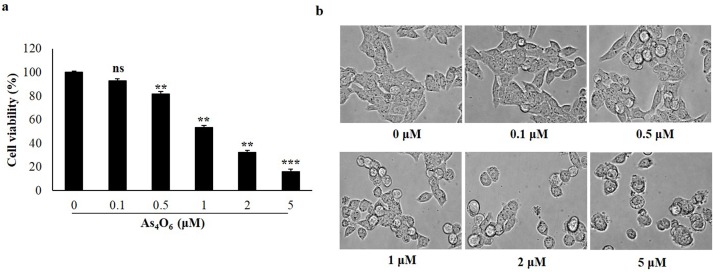
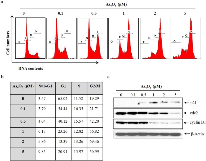
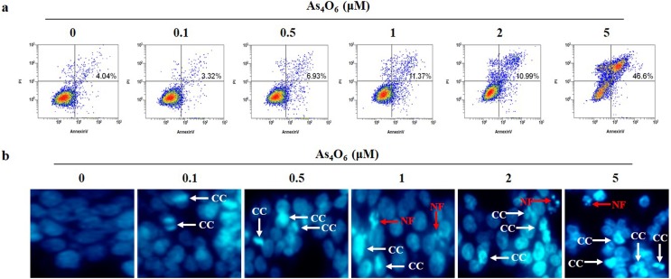
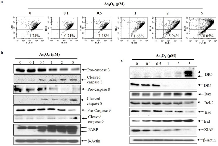
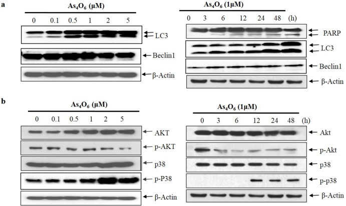
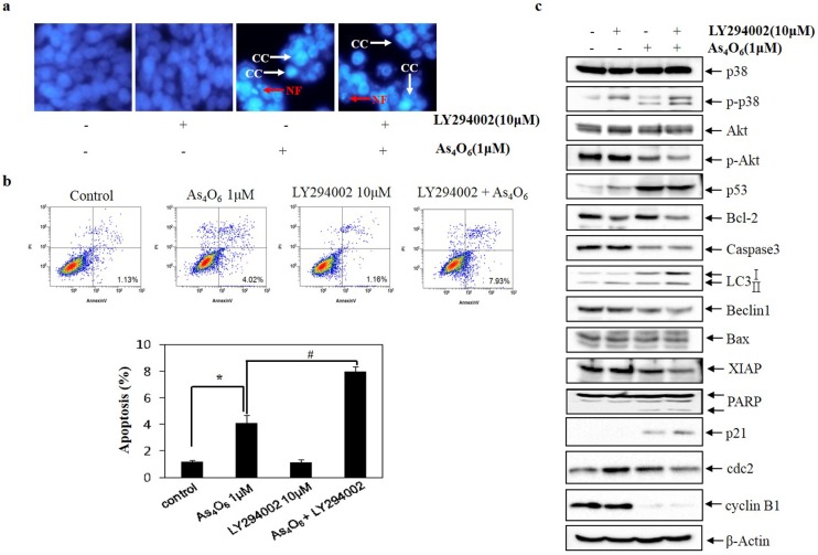
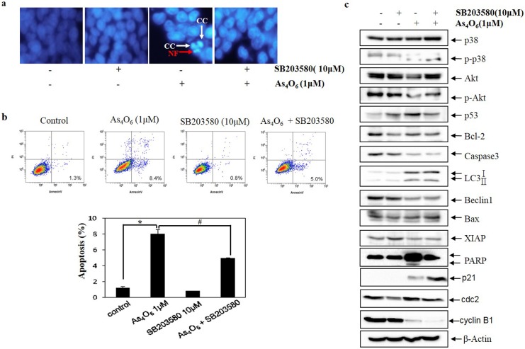
Tetraarsenic hexoxide (As4O6) has been used in Korean folk medicines for the treatment of cancer, however its anti-cancer mechanisms remain obscured. Here, this study investigated the anti-cancer effect of As4O6 on SW620 human colon cancer cells. As4O6 has showed a dose-dependent inhibition of SW620 cells proliferation. As4O6 significantly increased the sub-G1 and G2/M phase population, and Annexin V-positive cells in a dose-dependent manner. G2/M arrest was concomitant with augment of p21 and reduction in cyclin B1, cell division cycle 2 (cdc 2) expressions. Nuclear condensation, cleaved nuclei and poly (adenosine diphosphate‑ribose) polymerase (PARP) activation were also observed in As4O6-treated SW620 cells. As4O6 induced depolarization of mitochondrial membrane potential (MMP, ΔΨm) but not reactive oxygen species (ROS) generation. Further, As4O6 increased death receptor 5 (DR5), not DR4 and suppressed the B‑cell lymphoma‑2 (Bcl-2) and X-linked inhibitor of apoptosis protein (XIAP) family proteins. As4O6 increased the formation of AVOs (lysosomes and autophagolysosomes) and promoted the conversion of microtubule-associated protein 1A/1B-light chain 3 (LC3)-I to LC3-II in a dose- and time- dependent manner. Interestingly, a specific phosphoinositide 3-kinase (PI3K)/Akt inhibitor (LY294002) augmented the As4O6 induced cell death; whereas p38 mitogen-activated protein kinases (p38 MAPK) inhibitor (SB203580) abrogated the cell death. Thus, the present study provides the first evidence that As4O6 induced G2/M arrest, apoptosis and autophagic cell death through PI3K/Akt and p38 MAPK pathways alteration in SW620 cells.


Conflict of interest statement

Competing Interests: The authors have declared that no competing interests exist.

Figures

Fig 1
Fig 1. As4O6 inhibited proliferation of SW620 cells.
The cells were seeded at the density of 5x10⁴ cells per ml. (a) The inhibition of cell proliferation was measured by MTT assay. The cells were treated with As4O6 at 0, 0.1, 0.5, 1, 2 and 5 μM concentrations for 24 h. The antiproliferation of As4O6 is shown in a dose-dependent manner. (b) Cellular morphology was observed under light microscope (Magnification, X 400). The data are shown as means ± SD of three independent experiments. ‘ns’ represents not significant; ‘*’ represents significance (**p<0.01 and *** p<0.001between the treated and the untreated control group).
Fig 2
Fig 2. As4O6 induced G2/M cell cycle arrest of SW620 cells.
The cells were seeded at the density of 5x10⁴ cells per ml. (a) To quantify the cell cycle phase distribution, the cells were treated with As4O6 at 0, 0.1, 0.5, 1, 2 and 5 μM concentrations for 24 h and stained with PI followed by flow cytometry was performed. (b) Quantitative data represents G2/M arrest was induced by As4O6 on SW620 cells in a dose dependent manner. (c) The cells were treated with As4O6 at 0, 0.1, 0.5, 1, 2 and 5 μM concentrations for 24 h. Total cell lysates were resolved by SDS-polyacrylamide gels and transferred onto nitrocellulose membranes. The membranes were probed with the p21, cyclin B1, cdc2 antibodies. To confirm equal loading, the blot was stripped of the bound antibody and reprobed with the anti β-actin antibody. The proteins were visualized using an ECL detection system. The data are shown of three independent experiments.
Fig 3
Fig 3. As4O6-induced apoptosis in SW620 cells.
The cells were seeded at the density of 5x10⁴ cells per ml. (a) To quantify the extent of As4O6-induced apoptosis, the cells were treated with As4O6 at 0, 0.1, 0.5, 1, 2 and 5 μM concentrations for 24 h and Annexin V was analyzed by flow cytometry. (b) After fixation, the cells were stained with DAPI solution to observe apoptotic body. Stained nuclei were then observed under fluorescent microscope using a blue filter (Magnification, X 400). CC represents chromatin condensation; NF represents nuclear fragmentation. The data are shown of three independent experiments.
Fig 4
Fig 4. As4O6 induces MMP (ΔΨm) depolarization, activates of caspases and cleavage of PARP, and DR5 as well as regulation of Bcl-2, IAP family.
The cells were incubated at 0, 0.1, 0.5, 1, 2 and 5 μM concentrations of As4O6 for 24 h. (a) The cells were stained with JC-1 and incubated at 37°C for 30 min. The mean JC-1 fluorescence intensity was assessed by a flow cytometer. As4O6 induced depolarization of MMP (ΔΨm) at 2 and 5 μM concentrations. (b) Total cell lysates were resolved by SDS-polyacrylamide gels and transferred onto nitrocellulose membranes. The membranes were probed with the anti-caspase-3, anti-caspase-8, anti-caspase-9 and anti-PARP antibodies. (c) The membranes were probed with the antibodies against DR4, DR5, Bcl-2 and IAP family members. To confirm equal loading, the blot was stripped of the bound antibody and reprobed with the anti ß-actin antibody. The proteins were visualized using an ECL detection system. The data are shown of three independent experiments.
Fig 5
Fig 5. Modulation of autophagy markers, Akt, and p38 MAPK expressions in As4O6-treated SW620 cells.
The cells were treated with As4O6 at 0, 0.1, 0.5, 1, 2 and 5 μM concentrations for the indicated time intervals. For western blot analysis of (a) Beclin1, LC3, PARP and (b) Akt, and p38 MAPK, equal amounts of cell lysate (30 μg) were resolved by SDS-polyacrylamide gels and transferred onto nitrocellulose membranes. To confirm equal loading, the blot was stripped of the bound antibody and reprobed with the anti ß-actin antibody. The results are from at least three independent experiments that showed similar patterns.
Fig 6
Fig 6. Role of Akt in As4O6 induced cell death in SW620 cells.
The cells were treated with LY294002 (10 μM) 30 minute before treatment with As4O6 (1 μM) for 48 h. (a) To confirm apoptosis, the cells were stained with DAPI solution after fixation. Stained nuclei were then observed under fluorescent microscope using a blue filter (Magnification, X 400). CC represents chromatin condensation; NF represents nuclear fragmentation. (b) Apoptosis was assessed by Annexin V/PI flow cytometry assay. (c) Equal amounts of cell lysate (30 μg) were resolved by SDS-polyacrylamide gels and transferred onto nitrocellulose membranes. To confirm equal loading, the blot was stripped of the bound antibody and reprobed with the anti ß-actin antibody. The data are shown as mean ± SD of three independent experiments. * p<0.05 between the treated and the untreated control group.; # p<0.05 compared between the combination treatment group (LY294002 and As4O6) and As4O6 alone treatment group.
Fig 7
Fig 7. The role of p38 MAPK in As4O6-induced cell death in SW620 cells.
SW620 cells were treated with SB203580 (10 μM) before As4O6 (1 μM) for 48 h. (a) To confirm apoptosis, the cells were stained with DAPI solution after fixation. Stained nuclei were then observed under fluorescent microscope using a blue filter (Magnification, X 400). CC represents chromatin condensation; NF represents nuclear fragmentation. (b) Apoptosis was assessed by Annexin V/PI flow cytometry assay. (c) The cells were lysed and equal amount of the lysate was separated by SDS-polyacrylamide gels, and then transferred to nitrocellulose membranes. The membranes were probed with the indicated antibodies, and detected by an ECL detection system. To confirm equal loading, the blot was stripped of the bound antibody and reprobed with the anti ß-actin antibody. The data are shown as mean ± SD of three independent experiments. * p<0.05between the As4O6-treated and the untreated control group; # p<0.05 compared between the combination treatment group (SB203580 and As4O6) and As4O6 alone treatment group.

References

    1. Siegel RL, Miller KD, Jemal A. Cancer statistics, 2015. CA Cancer J Clin. 2015;65(1):5–29. Epub 2015/01/07. 10.3322/caac.21254 - DOI - PubMed
    1. Jung KW, Won YJ, Oh CM, Kong HJ, Cho H, Lee DH, et al. Prediction of cancer incidence and mortality in Korea, 2015. Cancer Res Treat. 2015;47(2):142–8. Epub 2015/03/18.; PubMed Central PMCID: PMC4398104. 10.4143/crt.2015.066 - DOI - PMC - PubMed
    1. Van Cutsem E, Cervantes A, Nordlinger B, Arnold D. Metastatic colorectal cancer: ESMO Clinical Practice Guidelines for diagnosis, treatment and follow-up. Ann Oncol. 2014;25 Suppl 3:iii1–9. Epub 2014/09/06. - PubMed
    1. Nikoletopoulou V, Markaki M, Palikaras K, Tavernarakis N. Crosstalk between apoptosis, necrosis and autophagy. Biochim Biophys Acta. 2013;1833(12):3448–59. Epub 2013/06/19. 10.1016/j.bbamcr.2013.06.001 - DOI - PubMed
    1. Thorburn A. Apoptosis and autophagy: regulatory connections between two supposedly different processes. Apoptosis. 2008;13(1):1–9. Epub 2007/11/09. PubMed Central PMCID: PMC2601595. 10.1007/s10495-007-0154-9 - DOI - PMC - PubMed

MeSH terms

LinkOut - more resources