. 2017 Mar 29;12(3):e0174596.
doi: 10.1371/journal.pone.0174596. eCollection 2017.

Mild hypothermia alleviates brain oedema and blood-brain barrier disruption by attenuating tight junction and adherens junction breakdown in a swine model of cardiopulmonary resuscitation

Affiliations

Mild hypothermia alleviates brain oedema and blood-brain barrier disruption by attenuating tight junction and adherens junction breakdown in a swine model of cardiopulmonary resuscitation

Jiebin Li et al. PLoS One. .

Abstract

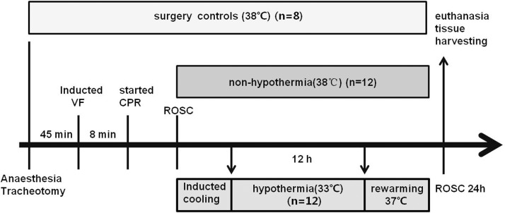
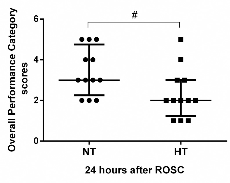
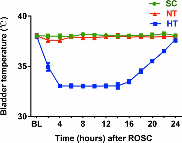
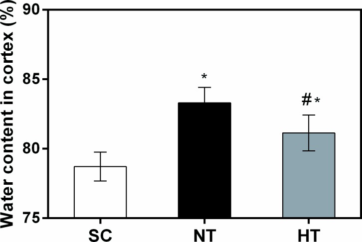
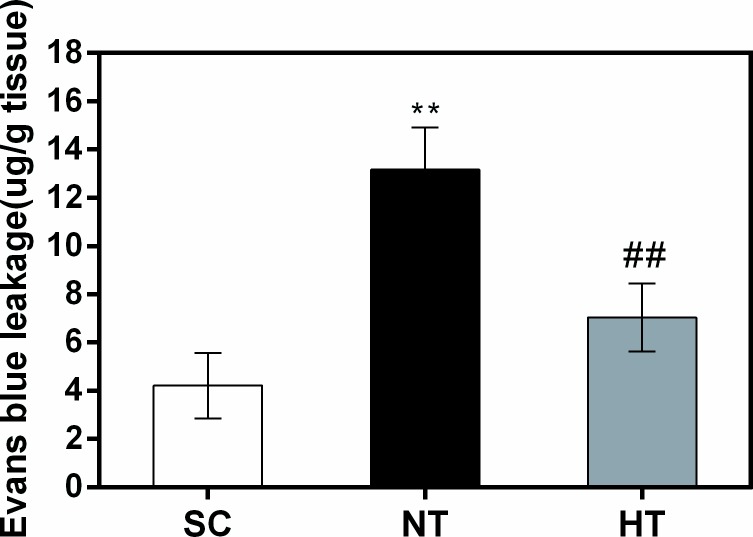
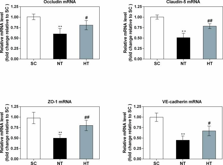
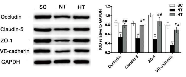
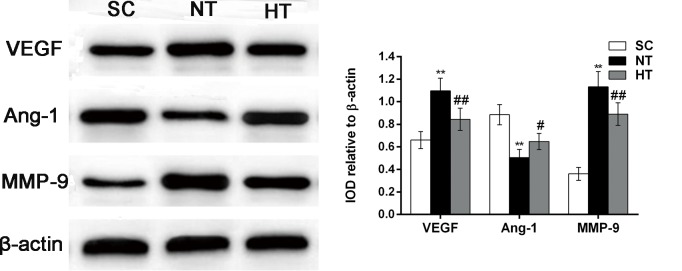
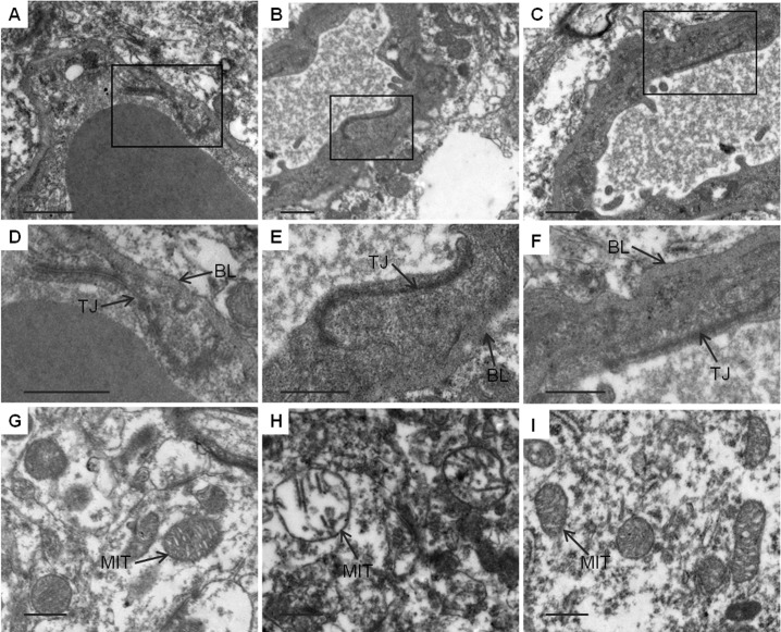
Mild hypothermia improves survival and neurological recovery after cardiac arrest (CA) and cardiopulmonary resuscitation (CPR). However, the mechanism underlying this phenomenon is not fully elucidated. The aim of this study was to determine whether mild hypothermia alleviates early blood-brain barrier (BBB) disruption. We investigated the effects of mild hypothermia on neurologic outcome, survival rate, brain water content, BBB permeability and changes in tight junctions (TJs) and adherens junctions (AJs) after CA and CPR. Pigs were subjected to 8 min of untreated ventricular fibrillation followed by CPR. Mild hypothermia (33°C) was intravascularly induced and maintained at this temperature for 12 h, followed by active rewarming. Mild hypothermia significantly reduced cortical water content, decreased BBB permeability and attenuated TJ ultrastructural and basement membrane breakdown in brain cortical microvessels. Mild hypothermia also attenuated the CPR-induced decreases in TJ (occludin, claudin-5, ZO-1) and AJ (VE-cadherin) protein and mRNA expression. Furthermore, mild hypothermia decreased the CA- and CPR-induced increases in matrix metalloproteinase-9 (MMP-9) and vascular endothelial growth factor (VEGF) expression and increased angiogenin-1 (Ang-1) expression. Our findings suggest that mild hypothermia attenuates the CA- and resuscitation-induced early brain oedema and BBB disruption, and this improvement might be at least partially associated with attenuation of the breakdown of TJ and AJ, suppression of MMP-9 and VEGF expression, and upregulation of Ang-1 expression.


Conflict of interest statement

Competing Interests: The authors have declared that no competing interests exist.

Figures

Fig 1
Fig 1. Experimental procedure.
VF, ventricular fibrillation; CPR, cardiopulmonary resuscitation; ROSC, restoration of spontaneous circulation.
Fig 2
Fig 2. Overall performance category scores.
Neurologic outcome was evaluated based on Overall Performance Category scores in the NT (n = 12) and HT (n = 12) groups. A score of 1 represents normal, whereas a score of 5 indicates brain death. Values are given as the median ± interquartile range. ROSC, restoration of spontaneous circulation; NT, non-hypothermia group; HT, mild hypothermia group. #P<0.05 versus NT group.
Fig 3
Fig 3. Bladder temperature.
BL, baseline; SC, surgery control group; NT, non-hypothermia group; HT, mild hypothermia group.
Fig 4
Fig 4. Cortical tissue water content at 24 hours after CA and resuscitation.
Brain oedema was calculated as cortical tissue water content in the SC (n = 4), NT (n = 5), and HT (n = 7) groups. Data are presented as means ± sd. SC, surgery control group; NT, non-hypothermia group; HT, mild hypothermia group. *P<0.05 versus SC group; #P<0.05 versus NT group.
Fig 5
Fig 5. Blood–brain barrier permeability in cortical tissues at 24 hours after CA and resuscitation.
Blood–brain barrier permeability was quantitatively evaluated via leakage of Evans blue in the SC (n = 4), NT (n = 4), and HT (n = 4) groups. Data are presented as means ± sd. SC, surgery control group; NT, non-hypothermia group; HT, mild hypothermia group. **P<0.01 versus SC group; ##P<0.01 versus NT group.
Fig 6
Fig 6. Occludin, claudin-5, ZO-1, and VE-cadherin mRNA expression in cortical tissues at 24 hours after CA.
The mRNA levels were quantified via real-time PCR in the SC (n = 4), NT (n = 5), and HT (n = 7) groups, and the data were normalized to GAPDH levels. The fold changes in occludin, claudin-5, ZO-1, and VE-cadherin mRNA expression were calculated relative to the expression levels in the SC group, and the results are presented as the mean fold changes ± sd relative to the SC group. SC, surgery control group; NT, non-hypothermia group; HT, mild hypothermia group. *P<0.05, **P<0.01 versus SC group; #P<0.05, ##P<0.01 versus NT group.
Fig 7
Fig 7. Occludin, claudin-5, ZO-1, and VE-cadherin protein expression in cortical tissues at 24 hours after CA.
Western blotting (left) was used to measure occludin, claudin-5, ZO-1, and VE-cadherin protein levels in the SC (n = 4), NT (n = 5), and HT (n = 7) groups. The IOD of each band was measured using Gel-pro Analyzer software. Occludin, claudin-5, ZO-1, and VE-cadherin levels were normalized to GAPDH levels, and the results are presented as the mean ± sd. SC, surgery control group; NT, non-hypothermia group; HT, mild hypothermia group. **P<0.01 versus SC group; ##P<0.01 versus NT group.
Fig 8
Fig 8. VEGF, Ang-1 and MMP-9 protein expression in cortical tissues at 24 hours after CA.
The expression of VEGF, Ang-1 and MMP-9 was measured by western blotting (left) in the SC (n = 4), NT (n = 5), and HT (n = 7) groups. VEGF, Ang-1 and MMP-9 levels were normalized to β-actin and are presented as the mean ± sd. SC, surgery control group; NT, non-hypothermia group, HT, mild hypothermia group; VEGF, vascular endothelial growth factor; Ang-1, angiogenin-1; MMP-9, matrix metalloproteinase-9. *P<0.05, **P<0.01 versus SC group; #P<0.05, ##P<0.01 versus NT group.
Fig 9
Fig 9. TEM observation of ultrastructural alterations in cortical tissues 24 hours after CA and resuscitation.
Normal TJs, basement membranes and neuronal mitochondria were observed in the SC group (A, D, G). In the NT group, the TJs were obscure, and the basement membranes were disrupted; moreover, neuronal mitochondria were markedly swollen, with disrupted cristae (B, E, H). In the HT group, the TJs and basement membrane were continuous, and neuronal mitochondria were slightly damaged, with normal cristae (C, F, I). SC, surgery control group; NT, non-hypothermia group; HT, mild hypothermia group; TJ, tight junction; BL, basement membrane; MIT, mitochondria. Scale bars = 2 μm.

Similar articles

Cited by

References

    1. Xiao F, Zhang S, Arnold TC, Alexander JS, Huang J, Carden DL, et al. Mild hypothermia induced before cardiac arrest reduces brain edema formation in rats. Acad Emerg Med. 2002;9: 105–114. - PubMed
    1. Morimoto Y, Kemmotsu O, Kitami K, Matsubara I, Tedo I. Acute brain swelling after out-of-hospital cardiac arrest: pathogenesis and outcome. Crit Care Med. 1993;21: 104–110. - PubMed
    1. Xiao F. Bench to bedside: brain edema and cerebral resuscitation: the present and future. Acad Emerg Med. 2002;9: 933–946. - PubMed
    1. Bernard SA, Gray TW, Buist MD, Jones BM, Silvester W, Gutteridge G, et al. Treatment of comatose survivors of out-of-hospital cardiac arrest with induced hypothermia. N Engl J Med. 2002;346: 557–563. 10.1056/NEJMoa003289 - DOI - PubMed
    1. Zhao H, Li CS, Gong P, Tang ZR, Hua R, Mei X, et al. Molecular mechanisms of therapeutic hypothermia on neurological function in a swine model of cardiopulmonary resuscitation. Resuscitation. 2012;83: 913–920. 10.1016/j.resuscitation.2012.01.001 - DOI - PubMed

MeSH terms