. 2017 Mar 28;112(6):1063-1076.
doi: 10.1016/j.bpj.2017.01.022.

Volume Transitions of Isolated Cell Nuclei Induced by Rapid Temperature Increase

Affiliations

Volume Transitions of Isolated Cell Nuclei Induced by Rapid Temperature Increase

Chii J Chan et al. Biophys J. .

Abstract

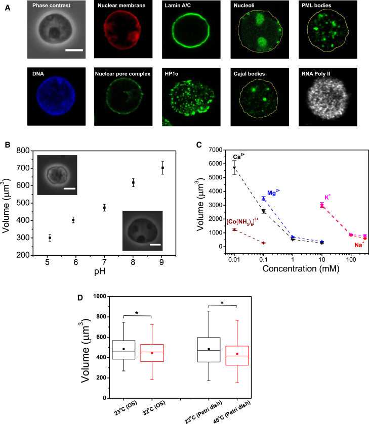
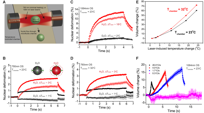
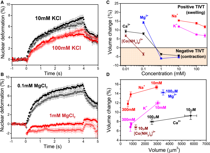
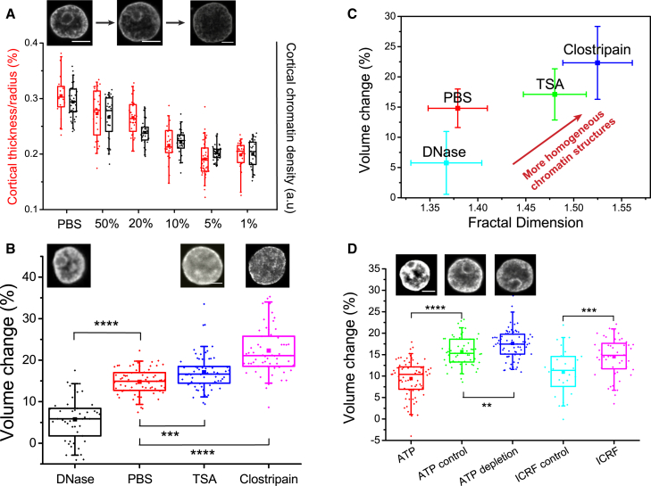
Understanding the physical mechanisms governing nuclear mechanics is important as it can impact gene expression and development. However, how cell nuclei respond to external cues such as heat is not well understood. Here, we studied the material properties of isolated nuclei in suspension using an optical stretcher. We demonstrate that isolated nuclei regulate their volume in a highly temperature-sensitive manner. At constant temperature, isolated nuclei behaved like passive, elastic and incompressible objects, whose volume depended on the pH and ionic conditions. When the temperature was increased suddenly by even a few degrees Kelvin, nuclei displayed a repeatable and reversible temperature-induced volume transition, whose sign depended on the valency of the solvent. Such phenomenon is not observed for nuclei subjected to slow heating. The transition temperature could be shifted by adiabatic changes of the ambient temperature, and the magnitude of temperature-induced volume transition could be modulated by modifying the chromatin compaction state and remodeling processes. Our findings reveal that the cell nucleus can be viewed as a highly charged polymer gel with intriguing thermoresponsive properties, which might play a role in nuclear volume regulation and thermosensing in living cells.


Figures

Figure 1
Figure 1
Nuclear volume is sensitive to changes in pH, salt conditions, and ambient temperature. (A) Nuclear compartmentalization is conserved after isolation. Immunofluorescence images (midsection confocal slices) of isolated HL60 nuclei stained for DNA (Hoechst), nuclear membrane (DID), nuclear pore complex (anti-nucleoporin), lamin A/C (anti-lamin A/C), HP1α (anti-HP1α), nucleoli (NUCLEOLAR-ID Green Detection Reagent), Cajal bodies (anti-coilin), PML bodies (anti-PML), and RNA polymerase II (anti-pol II). (Yellow lines) Approximate nuclear outlines. (B) Volume versus pH (mean ± SE) for optically trapped nuclei (trap power = 0.2 W, negligible heating). n = 26, 52, 47, 58, and 43 for pH = 5, 6, 7, 8, and 9, respectively. (Insets) Phase contrast images of nuclei at low and high pH. (C) Nuclear volume versus ionic concentrations of various valencies (mean ± SE). n > 30 for each salt condition. (D) Nuclear volume measured at different ambient temperatures in 780 nm OS (trap power = 0.2 W, n = 95 and 126 for 23°C and 32°C, respectively) and a heating Petri dish (n = 98, 83 for 23 and 45°C, respectively). p<0.05. Scale bars, 5 μm. To see this figure in color, go online.
Figure 2
Figure 2
Temperature-induced volume transitions in isolated HL60 nuclei. (A) Schematic of OS operating with 1064 nm (significant heating) or 780 nm wavelength (reduced heating). Nuclei flowing through the microfluidic channel were serially trapped and stretched/heated by two counterpropagating laser beams. (B) Deformation curves for nuclei stretched in a 780 nm OS at 1.6 W at 23°C. In heavy water (black, n = 104), nuclei showed positive (and negative) deformation along the major (solid circle) and minor axes (open circle), respectively, indicating volume conservation. Replacing heavy water with deionized water led to an increase in temperature jump and swelling along both the major and minor axes (red, n = 108). (C) Deformation curves for nuclei stretched in a 1064 nm OS at 1.6 W at 23°C, in heavy (black, n = 85) and deionized water (red, n = 53). (D) Deformation curves for nuclei stretched in a 780 nm OS at 1.6 W at 32°C. Similar volume conservation in heavy water (black, n = 53) and swelling in deionized water (red, n = 60) were observed. (E) Temperature-induced volume change of nuclei at different ambient temperatures, based on collective data from both 780 and 1064 nm OS. n > 40 for each data point. (F) Volume changes for nuclei in response to linearly increasing laser powers in 1064 nm OS at 23°C. Maximum power reached was 2.2 W (Tlaser=24°C). Heat ramp was performed over 1 s (black, n = 24), 5 s (red, n = 37), 12 s (blue, n = 22), and 60 s (magenta, n = 10), with the respective rates of heat increase indicated in the legend. All error bars denote SE. To see this figure in color, go online.
Figure 3
Figure 3
Temperature-induced volume transitions are sensitive to ionic conditions. (A) Deformation curves for HL60 nuclei in a 1064 nm OS at 23°C in 10 mM (black, n = 30), and 100 mM KCl (red, n = 42). (B) Deformation curves for HL60 nuclei in 0.1 mM (black, n = 46) and 1 mM MgCl2 (red, n = 52). In both (A) and (B), the major deformation is denoted by the solid circles and the minor deformation by the open circles. (C) Phase diagram for TIVT in various salt conditions, depicting the transition to negative volume transitions in sufficiently high amounts of multivalent salts. (D) Volume changes of nuclei in salts of various valencies and concentrations as a function of initial nuclear volume. Temperature-induced swelling is sensitive to the valency and ionic radii of the salts. In all experiments, 1.7 W stretch power (Tlaser=18°C) was applied between t = 0 and 4 s. Volume changes were measured at t = 1 s into the stretch. n > 30 for each data point in (C) and (D). All error bars denote SE. To see this figure in color, go online.
Figure 4
Figure 4
Temperature-induced volume transitions depend on chromatin compaction and remodeling dynamics. (A) Quantification of the relative thickness (red) and the average chromatin density (black) of the cortical chromatin shell for nuclei swollen to various degrees by PBS dilution. (Top row) DNA-stained nuclei in 100% PBS, 20% PBS, and 5% PBS. (B) Temperature-induced volume changes for nuclei under DNA degradation and histone modifications. (Top row) DNA-stained nuclei under corresponding pharmacological treatments. Nuclei treated with TSA (n = 69) and clostripain (n = 63) were significantly softer with larger TIVT than controls in PBS (n = 72). (C) Temperature-induced volume changes of nuclei versus fractal dimension. Addition of TSA (n = 44) and clostripain (n = 27) led to higher fractal dimension compared to control nuclei in PBS (n = 21) and DNase (n = 17). Error bars represent SD. (D). Temperature-induced volume changes of nuclei mediated by chromatin remodeling. Nuclei subjected to ATP addition (n = 73) or depletion (n = 96) showed less and more swelling effect, respectively, compared to the controls (n = 61). Topo II inhibition (n = 48) led to nuclear softening compared to the controls (n = 26). (Top row) DNA-stained nuclei under ATP addition or depletion, and in controls. All volume changes were measured at t = 1 s after optical stretching (laser powers = 1.7 W, Tlaser=18°C, 1064 nm). ∗∗∗∗p<0.0001, ∗∗∗p<0.001, and ∗∗p<0.01. Scale bars represent 5 μm. To see this figure in color, go online.

Similar articles

Cited by

References

    1. Lammerding J., Hsiao J., Lee R.T. Abnormal nuclear shape and impaired mechanotransduction in emerin-deficient cells. J. Cell Biol. 2005;170:781–791. - PMC - PubMed
    1. Swift J., Ivanovska I.L., Discher D.E. Nuclear lamin-A scales with tissue stiffness and enhances matrix-directed differentiation. Science. 2013;341:1240104. - PMC - PubMed
    1. Guilak F., Tedrow J.R., Burgkart R. Viscoelastic properties of the cell nucleus. Biochem. Biophys. Res. Commun. 2000;269:781–786. - PubMed
    1. Maniotis A.J., Chen C.S., Ingber D.E. Demonstration of mechanical connections between integrins, cytoskeletal filaments, and nucleoplasm that stabilize nuclear structure. Proc. Natl. Acad. Sci. USA. 1997;94:849–854. - PMC - PubMed
    1. Versaevel M., Grevesse T., Gabriele S. Spatial coordination between cell and nuclear shape within micropatterned endothelial cells. Nat. Commun. 2012;3:671. - PubMed

LinkOut - more resources