. 2017 Mar 28;18(13):3204-3218.
doi: 10.1016/j.celrep.2017.03.018.

Identification of Interleukin-1 by Functional Screening as a Key Mediator of Cellular Expansion and Disease Progression in Acute Myeloid Leukemia

Affiliations

Identification of Interleukin-1 by Functional Screening as a Key Mediator of Cellular Expansion and Disease Progression in Acute Myeloid Leukemia

Alyssa Carey et al. Cell Rep. .

Abstract

Secreted proteins in the bone marrow microenvironment play critical roles in acute myeloid leukemia (AML). Through an ex vivo functional screen of 94 cytokines, we identified that the pro-inflammatory cytokine interleukin-1 (IL-1) elicited profound expansion of myeloid progenitors in ∼67% of AML patients while suppressing the growth of normal progenitors. Levels of IL-1β and IL-1 receptors were increased in AML patients, and silencing of the IL-1 receptor led to significant suppression of clonogenicity and in vivo disease progression. IL-1 promoted AML cell growth by enhancing p38MAPK phosphorylation and promoting secretion of various other growth factors and inflammatory cytokines. Treatment with p38MAPK inhibitors reversed these effects and recovered normal CD34+ cells from IL-1-mediated growth suppression. These results highlight the importance of ex vivo functional screening to identify common and actionable extrinsic pathways in genetically heterogeneous malignancies and provide impetus for clinical development of IL-1/IL1R1/p38MAPK pathway-targeted therapies in AML.

Keywords: AML; IL1R1; bone marrow microenvironment; functional screening; interleukin-1; p38MAPK.


Figures

Figure 1
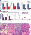
Figure 1. IL-1 promotes the growth of a subset of primary AML patient samples
(A) Primary mononuclear cells derived from bone marrow and peripheral blood of 60 AML patient samples were cultured for 3 days in RPMI-1640 supplemented with 5% FBS. The effect of individual cytokines on cell growth was measured by MTS assay (screen layout in Figure S1A). Absorbance values were normalized against the growth of the cells without cytokines. Cytokines causing increased growth equal to or more than the growth response to HS-5 conditioned media were designated as growth-promoting cytokines; those causing decreased growth compared to the no-cytokine control were designated as growth-suppressive cytokines. The percentage of primary samples responding to individual cytokines is shown. (B) Heatmap representation of cytokine response clustering with IL-1 (full heatmap in Figure S1B). Red indicates increased growth; blue indicates decreased growth. The growth response for selective cytokines is show in Figure S2 and correlation of IL-1-mediated growth response with clinical, demographic, and genetic features of AML patients is shown in Figure S3 and Table S2. (C) Impact of graded concentrations of IL-1α and IL-1β on the growth of primary AML mononuclear cells (MNCs) from each patient sample screened in Figure 1A by colorimetric MTS assay. In this assay, the absorbance value for untreated cells were considered as maximum viability and fold change over the untreated controls were calculated. (D) Purified CD34+ cells derived from AML patient samples bone marrow were cultured in RPMI-1640 supplemented with 5% FBS with graded concentrations of IL-1α and IL-1β and the effect on growth was measured by MTS and colony formation assays in cytokine free MethoCult supplemented with 10 ng/ml IL-3 and 50 ng/ml SCF. (E) Purified normal CD34+ cells derived from healthy donors (NL) bone marrow were cultured with graded concentrations of IL-1β and effect on growth was measured by colony formation assay in cytokine-free MethoCult supplemented with 10 ng/ml IL-3 and 50 ng/ml SCF. Levels of statistical significance: * p≤0.05, ** p≤0.01, *** p≤0.001.
Figure 2
Figure 2. IL-1-sensitive AML patient samples have increased IL-1β and IL-1 receptor expression in the bone marrow, and IL-1-secreting monocytes regulate the growth of AML cells in paracrine manner
(A) IL-1β levels were measured by ELISA in plasma from peripheral blood (PB) from healthy individuals, and from AML peripheral blood and bone marrow samples (BM). (B) IL-1β level by ELISA and expression by quantitative PCR were measured from IL-1β-sensitive (S) and nonsensitive (NS) AML peripheral blood and bone marrow samples. (C) IL1R1 expression was measured by quantitative PCR from IL-1β-sensitive (S) and nonsensitive (NS) AML peripheral blood and bone marrow samples. (D,E) IL-1 receptor (IL1R1, IL1RAcP) expression and intracellular IL-1β levels were measured by flow cytometry in various subpopulations of bone marrow cells from healthy individuals and IL-1β-sensitive and nonsensitive AML patients. Percentage of positive cells for each subpopulation is shown as mean+SEM from three independent bone marrow samples. The representative gating strategy and FMO controls are shown in Figure S4D. (F) Representative plots showing percentage of IL-1β-positive cells in various subpopulations in bone marrow of healthy individuals and IL-1β-sensitive and nonsensitive AML patients. See also Figure S4 for the percentage of CD14+, CD34+ and CD33+ cells. (G) CD14+ cells were depleted from IL-1β-sensitive primary AML MNCs bone marrow, and the effect of depleting CD14+ cells and adding them back to MNCs was measured by MTS assay. * p≤0.05, ** p≤0.01, *** p≤0.001.
Figure 3
Figure 3. The absence of IL1R1 attenuates the growth of primary AML cells as well as colony-forming ability and disease progression in a murine AML bone marrow transduction/transplantation model
(A) Primary AML CD34+ cells purified from bone marrow were infected with a combination of two inducible IL1R1 shRNA hairpins (see also Figure S6). After 48 h of infection, cells were sorted for GFP positivity, and those cells stably expressing IL1R1 shRNA were treated with doxycycline. The effect of knockdown was determined on IL1R1 expression by qPCR after 48 h, cell growth by MTS assay after 4 days after culturing cells in IMDM supplemented with10%FBS and 104 M β-ME, and colony formation assay in cytokine-free MethoCult supplemented with 10 ng/ml IL-3 and 50 ng/ml SCF. The experiment is representative of 3 independent experiments. (B) Myeloid colony formation assay using mouse bone marrow cells from wild-type and IL1R1-null mice transduced with AML1-ETO9a/NRASG12D or empty vector and plated with IL-1α and IL-1β together (2 ng/ml, each) with either cytokine-free or cytokine-supplemented MethoCult. Colonies were scored on day 8 (see also Figure S7). (C) Lethally irradiated wild-type mice were injected retro-orbitally with equal numbers of AML1-ETO9a/NRASG12D-transduced GFP+ bone marrow from wild-type and IL1R1-null mice and evaluated for survival using Kaplan-Meier statistics. (D) Hematoxylin and eosin (H&E) histopathological analysis of mouse tissues from IL1R1-null and wild-type leukemic mice. Insets represent megakaryocytic and myeloid infiltrates. * p≤0.05, ** p≤0.01, *** p≤0.001.
Figure 4
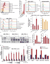
Figure 4. IL-1β promotes the growth of AML cells by increasing p38MAPK phosphorylation and IL-1-dependent growth of AML cells is inhibited by a p38MAPK inhibitor
(A) Basal p38MAPK phosphorylation of CD34+ cells purified from the bone marrow of IL-1-sensitive and -nonsensitive primary AML samples, as well as healthy controls, as measured by phosphoflow analysis. The bar graph shows relative mean fluorescent intensity (MFI) of p38MAPK phosphorylation for 3 IL-1-sensitive and 3 IL-1-nonsensitive AML samples and 2 healthy controls. (B,C) CD34+ cells purified from primary AML samples and normal CD34+ cells from a healthy individual were stimulated with 10 ng/ml IL-1β or a non-IL-1β inclusive cocktail of various cytokines (20 ng/ml IL-6, 10 ng/ml IL-11, 50 ng/ml FLT-3L, and 100 ng/ml SCF). The effect of 500 nM of the p38MAPK inhibitor doramapimod (Dora) was determined by measuring p38MAPK phosphorylation by phosphoflow analysis. The results from IL-1-sensitive and -nonsensitive primary mononuclear cells (MNCs) and AML cell lines are shown in Figure S7A,B and S7E. (D) CD34+ cells purified from primary AML samples and normal CD34+ cells from a healthy individual were stimulated with 10 ng/ml IL-1β. The effect of 500 nM of the p38MAPK inhibitor ralimetinib (Ralim) was determined by measuring p38MAPK phosphorylation by phosphoflow analysis. (E) The average mean fluorescent intensity (MFI) of p38MAPK phosphorylation, measured by phosphoflow analysis, for 4 IL-1-sensitive AML samples and 2 healthy controls. (F) Primary CD34+ cells derived from 3 IL-1-sensitive AML bone marrow samples were stimulated with 10 ng/ml IL-1β with and without 500 nM doramapimod for the indicated times. The effect of IL-1 stimulation on downstream signaling by measured by immunoblotting. Relative expression of phospho-p38MAPK and phospho-IRAK-1 normalized to GAPDH controls is shown as the mean+SEM from 3 samples. (G) CD34+ AML cells were cultured in the indicated concentration of IL-1α or IL-1β (ng/mL) with and without 500 nM doramapimod or ralimetinib, and cell viability was measured by MTS assay. Results from IL-1-sensitive and -nonsensitive primary MNCs are shown in Figure S7C-D. (H) Primary AML MNC, purified CD34+, and FACS sorted CD34+ IL1R1+ and CD34+IL1R1 cells were cultured in 10 ng/mL IL-1β with and without 500 nM doramapimod, for 3 days and cell viability was measured by MTS assay (see also Figure S7G). * p≤0.05, ** p≤0.01, *** p≤0.001.
Figure 5
Figure 5. IL-1-promotes the proliferation, survival, and colony formation ability of AML cells which are inhibited by a p38MAPK inhibitor
(A) Purified CD34+ progenitors from the bone marrow of healthy donors and AML patients were cultured in 10 ng/mL IL-1β with and without 500 nM doramapimod, for 6 days in a culture media containing IMDM+10%FBS with 20 ng/ml IL-6, 10 ng/ml IL-11, 50 ng/ml FLT-3L, and 100 ng/ml SCF. Fresh doses of cytokine and/or inhibitor were added to the culture every 3 days. Cells for counted alternate day and total cell number is shown over time. (B) CD34+ cells were labeled with a Violet cell tracer dye and dilution of dye was measured by FACS after 3 days of culturing in above indicated media. % proliferation is shown from a representative sample from three independent experiments from the bone marrow of healthy donors and IL-1 sensitive AML patients. (See also Figure S7H for IL-1 nonsensitive AML sample) (C) CD34+ AML and healthy bone marrow cells were cultured in 10 ng/mL IL-1β with and without 500 nM doramapimod and the effect on apoptosis was determined by measuring annexin V positivity after 2 days of treatment. (D) The effect of IL-1 and doramapimod on the clonogenic growth of CD34+ cells from primary AML patients and healthy bone marrow was measured by colony formation assay. Cytokine-free MethoCult was supplemented with 10 ng/ml IL-3 and 50 ng/ml SCF, and colonies were scored on day 14. (E) AML and healthy cells were cultured as described in Figure 5A and the effect of treatments were measured on differentiation using indicated surface markers. Histograms are showing mean flouroscence intensity (MFI) for a representative sample and bar graphs are showing mean+SEM for MFI and % of total cells from three samples. * p≤0.05, ** p≤0.01, *** p≤0.001.
Figure 6
Figure 6. IL-1β promotes the secretion of multiple growth-promoting inflammatory mediators from AML progenitors and their secretion is blocked by p38 kinase inhibition
Comprehensive evaluation of cytokines produced by IL-1-sensitive and non-sensitive AML CD34+ bone marrow progenitors using the 30-plex human cytokines Luminex platform. Purified CD34+ progenitors from IL-1-sensitive and non-sensitive AML samples (N=3) were treated with 10ng/ml IL-1β and 500nM doramapimod and cultured in 10% FBS+ RPMI media for 48 hrs and conditioned media were evaluated for cytokine production. Standard curves for each cytokine evaluated met quality control assessment. (A) Heat map showing the relative cytokine production for the 30 cytokines compared to untreated controls. (B) The mean absolute values of cytokines showing the profound response to IL-1 stimulation in AML progenitors are shown on a log10 scale as the mean of three independent samples. (See also figure S7I showing the effect of blocking individual cytokine on cell viability)
Figure 7
Figure 7. Summary model of IL-1-mediated promotion of AML cell growth and suppression of normal hematopoiesis
(A) In AML, increased IL-1β is secreted in the bone marrow by monocytes and leukemic cells. Increased IL-1β promotes the expansion of AML myeloid progenitors while suppressing the growth of normal myeloid progenitors due to the aberrant activation of the IL-1/p38MAPK pathway. This leads to disease progression in AML. (B) Knocking down IL1R1 and blocking IL-1 signaling with a p38MAPK inhibitors suppresses the growth of AML cells and rescues normal hematopoiesis.

References

    1. Agarwal A, Bumm TG, Corbin AS, O’Hare T, Loriaux M, VanDyke J, Willis SG, Deininger J, Nakayama KI, Druker BJ, Deininger MW. Absence of SKP2 expression attenuates BCR-ABL-induced myeloproliferative disease. Blood. 2008;112:1960–1970. - PMC - PubMed
    1. Agarwal A, Mackenzie RJ, Besson A, Jeng S, Carey A, LaTocha DH, Fleischman AG, Duquesnes N, Eide CA, Vasudevan KB, et al. BCR-ABL1 promotes leukemia by converting p27 into a cytoplasmic oncoprotein. Blood. 2014a;124:3260–3273. - PMC - PubMed
    1. Agarwal A, MacKenzie RJ, Eide CA, Davare MA, Watanabe-Smith K, Tognon CE, Mongoue-Tchokote S, Park B, Braziel RM, Tyner JW, Druker BJ. Functional RNAi screen targeting cytokine and growth factor receptors reveals oncorequisite role for interleukin-2 gamma receptor in JAK3-mutation-positive leukemia. Oncogene 2014b - PMC - PubMed
    1. Agerstam H, Hansen N, von Palffy S, Sanden C, Reckzeh K, Karlsson C, Lilljebjorn H, Landberg N, Askmyr M, Hogberg C, et al. IL1RAP antibodies block IL-1-induced expansion of candidate CML stem cells and mediate cell killing in xenograft models. Blood. 2016;128:2683–2693. - PubMed
    1. Anur P, Yates J, Garbati MR, Vanderwerf S, Keeble W, Rathbun K, Hays LE, Tyner JW, Svahn J, Cappelli E, et al. p38 MAPK inhibition suppresses the TLR-hypersensitive phenotype in FANCC- and FANCA-deficient mononuclear phagocytes. Blood. 2012;119:1992–2002. - PMC - PubMed

Publication types

MeSH terms

Substances