. 2017 Apr 19;139(15):5588-5595.
doi: 10.1021/jacs.7b01791. Epub 2017 Apr 10.

Diverse Redoxome Reactivity Profiles of Carbon Nucleophiles

Affiliations

Diverse Redoxome Reactivity Profiles of Carbon Nucleophiles

Vinayak Gupta et al. J Am Chem Soc. .

Abstract

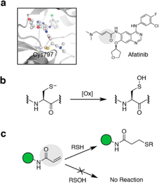
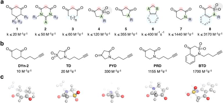
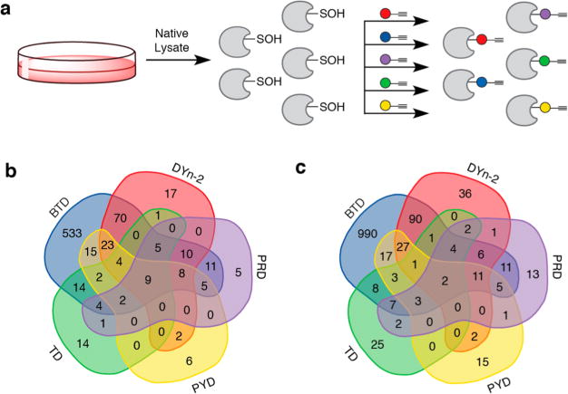
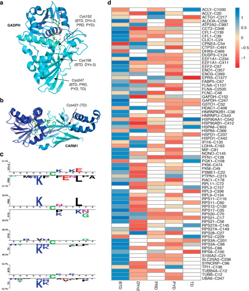
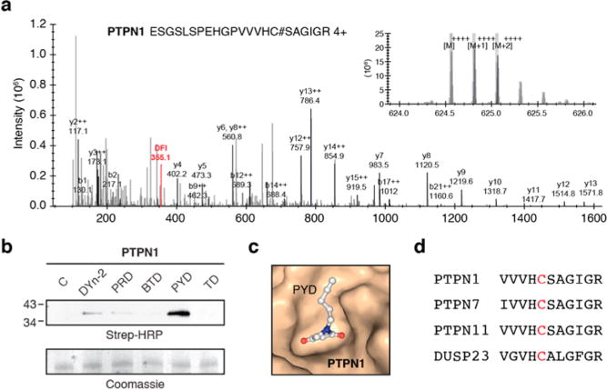
Targeted covalent inhibitors have emerged as a powerful approach in the drug discovery pipeline. Key to this process is the identification of signaling pathways (or receptors) specific to (or overexpressed in) disease cells. In this context, fragment-based ligand discovery (FBLD) has significantly expanded our view of the ligandable proteome and affords tool compounds for biological inquiry. To date, such covalent ligand discovery has almost exclusively employed cysteine-reactive small-molecule fragments. However, functional cysteine residues in proteins are often redox-sensitive and can undergo oxidation in cells. Such reactions are particularly relevant in diseases, like cancer, which are linked to excessive production of reactive oxygen species. Once oxidized, the sulfur atom of cysteine is much less reactive toward electrophilic groups used in the traditional FBLD paradigm. To address this limitation, we recently developed a novel library of diverse carbon-based nucleophile fragments that react selectively with cysteine sulfenic acid formed in proteins via oxidation or hydrolysis reactions. Here, we report analysis of sulfenic acid-reactive C-nucleophile fragments screened against a colon cancer cell proteome. Covalent ligands were identified for >1280 S-sulfenylated cysteines present in "druggable" proteins and orphan targets, revealing disparate reactivity profiles and target preferences. Among the unique ligand-protein interactions identified was that of a pyrrolidinedione nucleophile that reacted preferentially with protein tyrosine phosphatases. Fragment-based covalent ligand discovery with C-nucleophiles affords an expansive snapshot of the ligandable "redoxome" with significant implications for covalent inhibitor pharmacology and also affords new chemical tools to investigate redox-regulation of protein function.


Conflict of interest statement

Notes

The authors declare no competing financial interest.

Figures

Figure 1
Figure 1
Targeting the cysteine thiol (Cys-SH) with covalent inhibitors. (a) FDA-approved Afatinib forms a covalent adduct with EGFR 797Cys-SH via Michael addition. (b) Oxidation of Cys-SH generates cysteine sulfenic acid (Cys-SOH). (c) Covalent inhibitors with electrophilic warheads (such as the Michael acceptor highlighted in gray) undergo facile reaction with Cys-SH but not with Cys-SOH under physiological conditions.
Figure 2
Figure 2
Novel classes of nucleophile probes to profile cysteine oxidation (a) Structural classes of cyclic C-nucleophiles (18). In each structure, the nucleophilic carbon is highlighted in the red circle. Rate constants from ref . (b) From the pool of cyclic C-nucleophiles, DYn-2 and four new C-nucleophiles were selected on the basis of a range of reaction rate constants and scaffold diversity. (c) Energy-minimized 3-dimensional representation of the probes.
Figure 3
Figure 3
Application of new nucleophile probes for studying cellular sulfenylomes. (a) RKO whole cell lysates were prepared under nondenaturing conditions. Native lysate was divided into equal portions and incubated with one of five nucleophile probes (or vehicle) in separate reactions. Labeled proteins from each reaction were digested separately and the resulting peptides subjected to click reaction with UV-cleavable biotin-N3. Enrichment and photorelease afforded different probe-labeled peptides samples for proteomic analysis. (b) Protein Venn diagram of unique sulfenylated proteins. (c) Site Venn diagram of unique sulfenylated peptides.
Figure 4
Figure 4
Chemoproteomic analysis of RKO cellular S-sulfenylome. (a) Crystal structures of GAPDH (PDB: 4WNC) showing Cys152, Cys156, and Cys247 labeled by different probes. (b) CARM1 (PDB: 5U4X) showing Cys421 that is labeled by TD. (c) Comparative sequence analysis of peptides labeled with different probes. (d) Heat map of Z-score normalized peak intensities of S-sulfenylated sites labeled with various probes. Values in each row direction have been mean centered and scaled.
Figure 5
Figure 5
PTPs as privileged targets of PYD. (a) Representative MS/MS spectra of PTPN1 (PTP1B) labeled by PYD probe. (b) PYD probe preferentially modifies the recombinant oxidized form of PTP1B. (c) Molecular docking (using AutoDock Vina) of PYD into catalytic pocket of PTPN1 (PDB: 1OET). (d) Abbreviated sequence alignment of PTPs that harbor a Cys-SOH labeled by PYD probe.

Similar articles

Cited by

References

    1. Singh J, Petter RC, Baillie TA, Whitty A. Nat Rev Drug Discovery. 2011;10:307. - PubMed
    1. Baillie TA. Angew Chem, Int Ed. 2016;55:13408. - PubMed
    1. Liu Q, Sabnis Y, Zhao Z, Zhang T, Buhrlage SJ, Jones LH, Gray NS. Chem Biol. 2013;20:146. - PMC - PubMed
    1. Paulsen CE, Carroll KS. Chem Rev. 2013;113:4633. - PMC - PubMed
    1. Reddie KG, Carroll KS. Curr Opin Chem Biol. 2008;12:746. - PubMed

Publication types