

A genome-wide association study reveals a locus for bilateral iridal hypopigmentation in Holstein Friesian cattle
- PMID: 28356055
- PMCID: PMC5372310
- DOI: 10.1186/s12863-017-0496-4
A genome-wide association study reveals a locus for bilateral iridal hypopigmentation in Holstein Friesian cattle
Abstract
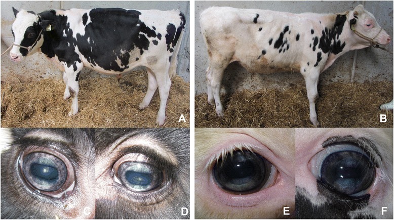
Background: Eye pigmentation abnormalities in cattle are often related to albinism, Chediak-Higashi or Tietz like syndrome. However, mutations only affecting pigmentation of coat color and eye have also been described. Herein 18 Holstein Friesian cattle affected by bicolored and hypopigmented irises have been investigated.
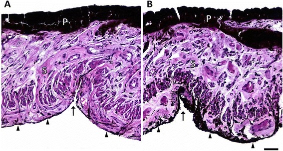
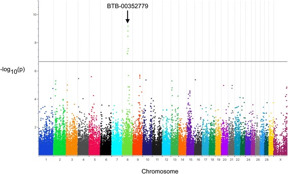
Results: Affected animals did not reveal any ophthalmological or neurological abnormalities besides the specific iris color differences. Coat color of affected cattle did not differ from controls. Histological examination revealed a reduction of melanin pigment in the iridal anterior border layer and stroma in cases as cause of iris hypopigmentation. To analyze the genetics of the iris pigmentation differences, a genome-wide association study was performed using Illumina BovineSNP50 BeadChip genotypes of the 18 cases and 172 randomly chosen control animals. A significant association on bovine chromosome 8 (BTA8) was identified at position 60,990,733 with a -log10(p) = 9.17. Analysis of genotypic and allelic dependences between cases of iridal hypopigmentation and an additional set of 316 randomly selected Holstein Friesian cattle controls showed that allele A at position 60,990,733 on BTA8 (P = 4.0e-08, odds ratio = 6.3, 95% confidence interval 3.02-13.17) significantly increased the chance of iridal hypopigmentation.
Conclusions: The clinical appearance of the iridal hypopigmentation differed from previously reported cases of pigmentation abnormalities in syndromes like Chediak-Higashi or Tietz and seems to be mainly of cosmetic character. Iridal hypopigmentation is caused by a reduced content of melanin pigment in the anterior border layer and iridal stroma. A single genomic position on BTA8 was detected to be significantly associated with iridal hypopigmentation in examined cattle. To our knowledge this is the first report about this phenotype in Holstein Friesian cattle.
Keywords: Albinism; Cattle; GWAS; Heterochromia iridis; Holstein Friesian; Iris hypopigmentation; Oculocutaneous hypopigmentation.
Figures
References
-
- Edwards M, Cha D, Krithika S, Johnson M, Cook G, Parra EJ. Iris pigmentation as a quantitative trait: variation in populations of European, East Asian and South Asian ancestry and association with candidate gene polymorphisms. Pigment Cell Melanoma Res. 2016;29(2):141–162. doi: 10.1111/pcmr.12435. - DOI - PubMed
Publication types
MeSH terms
LinkOut - more resources
Full Text Sources
Other Literature Sources
