

Corneal densitometry changes in a patient with interface fluid syndrome after small incision lenticule extraction
- PMID: 28356099
- PMCID: PMC5372330
- DOI: 10.1186/s12886-017-0428-0
Corneal densitometry changes in a patient with interface fluid syndrome after small incision lenticule extraction
Abstract
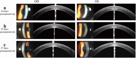
Background: To report a case of interface fluid syndrome (IFS) following small incision lenticule extraction (SMILE) evaluated with corneal densitometry and optical coherence tomography (OCT).
Case presentation: An 18-year-old man reported sudden vision loss 24 days after SMILE procedure. Intraocular pressure (IOP) was 36.3 mmHg (OD) and 36.7 mmHg (OS) by noncontact tonometry. Moderate corneal edema, interface fluid pocket and haze were observed by OCT and confirmed by corneal densitometry values. Discontinuation of steroids and addition of hypotensive medication were offered immediately. The symptoms were cured after the medication. Changes of corneal densitometry were consistent with the clinical course of IFS.
Conclusion: This case illustrates that it is crucial to be aware that a history of SMILE can also cause IFS. Both OCT and corneal densitometry can serve as auxiliary means to evaluate the clinical course of IFS, and appropriate IOP management is an effective approach.
Keywords: Corneal densitometry; Interface fluid syndrome (IFS); Small incision lenticule extraction (SMILE).
Figures
References
-
- Moya Calleja T, Iribarne Ferrer Y, Sanz Jorge A, Sedo FS. Steroid-induced interface fluid syndrome after LASIK. J Refract Surg. 2009;25(2):235–239. - PubMed
Publication types
MeSH terms
LinkOut - more resources
Full Text Sources
Other Literature Sources
