. 2017 Mar 29;9(1):31.
doi: 10.1186/s13073-017-0420-6.

Migration of mitochondrial DNA in the nuclear genome of colorectal adenocarcinoma

Affiliations

Migration of mitochondrial DNA in the nuclear genome of colorectal adenocarcinoma

Vinodh Srinivasainagendra et al. Genome Med. .

Abstract

Background: Colorectal adenocarcinomas are characterized by abnormal mitochondrial DNA (mtDNA) copy number and genomic instability, but a molecular interaction between mitochondrial and nuclear genome remains unknown. Here we report the discovery of increased copies of nuclear mtDNA (NUMT) in colorectal adenocarcinomas, which supports link between mtDNA and genomic instability in the nucleus. We name this phenomenon of nuclear occurrence of mitochondrial component as numtogenesis. We provide a description of NUMT abundance and distribution in tumor versus matched blood-derived normal genomes.

Methods: Whole-genome sequence data were obtained for colon adenocarcinoma and rectum adenocarcinoma patients participating in The Cancer Genome Atlas, via the Cancer Genomics Hub, using the GeneTorrent file acquisition tool. Data were analyzed to determine NUMT proportion and distribution on a genome-wide scale. A NUMT suppressor gene was identified by comparing numtogenesis in other organisms.

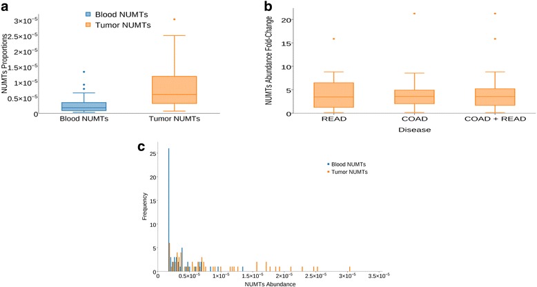
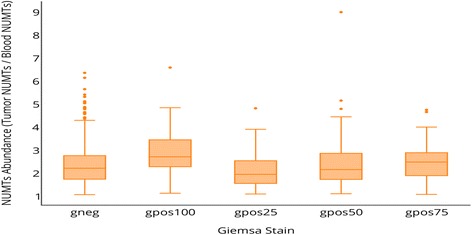
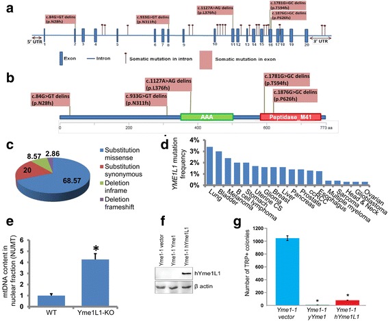
Results: Our study reveals that colorectal adenocarcinoma genomes, on average, contains up to 4.2-fold more somatic NUMTs than matched normal genomes. Women colorectal tumors contained more NUMT than men. NUMT abundance in tumor predicted parallel abundance in blood. NUMT abundance positively correlated with GC content and gene density. Increased numtogenesis was observed with higher mortality. We identified YME1L1, a human homolog of yeast YME1 (yeast mitochondrial DNA escape 1) to be frequently mutated in colorectal tumors. YME1L1 was also mutated in tumors derived from other tissues. We show that inactivation of YME1L1 results in increased transfer of mtDNA in the nuclear genome.

Conclusions: Our study demonstrates increased somatic transfer of mtDNA in colorectal tumors. Our study also reveals sex-based differences in frequency of NUMT occurrence and that NUMT in blood reflects NUMT in tumors, suggesting NUMT may be used as a biomarker for tumorigenesis. We identify YME1L1 as the first NUMT suppressor gene in human and demonstrate that inactivation of YME1L1 induces migration of mtDNA to the nuclear genome. Our study reveals that numtogenesis plays an important role in the development of cancer.

Keywords: Cancer; Colorectal cancer; Genetic instability; Mitochondria; Mitochondrial DNA; NUMT; Numtogenesis; Tumor; YME1L1; mtDNA transfer.


Figures

Fig. 1
Fig. 1
Quality control (QC) pipeline. Upstream QC pipeline conducted on aligned sequence data processed at Harvard Medical School (HMS-HK) was downloaded from TCGA CGHub. Clinical annotations for the matched tumor and blood-derived normal samples were downloaded from TCGA data matrix. COAD colon adenocarcinoma, READ rectum adenocarcinoma, HMS-HK Harvard Medical School
Fig. 2
Fig. 2
Distribution of NUMT proportions in tumor and normal genomes. a Distribution of NUMT proportions in 57 samples with matched tumor and blood-derived normal genomes. NUMT proportion is defined as the ratio between NUMT read count and total mapped read count on a genome-wide scale. The mean and standard deviation, respectively, across the 57 samples are 8.31 × 10−6 and 7.11 × 10−6 for tumor genomes and 2.65 × 10−6 and 2.49 × 10−6 for normal genomes. A two-tailed paired t-test conducted between the NUMT proportions for 57 (COAD + READ) samples revealed a P value of 1.63 × 10−5. When comparing the NUMT proportions between the cancer site group versus blood-derived group using a two-tailed unequal variance t-test, a P value of 1.43 × 10−5 was observed for COAD samples (N = 36) and 3.82 × 10−3 for READ samples (N = 21). b Fold change in the tumor NUMT proportions compared to blood-derived normal genomes across colon (COAD) and rectum (READ) cancer samples. Tumor genomes contained 4.42-fold more NUMTs than blood-derived normal genomes. A two-tailed unequal variance t-test conducted between the NUMT abundance of colon cancer samples (N = 36) and rectal cancer samples (N = 21) revealed a P value of 0.91, indicating no difference in the NUMT abundance between the two cancer sites, colon and rectum. c Right-skewed distribution, log transformed, and one-tailed paired t-test performed on tumor and matched blood-derived normal samples to determine the statistical significance (P value 8.79 × 10−13) of the log-transformed NUMT abundance levels
Fig. 3
Fig. 3
Log2-transformed NUMT proportions in tumor and blood-derived normal (Pij) genomes. a Log2-transformed NUMT proportions in tumor and normal genomes showcasing the difference in their means, indicating higher NUMT distribution in tumor genomes compared to matched blood-derived normal genomes. A two-tailed paired t-test conducted between the log2-transformed NUMT proportions for 57 samples revealed a P value of 1.87 × 10−11, showing significant difference in the log-transformed NUMT proportions between primary tumors and blood-derived normal genomes. b Relationship between abundance measures of blood-derived normal genome NUMTs and tumor genome NUMTs showing a positive relationship with R2 = 0.17 and P value = 0.0016
Fig. 4
Fig. 4
NUMT abundance across disease–sex combination. Colorectal tumors from women have higher NUMT abundance proportion (tumor NUMT/blood normal NUMT) than those from men. Women had a median NUMT fold change abundance of 4.52 (range 0.11 to 22.9) compared 3.1 for men (range 0.53 to 8.3). COAD colon adenocarcinoma, READ rectum adenocarcinoma. To investigate the sex difference in the NUMT abundances, an unequal variance t-test was performed on the raw NUMT proportions observed by the members of the two sex groups stratified by “blood-normal” and “primary tumor” classifications. The P value was 0.03 between males (N = 23) and females (N = 34) for blood-derived normal samples and 0.08 for tumor samples
Fig. 5
Fig. 5
NUMT abundance distribution according to vital status. a Increased NUMT abundance in deceased individuals with colorectal tumors. Although a Mann–Whitney U test showed a P value of 0.04 between the NUMT abundances of the Alive and Deceased groups, the very small sample size (N = 4) of the deceased group warrants further investigation with datasets enriched for deceased vital status. b NUMT proportions categorized based on disease (colon and rectal) and sex combination. This vital status observation in combination with NUMT abundance among sex-specific samples appears to be an early indicator of death events among colorectal cancer women with higher proportions of NUMTs
Fig. 6
Fig. 6
NUMT density in tumor and normal genomes (sorted by disease (T2), sex (T1), and age at initial pathologic diagnosis (T3)). Each peripheral node represents a TCGA sample whose blood-derived normal and tumor genomes were used in this study. From the outside to inside, tracks are ordered from 1 to 7 (T1–T7). T1: Sample sex where red nodes represent female and blue nodes represent male. T2: Disease type information. Rectal adenocarcinoma (READ) is rendered as green bands and colon adenocarcinoma (COAD) as red bands. T3: Age at initial pathologic diagnosis ranging from 30 to 90 years. White and black filled bars represent white and black race, respectively. T4: Red columns represent NUMT proportion in tumor genomes and green columns represent blood-derived normal NUMT proportion. T5: Vital status of the patients where red indicates deceased individuals and green alive status. T6: Stage of tumor represented in grey-scale—stage I white, stage II grey, stage III dark grey, and stage IV black. T7: Fold-change in NUMT abundance. Samples at <1-fold are rendered as colored bands; 1–4-fold, blue; 4–8-fold, green; 8–12-fold, yellow; 12–20-fold, orange; and >20-fold, red
Fig. 7
Fig. 7
Correlation of NUMT abundance and GC content. Positive correlation between gene density and NUMT abundance. GC content is positively correlated with gene density; hence, regions of high GC content have higher relative gene density than regions of low GC content. Sample sizes (number of chromosome bands annotated by a certain Giemsa stain value) for the various Giemsa stain groups are as follows; gneg, N = 366; gpos100, N = 75; gpos25, N = 73; gpos50, N = 109; and gpos75, N = 82
Fig. 8
Fig. 8
Top fragment sites of the mitochondrial genome identified in the nuclear genome. Genes on the mitochondrial genome and their potential fragile sites involved in the process of numtogenesis. Bands from the outside to inside represent: mitochondrial gene names; gene segments; numtogenesis regions of blood-derived normal samples (green bands); numtogenesis regions of primary tumor samples (red bands); numtogenesis regions unique to tumor samples but not observed in blood-derived normal samples (blue bands)
Fig. 9
Fig. 9
Human YME1L1 inactivation leads to increased numtogenesis. TCGA tumor sample data used in this study were screened for mutations in the YME1L1 gene. A total of 24 mutations (five exonic, 17 intronic, and two in the 3′ UTR) in YME1L1 were identified. All five exonic mutations were frameshift mutations. a, b The position of these mutations in the YME1L1 gene (a) and protein (b). c The total colorectal cancer samples available in TCGA database were analyzed on 4 April 2016 for mutations in Yme1L1 and their types determined. d Mutations in Yme1L1 in other cancers were also analyzed using the cBioPortal database. Altered frequency of Yme1L1 mutations in different cancer types are represented. e mtDNA content in nuclear fractions, i.e., NUMT accumulation was analyzed in wild-type (WT) and YME1L1 knockout (Yme1L1-KO) human cell lines. NUMT accumulation was about fourfold increased in YME1L1-KO cells compared with wild-type cells. Data are expressed as mean ± standard error of the mean (sem); *P < 0.05, Student’s t-test. f, g The yeast PTY33-Yme1-1 (ρ+, TRP1) strain was transformed with empty plasmid and plasmids expressing yYme1 and hYme1L1 with URA marker as indicated. Transformed cell colonies were selected by synthetic dropout medium lacking URA. f Whole cell lysate from the Yme1-1 vector, Yme1-1 yYme1, and Yme1-1 hYme1L1 strains was subjected to SDS-PAGE and western blotting was performed with antibodies against hYme1L1 and β-actin. g Yme1-1 vector, Yme1-1 yYme1, and Yme1-1 hYme1L1 cells were spread on plates lacking tryptophan; the experiment was performed three times in triplicate. Data are expressed as mean ± sem; *P < 0.05, Student’s t-test
Fig. 10
Fig. 10
Mechanism underlying numtogenesis. Our observations support the role of Yme1 in numtogenesis. The role of Yme1 in the regulation of mitophagy is well established. Mitophagy is a stringent mechanism that controls the quality of mitochondria in cells by degrading dysfunctional mitochondria. Compromised mitophagy due to altered Yme1 function leads to accumulation of undigested mtDNA in the cytoplasm that ultimately ends up in the nucleus, a process we have named numtogenesis

References

    1. Hazkani-Covo E, Zeller RM, Martin W. Molecular poltergeists: mitochondrial DNA copies (numts) in sequenced nuclear genomes. PLoS Genet. 2010;6:e1000834. doi: 10.1371/journal.pgen.1000834. - DOI - PMC - PubMed
    1. Bakeeva LE, Skulachev VP, Sudarikova YV, Tsyplenkova VG. Mitochondria enter the nucleus (one further problem in chronic alcoholism) Biochemistry (Mosc) 2001;66:1335–41. doi: 10.1023/A:1013374410540. - DOI - PubMed
    1. Bloom GD. A nucleus with cytoplasmic features. J Cell Biol. 1967;35:266–8. doi: 10.1083/jcb.35.1.266. - DOI - PMC - PubMed
    1. Brandes D, Schofield BH, Anton E. Nuclear mitochondria? Science. 1965;149:1373–4. doi: 10.1126/science.149.3690.1373. - DOI - PubMed
    1. De Vos WH, Houben F, Kamps M, Malhas A, Verheyen F, Cox J, et al. Repetitive disruptions of the nuclear envelope invoke temporary loss of cellular compartmentalization in laminopathies. Hum Mol Genet. 2011;20:4175–86. doi: 10.1093/hmg/ddr344. - DOI - PubMed

Publication types

MeSH terms