. 2017 Jun 1;312(6):C733-C740.
doi: 10.1152/ajpcell.00021.2017. Epub 2017 Mar 29.

Hemoglobin inhibits albumin uptake by proximal tubule cells: implications for sickle cell disease

Affiliations

Hemoglobin inhibits albumin uptake by proximal tubule cells: implications for sickle cell disease

Megan L Eshbach et al. Am J Physiol Cell Physiol. .

Abstract

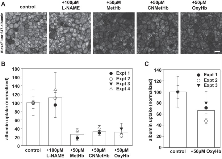
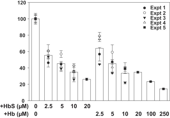
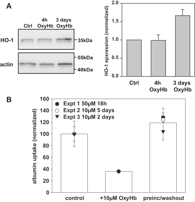
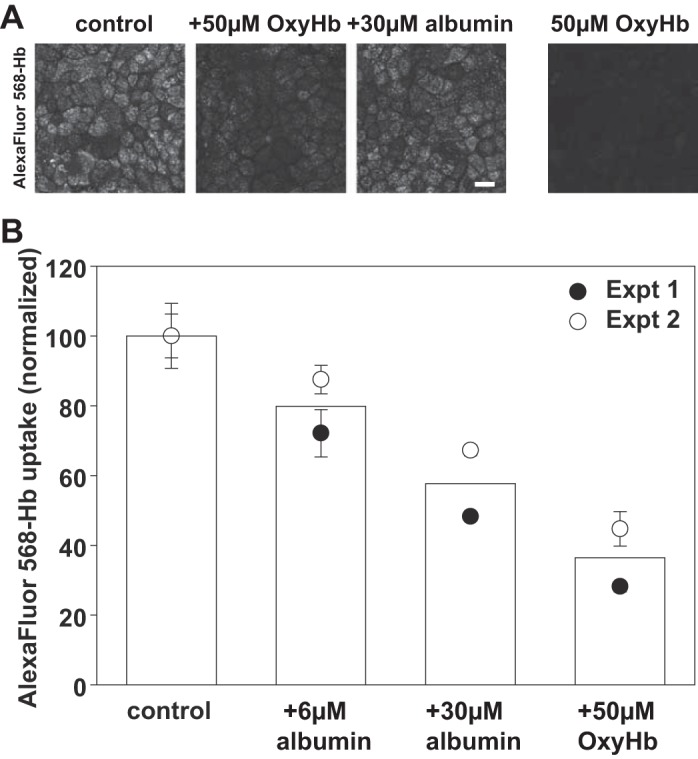
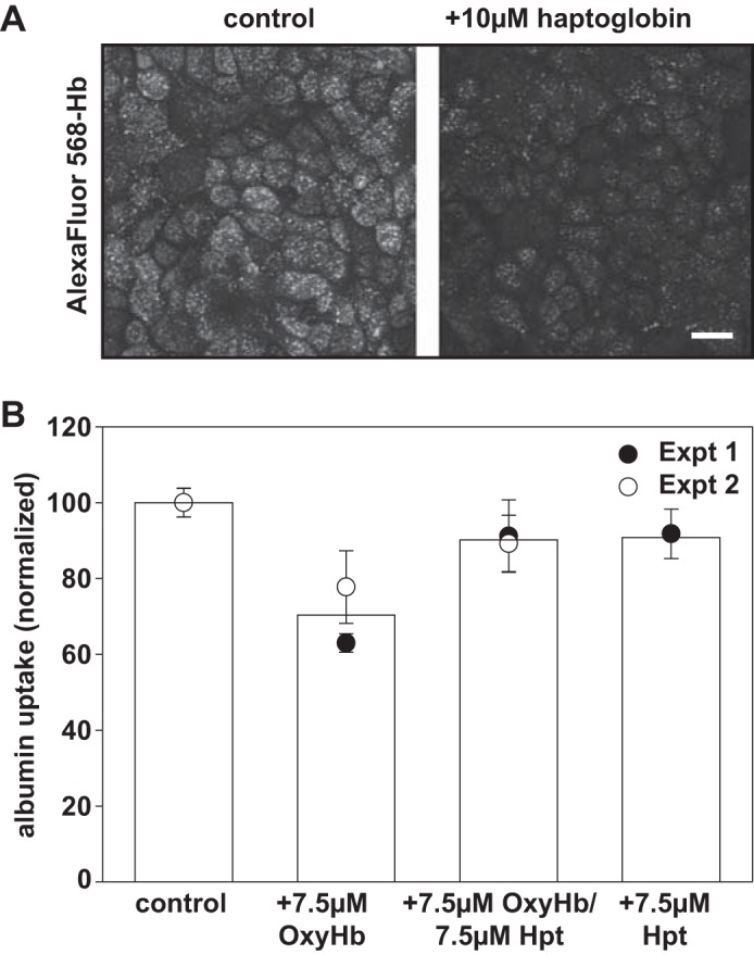
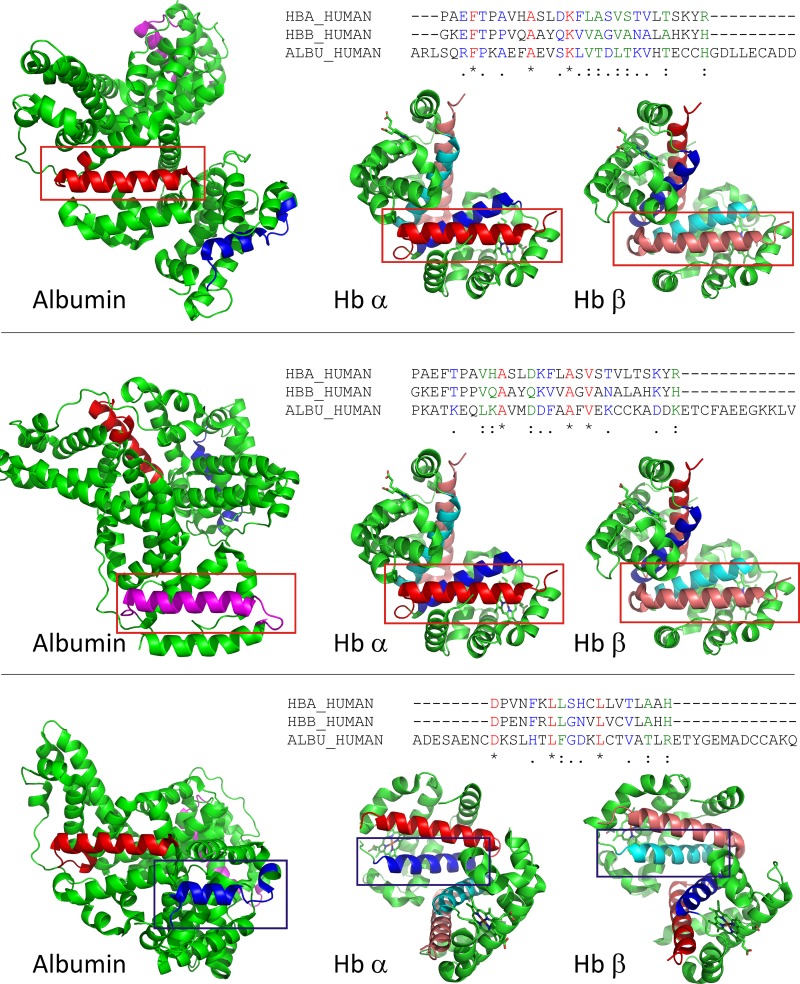
Proximal tubule (PT) dysfunction, including tubular proteinuria, is a significant complication in young sickle cell disease (SCD) that can eventually lead to chronic kidney disease. Hemoglobin (Hb) dimers released from red blood cells upon hemolysis are filtered into the kidney and internalized by megalin/cubilin receptors into PT cells. The PT is especially sensitive to heme toxicity, and tubular dysfunction in SCD is thought to result from prolonged exposure to filtered Hb. Here we show that concentrations of Hb predicted to enter the tubule lumen during hemolytic crisis competitively inhibit the uptake of another megalin/cubilin ligand (albumin) by PT cells. These effects were independent of heme reduction state. The Glu7Val mutant of Hb that causes SCD was equally effective at inhibiting albumin uptake compared with wild-type Hb. Addition of the Hb scavenger haptoglobin (Hpt) restored albumin uptake in the presence of Hb, suggesting that Hpt binding to the Hb αβ dimer-dimer interface interferes with Hb binding to megalin/cubilin. BLAST searches and structural modeling analyses revealed regions of similarity between Hb and albumin that map to this region and may represent sites of Hb interaction with megalin/cubilin. Our studies suggest that impaired endocytosis of megalin/cubilin ligands, rather than heme toxicity, may be the cause of tubular proteinuria in SCD patients. Additionally, loss of these filtered proteins into the urine may contribute to the extra-renal pathogenesis of SCD.

Keywords: megalin; proteinuria; proximal tubule; sickle cell disease; vitamin D.


Figures

Fig. 1.
Fig. 1.
Hemoglobin inhibits apical uptake of albumin by PT cells. A: filter-grown OK cells were preincubated for 30 min at 37°C with l-NAME (100 µM) where indicated and then exposed to 0.6 µM apically added Alexa Fluor 647-albumin in the presence or absence of 50 µM MetHb, CNMet-Hb, or OxyHb for 1 h at 37°C. After extensive washing, cells were fixed and processed for immunofluorescence to visualize cell-associated albumin. Representative fields are shown. Scale bar, 25 µm. B: cells incubated as above were solubilized, and cell-associated albumin was quantified by spectrofluorimetry. Mean albumin uptake in control cells was set at 100 to facilitate comparison between experiments. The results from several independent experiments (means ± SD of triplicate samples) are plotted, with each experiment represented by a different symbol. The mean uptake for a given condition is represented by the bar. C: filter-grown human HK-2 cells were incubated for 1 h at 37°C with 0.6 µM Alexa Fluor 647-albumin in the presence or absence of 50 µM OxyHb. Cell-associated albumin was quantified as above, and the mean ± SD of three independent experiments each performed in triplicate is plotted.
Fig. 2.
Fig. 2.
Dose response of hemoglobin and sickle cell hemoglobin S inhibition of albumin uptake. Triplicate filters of cells were preincubated with the indicated concentration of OxyHb or OxyHbS prior to addition of Alexa Fluor 647-albumin for 1 h and quantitation of albumin uptake. Cell-associated albumin in control untreated cells was normalized to 100 to enable combining data from multiple independent experiments, each represented by a distinct symbol. Based on this dose response, the half-maximal concentration at which OxyHb inhibits uptake of albumin is ~5 µM.
Fig. 3.
Fig. 3.
Hemoglobin acutely inhibits receptor-mediated endocytosis in proximal tubule cells. A: HK-2 cells were incubated with 50 µM OxyHb for 4 h or 3 days as indicated, then solubilized and blotted to detect heme oxygenase 1 (HO-1) and β-actin (as a loading control) Quantitation of HO-1 expression (avg ± range) relative to control and normalized to β-actin in two independent experiments is shown next to the blot. B: filter-grown OK cells were preincubated at 37°C with apically added OxyHb (10–50 µM) for 18 h to 5 days as indicated prior to extensive washout and subsequent addition of Alexa Fluor 647-albumin for 1 h at 37°C in the absence of OxyHb. As a positive control, OxyHb (10 µM) was included during the albumin uptake period; untreated cells incubated with Alexa Fluor 647-albumin were used as a negative control. Fluorescent cell-associated albumin was quantified by spectrofluorimetry. Data (means ± SD) from three independent experiments each performed in triplicate are shown.
Fig. 4.
Fig. 4.
Albumin inhibits endocytosis of hemoglobin by OK cells. A: OK cells were incubated with apically added 2 µM Alexa Fluor 568-Hb in the presence or absence of 50 µM unlabeled OxyHb or 30 µM albumin for 1 h at 37°C, then fixed and processed for immunofluorescence. As a control to confirm that OxyHb fluorescence is not detected in our studies, cells were incubated for 1 h with 50 µM unlabeled OxyHb (right-hand panel) Scale bar, 25 µm. B: OK cells were incubated with apically added 2 µM Alexa Fluor 568-Hb in the presence or absence of the incubated concentrations of albumin or unlabeled Hb for 1 h at 37°C prior to quantitation of cell-associated Hb fluorescence. Data from two experiments performed in triplicate are shown. Background fluorescence in cells incubated only with 50 µM unlabeled OxyHb was <10% that of the control values.
Fig. 5.
Fig. 5.
Haptoglobin inhibits hemoglobin uptake and restores albumin endocytosis by OK cells. A: OK cells were incubated with 2 µM Alexa Fluor 568-OxyHb for 1 h at 37°C in the presence or absence of 10 µM haptoglobin, then washed, fixed, and processed for immunofluorescence to visualize cell-associated Hb. Scale bar, 25 µm. B: filter-grown OK cells were incubated with 40 µg/ml Alexa Fluor 647-albumin in the presence of OxyHb (7.5 µM) and/or 7.5 µM haptoglobin (Hpt) as indicated. After extensive washing, cells were solubilized and fluorescent cell-associated albumin was quantified. Data (means ± SD) from two independent experiments each performed in triplicate are shown.
Fig. 6.
Fig. 6.
Sequence and structure comparison of potential megalin/cubilin binding regions in hemoglobin and albumin. Hb helical fragments involved in haptoglobin binding were aligned to human albumin sequence to search for comparable albumin motifs (see materials and methods for details). Three sequences in albumin were identified that have sequence and structural similarity to Hb. Top: alignment of Hb α- and β-helix H to albumin residues 244–271. The sequence alignment is shown on top. Aligned sequences are shown in the structures of albumin and Hb αβ-dimer and indicated by a red box. Helical elements are shown in red (albumin, hemoglobin α) and salmon (hemoglobin β); Middle: alignment of Hb α- and β-helix H to albumin residues 561–588. The sequence alignment is shown on top. Aligned sequences are shown in the structures of albumin and Hb αβ-dimer and indicated by a red box. Helical elements are shown in pink (albumin) red (hemoglobin α), and salmon (hemoglobin β) Bottom: alignment of Hb α- and β-helix G to albumin residues 87–105. The sequence alignment is shown on top. Aligned sequences are shown in the structures of albumin and Hb αβ-dimer and indicated by a blue box. Helical elements are shown in dark blue (albumin, hemoglobin α) and cyan (hemoglobin β).

References

    1. Alayash AI. Haptoglobin: old protein with new functions. Clin Chim Acta 412: 493–498, 2011. doi:10.1016/j.cca.2010.12.011. - DOI - PubMed
    1. Altschul SF, Madden TL, Schäffer AA, Zhang J, Zhang Z, Miller W, Lipman DJ. Gapped BLAST and PSI-BLAST: a new generation of protein database search programs. Nucleic Acids Res 25: 3389–3402, 1997. doi:10.1093/nar/25.17.3389. - DOI - PMC - PubMed
    1. Alvarez O, Montane B, Lopez G, Wilkinson J, Miller T. Early blood transfusions protect against microalbuminuria in children with sickle cell disease. Pediatr Blood Cancer 47: 71–76, 2006. doi:10.1002/pbc.20645. - DOI - PubMed
    1. Andersen CB, Torvund-Jensen M, Nielsen MJ, de Oliveira CL, Hersleth HP, Andersen NH, Pedersen JS, Andersen GR, Moestrup SK. Structure of the haptoglobin-haemoglobin complex. Nature 489: 456–459, 2012. doi:10.1038/nature11369. - DOI - PubMed
    1. Chiancone E, Alfsen A, Ioppolo C, Vecchini P, Agrò AF, Wyman J, Antonini E. Studies on the reaction of haptoglobin with haemoglobin and haemoglobin chains. I. Stoichiometry and affinity. J Mol Biol 34: 347–356, 1968. doi:10.1016/0022-2836(68)90258-1. - DOI - PubMed

MeSH terms