. 2017 Jun 1;312(6):C697-C706.
doi: 10.1152/ajpcell.00292.2016. Epub 2017 Mar 29.

Neurotrophin 3 upregulates proliferation and collagen production in human aortic valve interstitial cells: a potential role in aortic valve sclerosis

Affiliations

Neurotrophin 3 upregulates proliferation and collagen production in human aortic valve interstitial cells: a potential role in aortic valve sclerosis

Qingzhou Yao et al. Am J Physiol Cell Physiol. .

Abstract

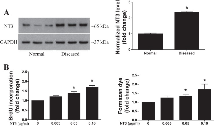
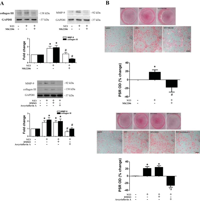
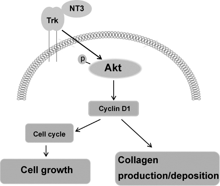
Calcific aortic valve disease (CAVD) is a leading cardiovascular disorder in the elderly. Diseased aortic valves are characterized by sclerosis (fibrosis) and nodular calcification. Sclerosis, an early pathological change, is caused by aortic valve interstitial cell (AVIC) proliferation and overproduction of extracellular matrix (ECM) proteins. However, the mechanism of aortic valve sclerosis remains unclear. Recently, we observed that diseased human aortic valves overexpress growth factor neurotrophin 3 (NT3). In the present study, we tested the hypothesis that NT3 is a profibrogenic factor to human AVICs. AVICs isolated from normal human aortic valves were cultured in M199 growth medium and treated with recombinant human NT3 (0.10 µg/ml). An exposure to NT3 induced AVIC proliferation, upregulated the production of collagen and matrix metalloproteinase (MMP), and augmented collagen deposition. These changes were abolished by inhibition of the Trk receptors. NT3 induced Akt phosphorylation and increased cyclin D1 protein levels in a Trk receptor-dependent fashion. Inhibition of Akt abrogated the effect of NT3 on cyclin D1 production. Furthermore, inhibition of either Akt or cyclin D1 suppressed NT3-induced cellular proliferation and MMP-9 and collagen production, as well as collagen deposition. Thus, NT3 upregulates cellular proliferation, ECM protein production, and collagen deposition in human AVICs. It exerts these effects through the Trk-Akt-cyclin D1 cascade. NT3 is a profibrogenic mediator in human aortic valve, and overproduction of NT3 by aortic valve tissue may contribute to the mechanism of valvular sclerosis.

Keywords: AVIC; Akt; Trk; fibrogenic response; neurotrophin 3.


Figures

Fig. 1.
Fig. 1.
Neurotrophin 3 (NT3) promotes human aortic valve interstitial cell (AVIC) proliferation. A: representative immunoblot and densitometric data show that AVICs of diseased valves have higher levels of NT3 protein in comparison with normal AVICs. Data are presented as means ± SE of 6 cell isolates from distinct donor valves in each group. Statistical analyses were performed using t-test and confirmed using nonparametric Mann-Whitney U-test. *P < 0.05 vs. normal. B: normal human AVICs were treated with different doses of recombinant human NT3 (0.005–0.10 µg/ml) for 3 days. 5-Bromo-2′-deoxyuridine (BrdU) and Cell Counting Kit (CCK)-8 (formazan dye formation) assays show that NT3 induces AVIC proliferation in a dose-dependent fashion. Data are presented as means ± SE of 6 cell isolates from distinct donor valves. Statistical analyses were performed using ANOVA with the post hoc Bonferroni/Dunn test and confirmed using nonparametric Kruskal-Wallis test. *P < 0.05 vs. control (without NT3).
Fig. 2.
Fig. 2.
NT3 upregulates collagen production in human AVICs. A: normal AVICs were treated with NT3 (0.10 µg/ml) for 3 days. Representative immunoblots of 5 experiments using cell isolates from distinct donor valves show that NT 3 elevates levels of cell-associated matrix metalloproteinase-9 (MMP-9) and collagen III but has no effect on MMP-2 and collagen I levels in the cells. B: normal AVICs were treated with NT3 for 28 days. Picro Sirius Red (PSR) staining was applied to stain collagens. Representative images (scale bar = 150 μm) and spectrophotometric analysis of eluted stain show that cells exposed to NT3 formed a greater amount of collagen deposits. Data are presented as means ± SE of 5 experiments using different cell isolates from distinct donor valves. Statistical analyses were performed using t-test and confirmed using nonparametric Mann-Whitney U-test. *P < 0.05 vs. control.
Fig. 3.
Fig. 3.
The proliferative effect of NT3 on human AVICs is mediated by the Trk receptors. Normal AVICs were treated with NT3 for 3 days in the presence or absence of Trk inhibitor K252a (0.20 µM) or isoform-selective inhibitors (Fc chimeras specific for TrkA, TrkB, and TrkC; 2.0 µg/ml each) for 3 days. Inhibition of Trk markedly reduces NT3-induced BrdU incorporation and formazan dye formation. Each isoform-selective inhibitor attenuates the BrdU incorporation and formazan dye formation induced by NT3. Data are presented as means ± SE of 5 experiments using different cell isolates from distinct donor valves. Statistical analyses were performed using ANOVA with the post hoc Bonferroni/Dunn test and confirmed using nonparametric Kruskal-Wallis test. *P < 0.05 vs. control; #P < 0.05 vs. NT3 or NT3 + DMSO.
Fig. 4.
Fig. 4.
NT3 upregulates collagen deposition through Trk receptors. A: normal AVICs were treated with NT3 in the presence or absence of the Trk inhibitor K252a (0.20 µM) for 3 days. Representative immunoblots and densitometric data show that inhibition of Trk suppresses NT3-induced expression of collagen III and MMP-9. Data are presented as means ± SE of 5 experiments using different cell isolates from distinct donor valves. Statistical analyses were performed using ANOVA with the post hoc Bonferroni/Dunn test and confirmed using nonparametric Kruskal-Wallis test. *P < 0.05 vs. control; #P < 0.05 vs. NT3 or NT3 + DMSO. B: normal AVICs were treated with NT3 in the presence or absence of K252a for 28 days. Representative images of PSR staining (scale bar = 150 μm) and spectrophotometric data show that collagen deposition induced by NT3 is markedly reduced by inhibition of Trk. Data are presented as means ± SE of 5 experiments using different cell isolates from distinct donor valves. Statistical analyses were performed using ANOVA with the post hoc Bonferroni/Dunn test and confirmed using nonparametric Kruskal-Wallis test. *P < 0.05 vs. control; #P < 0.05 vs. NT3 or NT3 + DMSO.
Fig. 5.
Fig. 5.
NT3 upregulates cyclin D1 levels through the Trk-Akt pathway. A: normal AVICs were treated with NT3 in the presence or absence of Trk inhibitor for 1 to 24 h. Representative immunoblots of 3 separate experiments show that NT3 induces the phosphorylation of Akt. Inhibition of Trk receptors suppresses NT3-induced Akt phosphorylation. B: normal AVICs were treated with NT3 in the presence or absence of Trk inhibitor (K252a; 0.20 µM). Representative immunoblots and densitometric data show that NT3 upregulates cyclin D1 levels at 24 h and inhibition of Trk suppresses NT3-induced cyclin D1 expression. Data are presented as means ± SE of 5 experiments using different cell isolates from distinct donor valves. Statistical analyses were performed using ANOVA with the post hoc Bonferroni/Dunn test and confirmed using nonparametric Kruskal-Wallis test. *P < 0.05 vs. control; #P < 0.05 vs. NT3 or NT3 + DMSO. C: normal AVICs were treated with NT3 in the presence or absence of Akt inhibitor (MK2206; 5.0 µM). Representative immunoblots and densitometric data show that inhibition of Akt reduces NT3-induced cyclin D1 expression. Data are presented as means ± SE of 5 experiments using different cell isolates from distinct donor valves. Statistical analyses were performed using ANOVA with the post hoc Bonferroni/Dunn test and confirmed using nonparametric Kruskal-Wallis test. *P < 0.05 vs. control; #P < 0.05 vs. NT3.
Fig. 6.
Fig. 6.
Inhibition of Akt or cyclin D1 suppresses NT3-induced AVIC proliferation. A: normal AVICs were treated with NT3 in the presence or absence of Akt inhibitor (MK2206, 5.0 µM) for 3 days. BrdU incorporation and formazan dye formation were reduced by MK2206. Data are presented as means ± SE of 5 experiments using different cell isolates from distinct donor valves. Statistical analyses were performed using ANOVA with the post hoc Bonferroni/Dunn test and confirmed using nonparametric Kruskal-Wallis test. *P < 0.05 vs. control; #P < 0.05 vs. NT3. B: normal AVICs were treated with NT3 in the presence or absence of cyclin D1 inhibitor (arcyriaflavin A; 0.25 µM) for 3 days. BrdU incorporation and formazan dye formation were reduced by Arcyriaflavin A. Data are presented as means ± SE of 5 experiments using different cell isolates from distinct donor valves. Statistical analyses were performed using ANOVA with the post hoc Bonferroni/Dunn test and confirmed using nonparametric Kruskal-Wallis test. *P < 0.05 vs. control; #P < 0.05 vs. NT3 or NT3 + DMSO.
Fig. 7.
Fig. 7.
Akt and cyclin D1 play a role in mediating NT3-induced collagen deposition. A: normal AVICs were treated with NT3 in the presence or absence of Akt inhibitor (MK2206; 5.0 µM) or cyclin D 1 inhibitor (arcyriaflavin A; 0.25 µM) for 3 days. Representative immunoblots and densitometric data show that inhibition of Akt or cyclin D1 suppresses NT3-induced expression of collagen III and MMP-9. Data are presented as means ± SE of 5 experiments using different cell isolates from distinct donor valves. Statistical analyses were performed using ANOVA with the post hoc Bonferroni/Dunn test and confirmed using nonparametric Kruskal-Wallis test. *P < 0.05 vs. control; #P < 0.05 vs. NT3 or NT3 + DMSO. B: normal AVICs were treated with NT3 in the presence or absence of Akt inhibitor or cyclin D1 inhibitor for 28 days. Representative images of PSR staining (scale bar = 150 µm) and spectrophotometric data show that collagen deposition induced by NT3 is markedly reduced by inhibition of Akt or cyclin D1. Data are presented as means ± SE of 5 experiments using different cell isolates from distinct donor valves. Statistical analyses were performed using ANOVA with the post hoc Bonferroni/Dunn test and confirmed using nonparametric Kruskal-Wallis test. *P < 0.05 vs. control; #P < 0.05 vs. NT3 or NT3 + DMSO.
Fig. 8.
Fig. 8.
Overview of the signaling pathways that mediate the profibrogenic effect of NT3 on human AVICs. NT3 promotes cellular proliferation, extracellular matrix (ECM) protein production and collagen deposition in human AVICs through Trk receptors. A Trk-Akt-cyclin D1 cascade mediates these effects of NT3 and may enhance valvular profibrogenic activity.

Similar articles

Cited by

References

    1. Bashey RI, Torii S, Angrist A. Age-related collagen and elastin content of human heart valves. J Gerontol 22: 203–208, 1967. doi:10.1093/geronj/22.2.203. - DOI - PubMed
    1. Chen WC, Lin HH, Tang MJ. Matrix-stiffness-regulated inverse expression of Krüppel-like factor 5 and Krüppel-like factor 4 in the pathogenesis of renal fibrosis. Am J Pathol 185: 2468–2481, 2015. doi:10.1016/j.ajpath.2015.05.019. - DOI - PubMed
    1. Cimini M, Boughner DR, Ronald JA, Aldington L, Rogers KA. Development of aortic valve sclerosis in a rabbit model of atherosclerosis: an immunohistochemical and histological study. J Heart Valve Dis 14: 365–375, 2005. - PubMed
    1. Cuevas CA, Gonzalez AA, Inestrosa NC, Vio CP, Prieto MC. Angiotensin II increases fibronectin and collagen I through the β-catenin-dependent signaling in mouse collecting duct cells. Am J Physiol Renal Physiol 308: F358–F365, 2015. doi:10.1152/ajprenal.00429.2014. - DOI - PMC - PubMed
    1. Dagnell C, Kemi C, Klominek J, Eriksson P, Sköld CM, Eklund A, Grunewald J, Olgart Höglund C. Effects of neurotrophins on human bronchial smooth muscle cell migration and matrix metalloproteinase-9 secretion. Transl Res 150: 303–310, 2007. doi:10.1016/j.trsl.2007.05.001. - DOI - PubMed

MeSH terms

Supplementary concepts

LinkOut - more resources