. 2017 Jun 1;312(6):C724-C732.
doi: 10.1152/ajpcell.00348.2016. Epub 2017 Mar 29.

Ulk1-mediated autophagy plays an essential role in mitochondrial remodeling and functional regeneration of skeletal muscle

Affiliations

Ulk1-mediated autophagy plays an essential role in mitochondrial remodeling and functional regeneration of skeletal muscle

Jarrod A Call et al. Am J Physiol Cell Physiol. .

Abstract

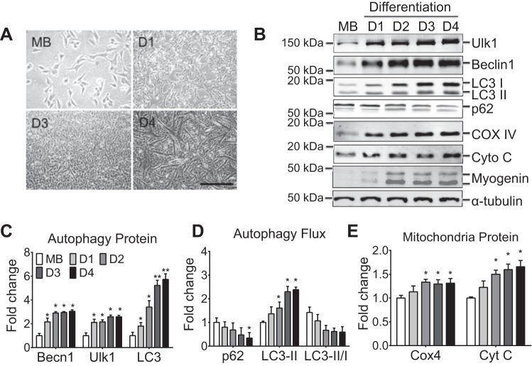
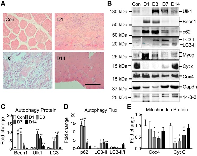
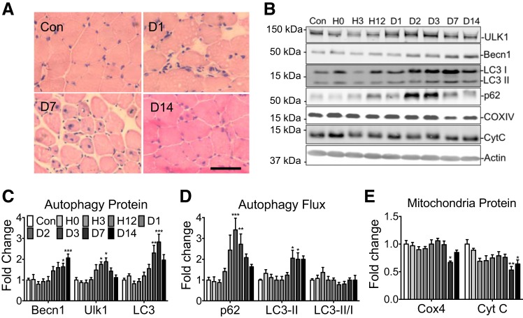
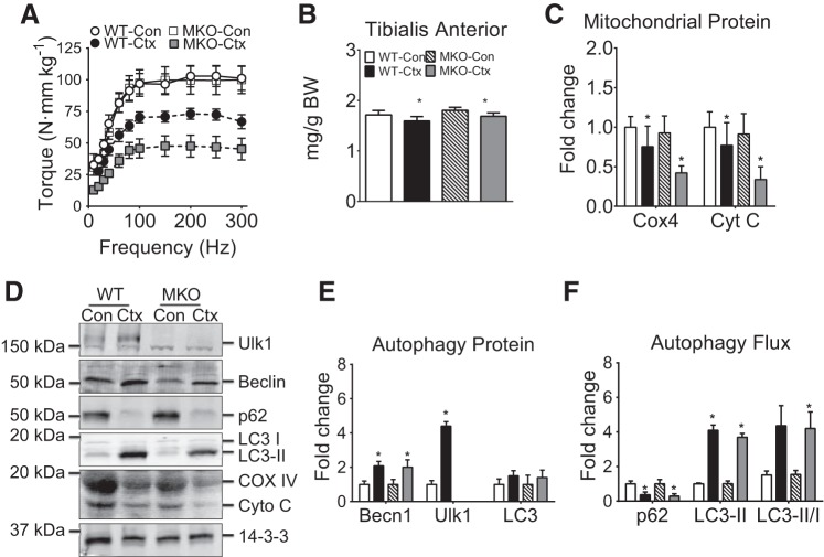
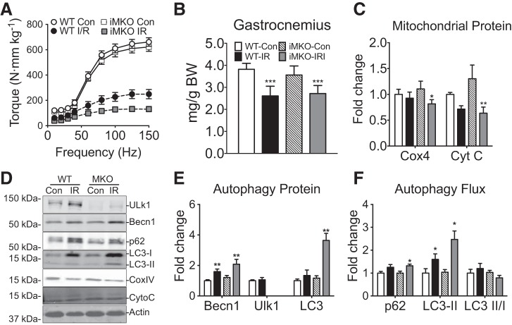
Autophagy is a conserved cellular process for degrading aggregate proteins and dysfunctional organelle. It is still debatable if autophagy and mitophagy (a specific process of autophagy of mitochondria) play important roles in myogenic differentiation and functional regeneration of skeletal muscle. We tested the hypothesis that autophagy is critical for functional regeneration of skeletal muscle. We first observed time-dependent increases (3- to 6-fold) of autophagy-related proteins (Atgs), including Ulk1, Beclin1, and LC3, along with reduced p62 expression during C2C12 differentiation, suggesting increased autophagy capacity and flux during myogenic differentiation. We then used cardiotoxin (Ctx) or ischemia-reperfusion (I/R) to induce muscle injury and regeneration and observed increases in Atgs between days 2 and 7 in adult skeletal muscle followed by increased autophagy flux after day 7 Since Ulk1 has been shown to be essential for mitophagy, we asked if Ulk1 is critical for functional regeneration in skeletal muscle. We subjected skeletal muscle-specific Ulk1 knockout mice (MKO) to Ctx or I/R. MKO mice had significantly impaired recovery of muscle strength and mitochondrial protein content post-Ctx or I/R. Imaging analysis showed that MKO mice have significantly attenuated recovery of mitochondrial network at 7 and 14 days post-Ctx. These findings suggest that increased autophagy protein and flux occur during muscle regeneration and Ulk1-mediated mitophagy is critical for recovery for the mitochondrial network and hence functional regeneration.

Keywords: Unc-51-like autophagy activating kinase 1; mitophagy; muscle repair; torque.


Figures

Fig. 1.
Fig. 1.
Autophagy-related protein content during myoblast differentiation. A and B: representative contrast light images of differentiating myoblast and immunoblot images of Ulk1, Beclin1, LC3-I, LC3-II, p62, cytochrome c oxidase complex IV (COX4), cytochrome c (Cyt C), myogenin (myog), and tubulin. C: quantification of Beclin1, Ulk1, and total LC3, collectively an index of autophagy capacity. D: quantification of p62, LC3-II, and LC3-II/I ratio collectively an index of autophagy flux. E: quantification of COX4 and Cyt C as a measure of mitochondrial content. Results are represented as means ± SE from n = 4 separate cell culture experiments per day; n = 4 for each time point. *P < 0.05, significantly different from day 1. **P < 0.05, significantly different from day 2.
Fig. 2.
Fig. 2.
Autophagy-related protein content during adult skeletal muscle regeneration. A: representative histological images of regenerating muscle at 1, 3, and 14 days postinjury relative to contralateral uninjured control: scale bar = 200 μm. B: representative immunoblot images of Ulk1, Beclin1, p62, LC3-I, LC3-II, myogenin, Cyt C, COX4, GAPDH, and 14-3-3- for cardiotoxin (Ctx)-injured muscles and contralateral control muscles at 1, 3, 7, and 14 days postinjury (D1, D3, D7, and D14). C: quantification of Beclin1, Ulk1, and total LC3, collectively an index of autophagy capacity, during muscle regeneration. D: quantification of p62, LC3-II, and LC3-II/I ratio collectively an index of autophagy flux, during muscle regeneration. E: quantification of COX4 and Cyt C as a measure of mitochondrial content. Results are represented as means ± SE from n = 5 mice for each day. *P < 0.05, **P < 0.01, ***P < 0.001, significantly different from contralateral control.
Fig. 3.
Fig. 3.
Autophagy-related protein content following hindlimb ischemia-reperfusion injury. A: representative histological images of regenerating plantaris muscle at 24 h and 7 and 14 days postinjury relative to uninjured control (Con): scale bar = 100 μm. B: representative immunoblot images of Ulk1, Beclin1, p62, LC3-I, LC3-II, Cyt C, COX4, and actin from the plantaris muscle at 0, 3, 12, 24, 48, and 72 h, and 7 and 14 days post-ischemia-reperfusion (I/R) injury and Con control. C: quantification of Beclin1, Ulk1, and total LC3, collectively an index of autophagy capacity, during muscle regeneration. D: quantification of p62, LC3-II collectively as an index of autophagy flux, during muscle regeneration. E: quantification of COX4 and Cyt C as a measure of mitochondrial content. Results are represented as means ± SE for n = 6 mice for each time point. *P < 0.05, **P < 0.01, ***P < 0.0001 significantly different from Con.
Fig. 4.
Fig. 4.
Muscle-specific Ulk1 deficiency blunts functional muscle regeneration following Ctx. A: quantification of tibialis anterior muscle mass and body mass for muscle-specific Ulk1 knockout mice (MKO) mice and littermate wild-type (WT) mice at day 7 postinjury. B: torque-frequency analysis shown relative to uninjured WT (WT-Con) at day 7 postinjury. Mass and torque results are represented as means ± SE for n = 5 WT-Con and WT-Ctx and n = 7 MKO-Con and MKO-Ctx. C: quantification of COX4 and Cyt C at day 7 postinjury. All immunoblot results are represented as means ± SE for n = 5 mice for each genotype/injury. D: representative immunoblot images of Ulk1, Beclin1, p62, LC3-I, LC3-II, Cyt C, COX4, and 14-3-3 for injured muscles (day 7 postinjury) and contralateral control for MKO mice and littermate WT mice. E: quantification of Beclin1, Ulk1, and total LC3, collectively an index of autophagy capacity, at day 7 postinjury. F: quantification of p62, LC3-II collectively an index of autophagy flux, at day 7 postinjury.
Fig. 5.
Fig. 5.
Muscle-specific conditional Ulk1 deficiency blunts functional muscle regeneration following I/R. A: quantification of gastrocnemius muscle mass and body mass for MKO and littermate WT mice at day 14 postinjury. B: torque frequency analysis of plantar flexors relative to WT Con. Mass and torque results are represented as means ± SE for n = 5–6 for each genotype/injury. C: quantification of COX4 and Cyt C at day 7 postinjury. All immunoblot results are represented as means ± SE for n = 5–6 for each genotype/injury. D: representative immunoblots of Ulk1, Beclin1, p62, LC3-I, LC3-II, COX4, Cyt C, and 14-3-3 for injured muscles (day 14 postinjury) and Con for MKO mice and littermate WT mice. E: quantification of Beclin1, Ulk1, and total LC3 indicant of autophagy capacity at day 14 postinjury. F: quantification of p62, LC3-II as an index of autophagy flux at day 14 postinjury.
Fig. 6.
Fig. 6.
Mitochondrial network organization during muscle regeneration. Confocal microscopy analysis of the MitoTimer reporter gene was performed for somatic gene transfer-transfected flexor digitorum longus muscle following muscle injury at 1, 7, and 14 days of recovery and compared with the contralateral uninjured muscle (n = 4 mice per time point).

References

    1. Baltgalvis KA, Call JA, Cochrane GD, Laker RC, Yan Z, Lowe DA. Exercise training improves plantar flexor muscle function in mdx mice. Med Sci Sports Exerc 44: 1671–1679, 2012. doi:10.1249/MSS.0b013e31825703f0. - DOI - PMC - PubMed
    1. Bernet JD, Doles JD, Hall JK, Kelly Tanaka K, Carter TA, Olwin BB. p38 MAPK signaling underlies a cell-autonomous loss of stem cell self-renewal in skeletal muscle of aged mice. Nat Med 20: 265–271, 2014. doi:10.1038/nm.3465. - DOI - PMC - PubMed
    1. Blau HM, Cosgrove BD, Ho AT. The central role of muscle stem cells in regenerative failure with aging. Nat Med 21: 854–862, 2015. doi:10.1038/nm.3918. - DOI - PMC - PubMed
    1. Bonheur JA, Albadawi H, Patton GM, Watkins MT. A noninvasive murine model of hind limb ischemia-reperfusion injury. J Surg Res 116: 55–63, 2004. doi:10.1016/S0022-4804(03)00232-4. - DOI - PubMed
    1. Call JA, Chain KH, Martin KS, Lira VA, Okutsu M, Zhang M, Yan Z. Enhanced skeletal muscle expression of extracellular superoxide dismutase mitigates streptozotocin-induced diabetic cardiomyopathy by reducing oxidative stress and aberrant cell signaling. Circ Heart Fail 8: 188–197, 2015. doi:10.1161/CIRCHEARTFAILURE.114.001540. - DOI - PMC - PubMed

MeSH terms

LinkOut - more resources