Review
. 2017 Jul 1;313(1):F55-F61.
doi: 10.1152/ajprenal.00046.2017. Epub 2017 Mar 29.

Saving the sweetness: renal glucose handling in health and disease

Affiliations
Review

Saving the sweetness: renal glucose handling in health and disease

Blythe D Shepard et al. Am J Physiol Renal Physiol. .

Abstract

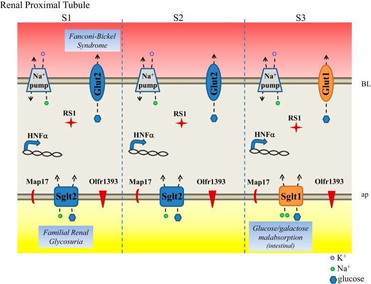
Glucose homeostasis is highly controlled, and the function of the kidney plays an integral role in this process. The exquisite control of blood glucose relies, in part, on renal glucose filtration, renal glucose reabsorption, and renal gluconeogenesis. Particularly critical to maintaining glucose homeostasis is the renal reabsorption of glucose; with ~162 g of glucose filtered by the kidney per day, it is imperative that the kidney have the ability to efficiently reabsorb nearly 100% of this glucose back in the bloodstream. In this review, we focus on this central process, highlighting the renal transporters and regulators involved in both the physiology and pathophysiology of glucose reabsorption.

Keywords: glucose reabsorption; glucose transporter 2; proximal tubule; sodium-glucose cotransporter 1; sodium-glucose cotransporter 2.


Figures

Fig. 1.
Fig. 1.
Glucose transporters and their regulators in the renal proximal tubule. Na+-glucose cotransporters Sglt1 and Sglt2 localize to the apical plasma (ap) membrane of the proximal tubule where they face the newly forming urine and use the existing Na+ gradient to reabsorb all filtered glucose. The Na+ gradient is maintained by the Na+-K+-ATPase (labeled Na+ pump) on the basolateral (BL) membrane. Glut1 and Glut2 localize to the BL plasma membrane where they function to release the reabsorbed and newly generated glucose back in circulation. Known regulators of the glucose transporters that are discussed in text are labeled in the proximal tubule segments in which they are known to be found. Diseases associated with the glucose transporters are also noted.

References

    1. Abbasi F, Azizi F, Javaheri M, Mosallanejad A, Ebrahim-Habibi A, Ghafouri-Fard S. Segregation of a novel homozygous 6 nucleotide deletion in GLUT2 gene in a Fanconi-Bickel syndrome family. Gene 557: 103–105, 2015. doi:10.1016/j.gene.2014.12.024. - DOI - PubMed
    1. Abdul-Ghani MA, DeFronzo RA, Norton L. Novel hypothesis to explain why SGLT2 inhibitors inhibit only 30-50% of filtered glucose load in humans. Diabetes 62: 3324–3328, 2013. doi:10.2337/db13-0604. - DOI - PMC - PubMed
    1. Albertoni Borghese MF, Majowicz MP, Ortiz MC, Passalacqua MR, Sterin Speziale NB, Vidal NA. Expression and activity of SGLT2 in diabetes induced by streptozotocin: relationship with the lipid environment. Nephron, Physiol 112: 45–52, 2009. doi:10.1159/000214214. - DOI - PubMed
    1. Alesutan I, Sopjani M, Dërmaku-Sopjani M, Munoz C, Voelkl J, Lang F. Upregulation of Na-coupled glucose transporter SGLT1 by Tau tubulin kinase 2. Cell Physiol Biochem 30: 458–465, 2012. doi:10.1159/000339039. - DOI - PubMed
    1. Augustin R. The protein family of glucose transport facilitators: It’s not only about glucose after all. IUBMB Life 62: 315–333, 2010. doi:10.1002/iub.315. - DOI - PubMed

Publication types

Substances