. 2017 May 1;216(5):1321-1335.
doi: 10.1083/jcb.201609071. Epub 2017 Mar 29.

The crossover conformational shift of the GTPase atlastin provides the energy driving ER fusion

Affiliations

The crossover conformational shift of the GTPase atlastin provides the energy driving ER fusion

James Winsor et al. J Cell Biol. .

Abstract

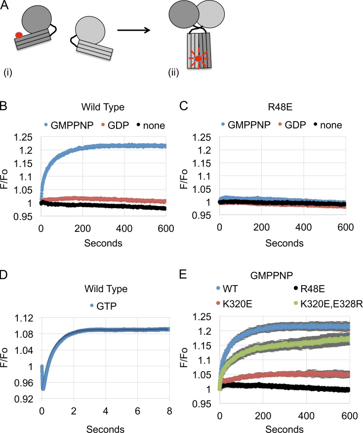
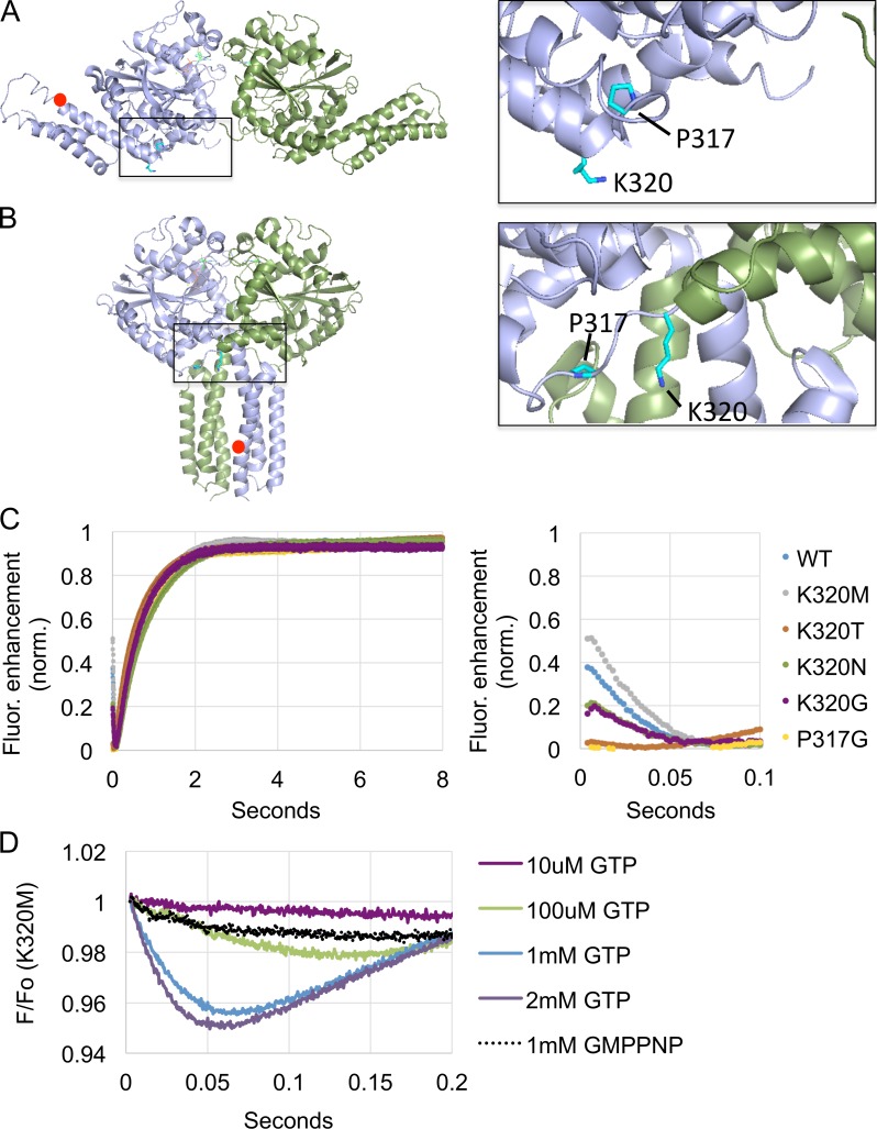
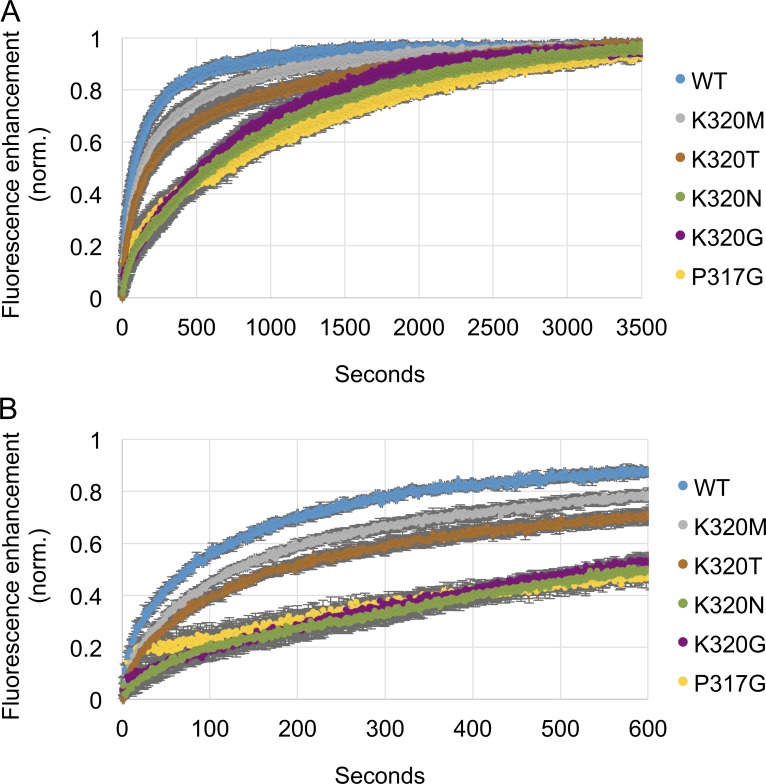
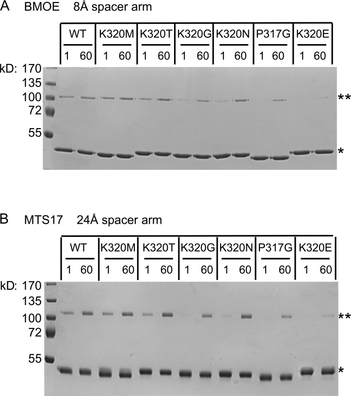
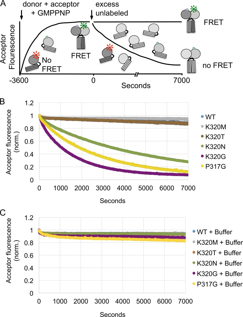
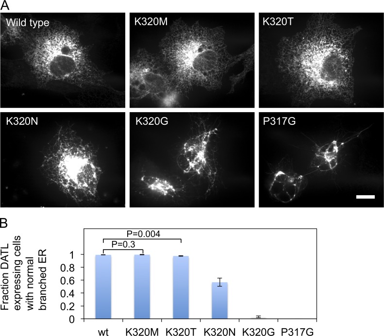
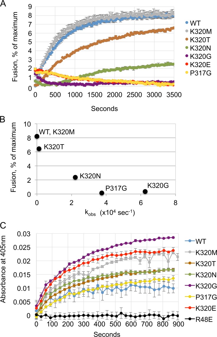
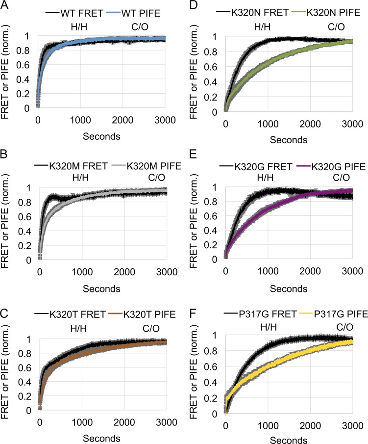
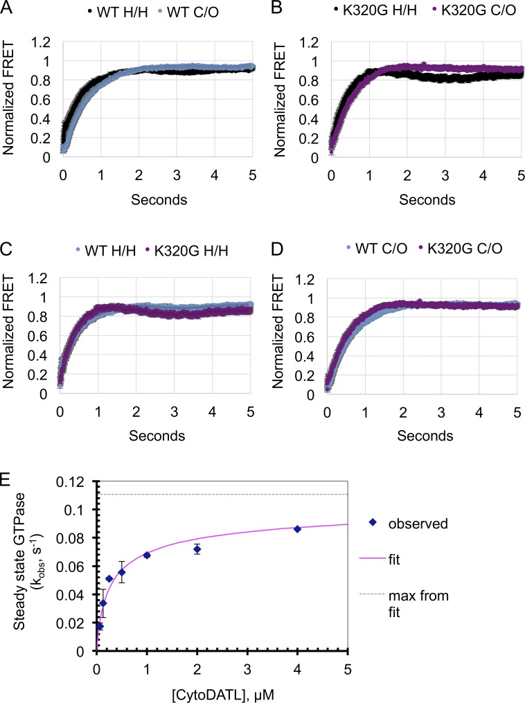
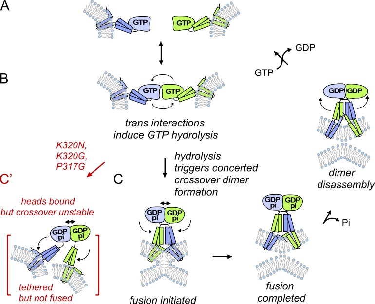
The homotypic fusion of endoplasmic reticulum membranes is catalyzed by the atlastin GTPase. The mechanism involves trans-dimerization between GTPase heads and a favorable crossover conformational shift, catalyzed by GTP hydrolysis, that converts the dimer from a "prefusion" to "postfusion" state. However, whether crossover formation actually energizes fusion remains unclear, as do the sequence of events surrounding it. Here, we made mutations in atlastin to selectively destabilize the crossover conformation and used fluorescence-based kinetic assays to analyze the variants. All variants underwent dimerization and crossover concurrently, and at wild-type rates. However, certain variants were unstable once in the crossover dimer conformation, and crossover dimer stability closely paralleled lipid-mixing activity. Tethering, however, appeared to be unimpaired in all mutant variants. The results suggest that tethering and lipid mixing are catalyzed concurrently by GTP hydrolysis but that the energy requirement for lipid mixing exceeds that for tethering, and the full energy released through crossover formation is necessary for fusion.


Figures

Figure 1.
Figure 1.
PIFE assay for crossover. (A) A schematic of Cy3 fluorescence enhancement as a cytoDATL monomer labeled with Cy3 on an engineered G343C residue in the 3HB (i) undergoes dimerization and crossover (ii). (B and C) PIFE, or fluorescence enhancement (F/Fo), over time for either wild type (B) or R48E (C) Cy3-cytoDATL mixed with the indicated nucleotides. (D) F/Fo over time when wild-type Cy3-cytoDATL is mixed with GTP. (E) F/Fo over time when wild type, R48E, K320E, or the double-mutant variant K320E, E328R is mixed with GMPPNP. For all assays, final concentrations after mixing were 2 µM Cy3-cytoDATL and 1 mM nucleotide. Either a single representative trace (B–D) or the mean of three replicates (±SEM) is shown (E). WT, wild type.
Figure 2.
Figure 2.
Mutant variants show no defects in GTP-catalyzed crossover formation. (A and B, left) The positions of K320 and P317 mutations made to target DATL crossover superimposed onto PyMOL renderings of (A) the hATL1 form2 extended dimer PDB 3QOF and (B) the hATL1 form3 crossover dimer PDB 4IDP. The position of the Cy3 dye in each structure is indicated with a red circle. (A and B, right) Enlargement of the boxed regions in A and B showing the K320 and P317 side chains highlighted in cyan. (C) Normalized PIFE over time when each of the indicated Cy3-cytoDATL mutant variants is mixed with GTP. (C, right) Zoomed-in view of the first 100 ms of the trace in C. The mean of seven runs is shown for each trace (SEM < 0.01) and all traces were repeated with independent protein preps with similar results. (D) A single representative PIFE trace over time when K320M Cy3-cytoDATL is mixed with the indicated concentrations of GTP or GMPPNP. The final concentrations after mixing were 2 µM Cy3-cytoDATL and 1 mM nucleotide unless indicated otherwise. WT, wild type.
Figure 3.
Figure 3.
Mutant variants show defects in GMPPNP-induced crossover. (A) Normalized PIFE (n = 3 replicates, ±SEM) over time when each of the indicated Cy3-cytoDATL variants is mixed with GMPPNP. (B) Zoomed-in view of the first 600 s of the trace in A. Final concentrations after mixing were 1 µM Cy3-cytoDATL and 1 mM nucleotide. All traces were repeated with independent protein preparations with similar results. WT, wild type.
Figure 4.
Figure 4.
Cross-linking confirms crossover defects for a subset of mutant variants. Each of the indicated cytoDATL variants was incubated at RT for either 1 min or 60 min in the presence of GMPPNP and then subjected to 20 s of cross-linking with either BMOE (8 Å spacer arm; A) or MTS17 (24 Å spacer arm; B). The structure of each cross-linker is shown to the right. Cross-linked dimers were resolved by SDS-PAGE and visualized with Coomassie blue. The single asterisk marks the monomer, and the double asterisk marks the cross-linked dimer. All variants had the G343C substitution. Concentrations before cross-linker addition were 2 µM CytoDATL and 1 mM nucleotide. The data shown are representative of at least two independent experiments. WT, wild type.
Figure 5.
Figure 5.
A subset of mutant variants has decreased crossover dimer stability. (A) Schematic of the assay. 2 µM of the indicated cytoDATL variants labeled with Alexa Fluor 488/647 at a 1:1 donor/acceptor ratio were incubated with 1 mM GMPPNP (final concentrations) for 60 min to form crossover dimers. After confirming that the FRET-induced acceptor fluorescence signal had plateaued, a fivefold molar excess of the corresponding unlabeled cytoDATL mutant variant was added and the subsequent decay in acceptor signal monitored over time. (B and C) Loss of acceptor fluorescence (n = 3 replicates, ±SEM) after addition of either the corresponding unlabeled competitor protein (B) or buffer (C). WT, wild type.
Figure 6.
Figure 6.
A subset of mutant variants causes abnormal ER network structure. (A) COS-7 cells transfected with each indicated variant of full-length Venus-tagged DATL were fixed and imaged 48 h later by confocal microscopy. Bar, 10 µm. (B) Quantification of the percentage of expressing cells displaying a normal branched ER (>100 cells per measurement; data represent mean of three independent measurements ± SD); *, P < 0.0001 (Student’s t test) with respect to wild type (wt).
Figure 7.
Figure 7.
Crossover dimer stability correlates more closely with fusion than tethering. (A) Mutant variants are variably defective in in vitro fusion activity. The full-length DATL version of each mutant variant was reconstituted into donor and acceptor vesicles at a 1:1,000 protein/lipid ratio. Fusion was monitored as the dequenching of MB-labeled lipid present in the donor vesicles (0.6 mM total lipid) over time after addition of 1 mM GTP (n = 3 replicates; ±SEM). (B) Fusion activity closely parallels crossover dimer stability. The apparent dissociation rate constant for each variant, calculated by fitting the mean of three traces (from Fig. 5 B) to an exponential decay equation (Materials and methods), is plotted against the mean percent fusion (SEM < 0.3%) in vitro (endpoint of A) achieved by the same variant. (C) Vesicle tethering activity does not correlate with crossover dimer stability. The full-length DATL version of each mutant variant was reconstituted into vesicles at a 1:1,000 protein/lipid ratio (0.6 mM total lipid). Tethering by each variant was monitored as the increase in 405-nm absorbance over time after addition of 1 mM GTP (n = 3 replicates; ±SEM). WT, wild type.
Figure 8.
Figure 8.
Head-to-head dimerization occurs before crossover when initiated with GMPPNP. Measurements of head-to-head dimerization monitored by FRET between wild type (A), K320M (B), K320T (C), K320N (D), K320G (E), and P317G (F) cytoDATL variants. Normalized FRET efficiency (E) = 1 − (IDA/ID), over time, from mixing 1 µM of the indicated variants with 1 mM GMPPNP, is shown relative to the normalized PIFE traces for each variant obtained in Fig. 3 A using the same concentrations of protein and nucleotide. The mean of three replicates (±SEM) is shown, and the entire set of traces was repeated with independent protein preparations with similar results. WT, wild type.
Figure 9.
Figure 9.
GTP hydrolysis is concentration dependent and stimulates simultaneous dimerization and crossover. Relative kinetics of head-to-head dimerization and crossover in wild type (A) and K320G cytoDATL (B) as monitored using FRET. Normalized FRET efficiency (E) = 1 − (IDA/ID), over time after mixing 1 µM of the appropriate FRET pairs, cytoDATL Alexa Fluor 488/647 for head-to-head dimerization and cytoDATL-mCer/SYFP for crossover, of the indicated variants with 1 mM GTP. Also shown are comparisons between the wild type and K320G with respect to head-to-head dimerization (C) and crossover (D), respectively. The mean of seven replicates (±SEM) is shown, and the entire set of traces was repeated with independent protein preparations with similar results. (E) CytoDATL steady-state GTPase activity varies with protein concentration. The observed GTPase activity (micromoles GDP s−1 per micromole cytoDATL) at the indicated concentrations of cytoDATL (n = 3 replicates; ±SD) are plotted and fit to a simple dimerization equation. The data shown are representative of two independent experiments. WT, wild type.
Figure 10.
Figure 10.
Working model for atlastin-catalyzed membrane fusion. GTP-bound atlastins on opposing membranes (A) encounter one another (B). This induces GTP hydrolysis, which triggers simultaneous tightening of the head-to-head interface and formation of the crossover dimer to initiate membrane fusion (C). When the crossover conformation is destabilized by the indicated mutations, fusion fails, resulting in a tethered state (C′).

References

    1. Bian X., Klemm R.W., Liu T.Y., Zhang M., Sun S., Sui X., Liu X., Rapoport T.A., and Hu J.. 2011. Structures of the atlastin GTPase provide insight into homotypic fusion of endoplasmic reticulum membranes. Proc. Natl. Acad. Sci. USA. 108:3976–3981. 10.1073/pnas.1101643108 - DOI - PMC - PubMed
    1. Byrnes L.J., and Sondermann H.. 2011. Structural basis for the nucleotide-dependent dimerization of the large G protein atlastin-1/SPG3A. Proc. Natl. Acad. Sci. USA. 108:2216–2221. 10.1073/pnas.1012792108 - DOI - PMC - PubMed
    1. Byrnes L.J., Singh A., Szeto K., Benvin N.M., O’Donnell J.P., Zipfel W.R., and Sondermann H.. 2013. Structural basis for conformational switching and GTP loading of the large G protein atlastin. EMBO J. 32:369–384. 10.1038/emboj.2012.353 - DOI - PMC - PubMed
    1. Carr C.M., Chaudhry C., and Kim P.S.. 1997. Influenza hemagglutinin is spring-loaded by a metastable native conformation. Proc. Natl. Acad. Sci. USA. 94:14306–14313. 10.1073/pnas.94.26.14306 - DOI - PMC - PubMed
    1. Chen Y.A., and Scheller R.H.. 2001. SNARE-mediated membrane fusion. Nat. Rev. Mol. Cell Biol. 2:98–106. 10.1038/35052017 - DOI - PubMed

LinkOut - more resources