. 2017 Jun;361(3):408-416.
doi: 10.1124/jpet.116.238113. Epub 2017 Mar 29.

Soluble Epoxide Hydrolase Pharmacological Inhibition Decreases Alveolar Bone Loss by Modulating Host Inflammatory Response, RANK-Related Signaling, Endoplasmic Reticulum Stress, and Apoptosis

Affiliations

Soluble Epoxide Hydrolase Pharmacological Inhibition Decreases Alveolar Bone Loss by Modulating Host Inflammatory Response, RANK-Related Signaling, Endoplasmic Reticulum Stress, and Apoptosis

Carlos Antonio Trindade-da-Silva et al. J Pharmacol Exp Ther. 2017 Jun.

Abstract

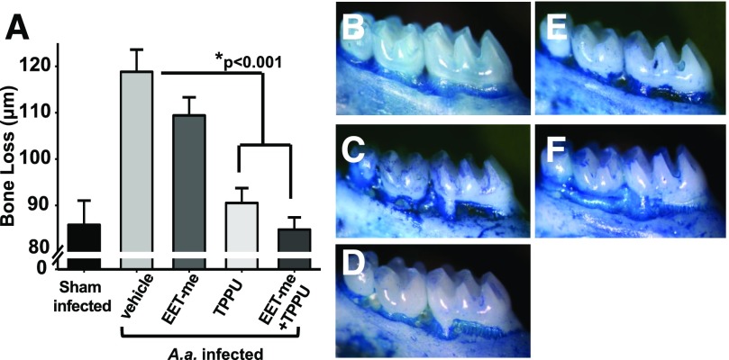
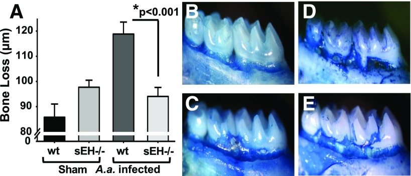
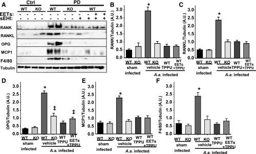
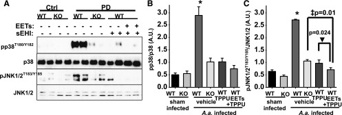
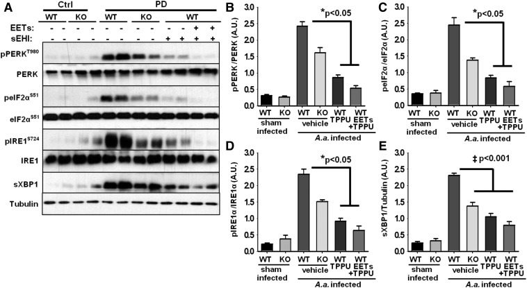
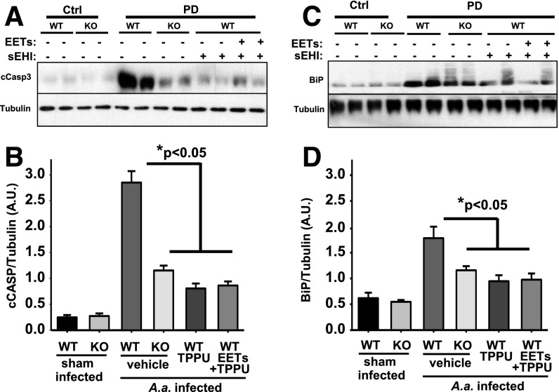
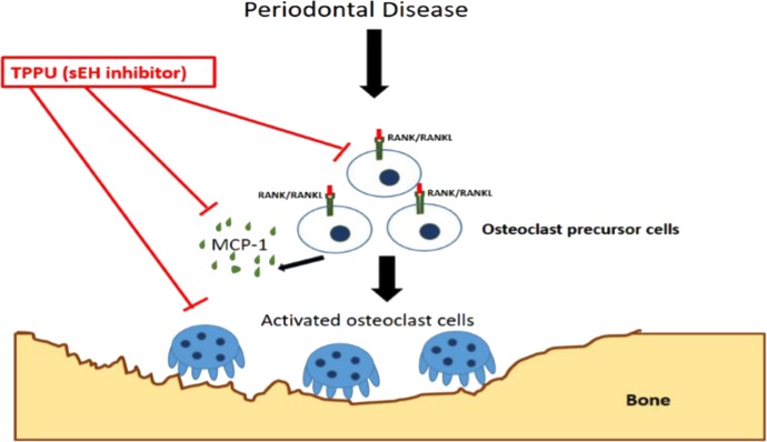
Epoxyeicosatrienoic acids (EETs), metabolites of arachidonic acid derived from the cytochrome P450 enzymes, are mainly metabolized by soluble epoxide hydrolase (sEH) to their corresponding diols. EETs but not their diols, have anti-inflammatory properties, and inhibition of sEH might provide protective effects against inflammatory bone loss. Thus, in the present study, we tested the selective sEH inhibitor, 1-trifluoromethoxyphenyl-3-(1-propionylpiperidin-4-yl) urea (TPPU), in a mouse model of periodontitis induced by infection with Aggregatibacter actinomycetemcomitans Oral treatment of wild-type mice with TPPU and sEH knockout (KO) animals showed reduced bone loss induced by A. actinomycetemcomitans This was associated with decreased expression of key osteoclastogenic molecules, receptor activator of nuclear factor-κB/RANK ligand/osteoprotegerin, and the chemokine monocyte chemotactic protein 1 in the gingival tissue without affecting bacterial counts. In addition, downstream kinases p38 and c-Jun N-terminal kinase known to be activated in response to inflammatory signals were abrogated after TPPU treatment or in sEH KO mice. Moreover, endoplasmic reticulum stress was elevated in periodontal disease but was abrogated after TPPU treatment and in sEH knockout mice. Together, these results demonstrated that sEH pharmacological inhibition may be of therapeutic value in periodontitis.


Figures

Fig. 1.
Fig. 1.
Inhibition of sEH with a potent and orally available small-molecule TPPU decreases bone loss. Mice were inoculated with freshly grown cultures of A. actinomycetemcomitans on 3 consecutive days, as described in Materials and Methods. The vehicle PEG400, TPPU (1 mg/kg), and EET methyl esters (1 µg/kg diluted in PEG400) were orally administered daily by gavage. Treatments continued until 15th day postinfection, and samples were obtained on the 16th day. Distance (µm) between the cement–enamel junction and the alveolar bone crest for all experimental groups was quantified. (A) The group vehicle alone developed significant bone loss, whereas groups treated with TPPU and TPPU + EET methyl esters displayed a marked reduction in bone loss. Mice receiving EET methyl esters were not different than vehicle-treated infected mice, suggesting the necessity of inhibiting sEH for EETs to display activity. (B–F) Panels display deboned and methylene blue–stained teeth from sham (B, n = 8), mice infected with A. actinomycetemcomitans (C, n = 14), EET methyl ester (1 µg/kg/daily) treated (D, n = 12), TPPU, 1 mg/kg treated (E, n = 13), and EET methyl ester + TPPU–treated groups (F, n = 13). The dark stained areas indicate sites of bone loss. The results are expressed as mean ± S.E.M. (*P < 0.001, one-way ANOVA followed by Student’s Newman–Keuls post hoc all pairwise comparison.)
Fig. 2.
Fig. 2.
Genetic inhibition of sEH by gene KO decreases bone loss similar to chemical inhibitor. sEH−/− and wild-type C57BL/6 mice were from a University of California, Davis–maintained colony. Mice at age 6 weeks were infected with A. actinomycetemcomitans three consecutive times, as described for Fig. 1, and at the end of the treatment period distance (µm) between the cement–enamel junction and the alveolar bone crest for all experimental groups was quantified. In parallel to the results with the sEH inhibitor TPPU, the genetic KO of sEH resulted in significantly reduced bone loss (Panel A). Panels display wild-type sham-infected (B, n = 8), sEH−/− sham-infected (C, n = 15), wild-type mice orally infected with A. actinomycetemcomitans (D, n = 14), and sEH−/− mice orally infected with A. actinomycetemcomitans (E, n = 14). The dark stained areas indicate sites of bone loss. The results are expressed as mean ± S.E.M. (*P < 0.001, one-way ANOVA followed by Student’s Newman–Keuls post hoc all pairwise comparison). wt, wild type.
Fig. 3.
Fig. 3.
The dysregulated RANK/RANKL/OPG system in PD is restored by chemical or genetic ablation of sEH. Protein expression levels of osteoclastogenesis-related factors in gingival tissues from all experimental groups were investigated by Western blotting. For quantification, band intensity was normalized to that of α-tubulin. Protein band intensity is represented as arbitrary units. Density quantification included all animals, and mean ± S.E.M. of each group (n = 6 per group) is displayed in the bar graphs. (A) Original blots displaying two randomly selected animals. (B) Bar graphs of mean band intensity for RANK (B), RANKL (C), OPG (D), MCP-1 (E) measured for all six mice and F4/80 (F) (*P < 0.001, P < 0.03, one-way ANOVA followed by Student’s Newman–Keuls post hoc all pairwise comparison). WT, wild type.
Fig. 4.
Fig. 4.
PD-mediated phosphorylation of proinflammatory p38 and JNK1/2 is reduced by chemical or genetic ablation of sEH. Phosphorylation and activation of p38 and JNK1/2 were quantified from all groups by normalizing band intensity to that of α-tubulin. (A) Original blots displaying two randomly selected animals. (B and C). Bar graphs of phosphorylation status of p38 and JNK1/2. Mean band intensity is measured for all six mice and is represented as arbitrary units (mean ± S.E.M.) (*P < 0.001, ‡P = 0.01, ▾P = 0.024, one-way ANOVA followed by Student’s Newman–Keuls post hoc all pairwise comparison). WT, wild type.
Fig. 5.
Fig. 5.
ER stress sensors are activated in gingival tissues of mice with PD and reversed by inhibition of sEH. (A) Original blots displaying two randomly selected animals for each group. (B–E) Bar graphs of phosphorylation status of protein kinase RNA-like ER kinase, eukaryotic initiation factor 2α, inositol-requiring enzyme 1, and expression level of spliced X-box binding protein 1 normalized to expression of α-tubulin. Mean band intensity is measured for all six mice and is represented as arbitrary units (mean ± S.E.M.) (‡P < 0.001, *P < 0.05, Kruskal–Wallis one-way analysis of variance on ranks, followed by Tukey’s all pairwise multiple comparison post hoc test). WT, wild type.
Fig. 6.
Fig. 6.
Genetic ablation or chemical inhibition of sEH reduces apoptosis in gingival tissue of mice infected with A. actinomycetemcomitans. Original blot displaying two randomly selected animals for each group. (A) Expression level of c-Caspase-3 and (C) immunoglobulin binding protein (BiP) is quantified by measuring band intensity for all six mice for each group and normalized to the expression level of a-tubulin. (B and D) Bar graph of mean caspase-3 and BiP band intensity respectively, represented as arbitrary units (mean 6 S.E.M.) (*P < 0.05, Kruskal-Wallis one-way analysis of variance on ranks, followed by Tukey’s all pairwise multiple comparison post hoc test). sEHI, soluble epoxide hydrolase inhibitor; WT, wild type.
Fig. 7.
Fig. 7.
Simplified schematic representation of sEH inhibition mediated suppression of inflammation and bone loss. Soluble epoxide hydrolase pharmacological inhibition decreases alveolar bone loss by modulating host inflammatory response, RANK-related signaling, ER stress, and apoptosis.

Similar articles

Cited by

References

    1. Bartold PM, Van Dyke TE. (2013) Periodontitis: a host-mediated disruption of microbial homeostasis: unlearning learned concepts. Periodontol 2000 62:203–217. - PMC - PubMed
    1. Bettaieb A, Chahed S, Bachaalany S, Griffey S, Hammock BD, Haj FG. (2015) Soluble epoxide hydrolase pharmacological inhibition ameliorates experimental acute pancreatitis in mice. Mol Pharmacol 88:281–290. - PMC - PubMed
    1. Bettaieb A, Nagata N, AbouBechara D, Chahed S, Morisseau C, Hammock BD, Haj FG. (2013) Soluble epoxide hydrolase deficiency or inhibition attenuates diet-induced endoplasmic reticulum stress in liver and adipose tissue. J Biol Chem 288:14189–14199. - PMC - PubMed
    1. Cao SS, Luo KL, Shi L. (2016) Endoplasmic reticulum stress interacts with inflammation in human diseases. J Cell Physiol 231:288–294. - PMC - PubMed
    1. Capdevila J, Chacos N, Werringloer J, Prough RA, Estabrook RW. (1981) Liver microsomal cytochrome P-450 and the oxidative metabolism of arachidonic acid. Proc Natl Acad Sci USA 78:5362–5366. - PMC - PubMed

Publication types

MeSH terms

Substances