. 2017 May;174(1):223-240.
doi: 10.1104/pp.16.01282. Epub 2017 Mar 29.

EXO70C2 Is a Key Regulatory Factor for Optimal Tip Growth of Pollen

Affiliations

EXO70C2 Is a Key Regulatory Factor for Optimal Tip Growth of Pollen

Lukáš Synek et al. Plant Physiol. 2017 May.

Abstract

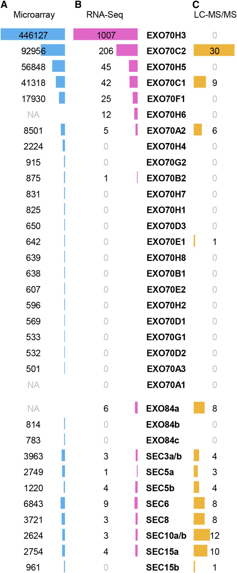
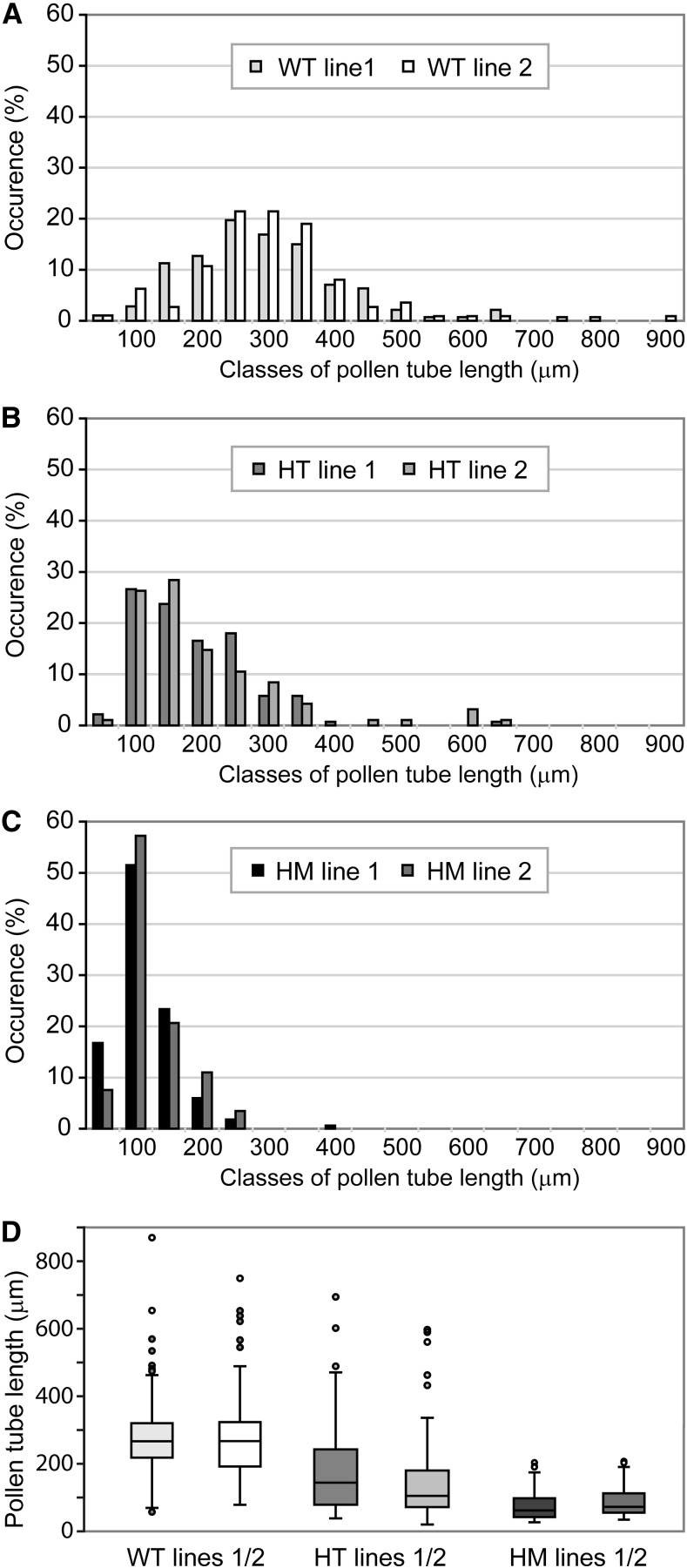
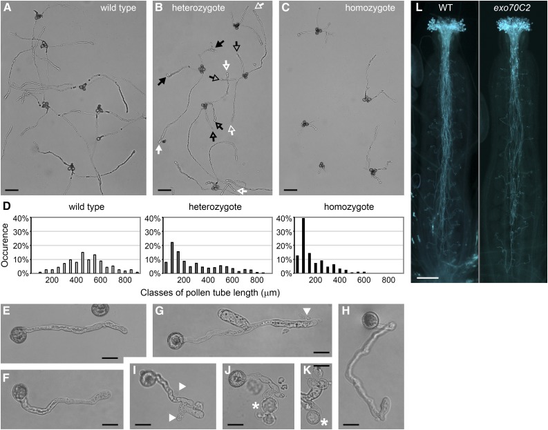
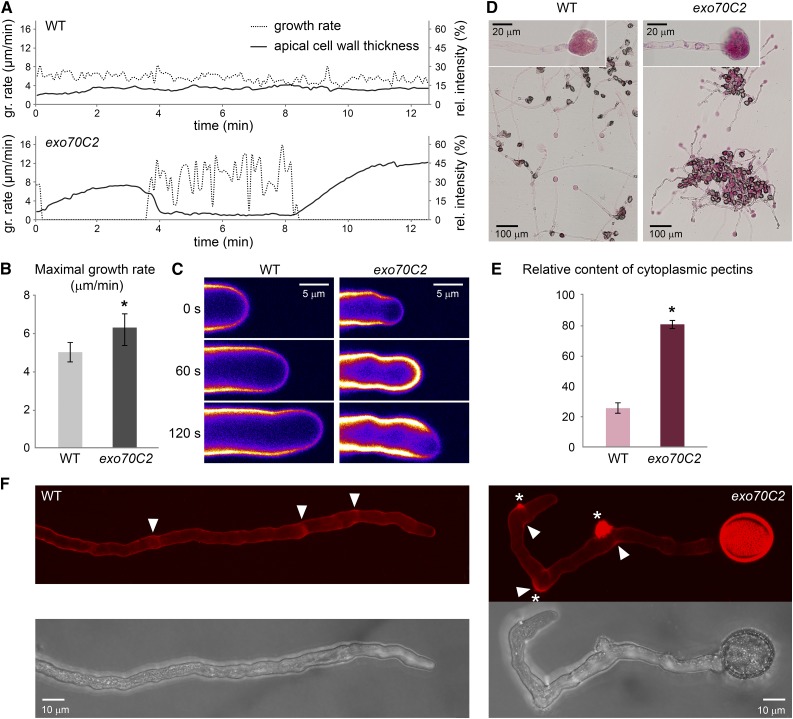
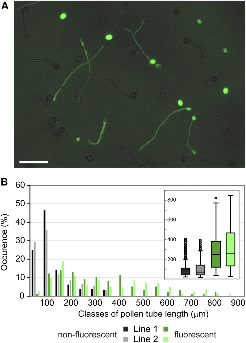
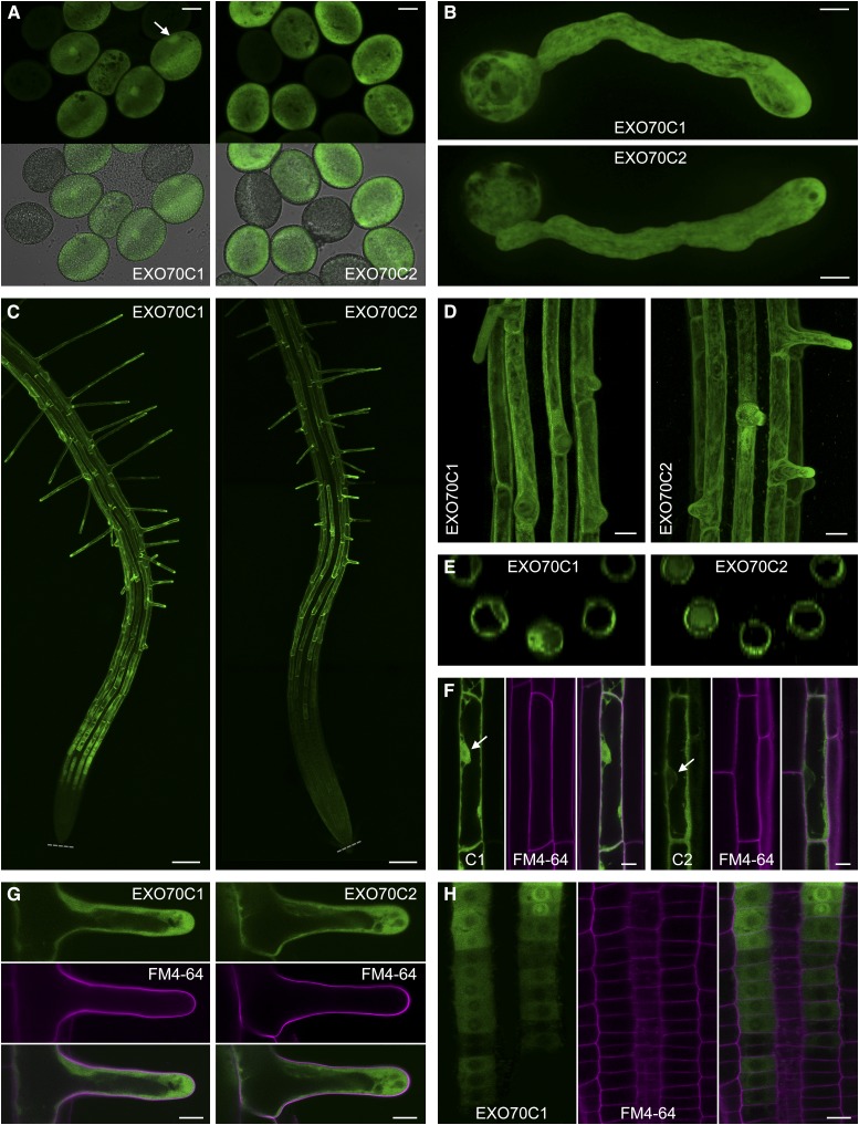
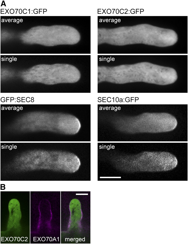
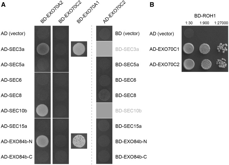
The exocyst, a eukaryotic tethering complex, coregulates targeted exocytosis as an effector of small GTPases in polarized cell growth. In land plants, several exocyst subunits are encoded by double or triple paralogs, culminating in tens of EXO70 paralogs. Out of 23 Arabidopsis thaliana EXO70 isoforms, we analyzed seven isoforms expressed in pollen. Genetic and microscopic analyses of single mutants in EXO70A2, EXO70C1, EXO70C2, EXO70F1, EXO70H3, EXO70H5, and EXO70H6 genes revealed that only a loss-of-function EXO70C2 allele resulted in a significant male-specific transmission defect (segregation 40%:51%:9%) due to aberrant pollen tube growth. Mutant pollen tubes grown in vitro exhibited an enhanced growth rate and a decreased thickness of the tip cell wall, causing tip bursts. However, exo70C2 pollen tubes could frequently recover and restart their speedy elongation, resulting in a repetitive stop-and-go growth dynamics. A pollen-specific depletion of the closest paralog, EXO70C1, using artificial microRNA in the exo70C2 mutant background, resulted in a complete pollen-specific transmission defect, suggesting redundant functions of EXO70C1 and EXO70C2. Both EXO70C1 and EXO70C2, GFP tagged and expressed under the control of their native promoters, localized in the cytoplasm of pollen grains, pollen tubes, and also root trichoblast cells. The expression of EXO70C2-GFP complemented the aberrant growth of exo70C2 pollen tubes. The absent EXO70C2 interactions with core exocyst subunits in the yeast two-hybrid assay, cytoplasmic localization, and genetic effect suggest an unconventional EXO70 function possibly as a regulator of exocytosis outside the exocyst complex. In conclusion, EXO70C2 is a novel factor contributing to the regulation of optimal tip growth of Arabidopsis pollen tubes.


Figures

Figure 1.
Figure 1.
Expression of exocyst genes in Arabidopsis pollen. A, Mean expression values in linear scale based on the ATH1 (Affymetrix) microarray data in Genevestigator. NA, Not available on the ATH1 DNA chip. B, Quantification of mRNA based on RNA-Seq data on mature pollen (Loraine et al., 2013). C, Number of peptides detected in the proteome of mature pollen using mass spectroscopy (LC-MS/MS; Grobei et al., 2009).
Figure 2.
Figure 2.
Pollen tube lengths of exo70C2 mutants. A to C, Distribution of pollen tube lengths in samples of in vitro-germinated pollen from the wild type (WT; A), exo70C2 heterozygotes (HT; B), and exo70C2 homozygotes (HM; C). Two independent lines of the same genotype are displayed in each graph. At least 120 pollen tubes were measured for each line. D, Box plots comparing the data from A to C.
Figure 3.
Figure 3.
Morphology of exo70C2 pollen tubes germinated in vitro. A to C, Germinated pollen of a wild-type plant (A), an exo70C2 heterozygous plant (B), and an exo70C2 homozygous plant (C) in the quartet-1 background. Putative wild-type or mutant pollen tubes are marked by white or black arrows, respectively; arrows of the same design correspond to the same tetrad. Bars = 100 μm. D, Distribution of pollen tube lengths in the samples above (A–C). E, A typical wild-type pollen tube. Bar = 20 μm. F to K, Mutant pollen tubes showing relatively normal morphology (F), branching (G and I), sharp bending (H), effusion of cytoplasm (G and I; marked by arrowheads), or the production of protoplast-like structures emerging from tube tips (J and K; marked by asterisks). Bars = 20 μm. L, Aniline Blue staining of pollinated pistils of the wild type (WT) and an exo70C2 homozygote visualizing callose in pollen tubes. Bar = 200 μm.
Figure 4.
Figure 4.
Growth rate and cell wall characteristics of exo70C2 and wild-type (WT) pollen tubes. A, Growth rate of typical wild-type and exo70C2 pollen tubes correlated with cell wall thickness (Calcofluor White fluorescence) at the tube apex. Images for measurement were captured at intervals of 5 s. B, The averaged maximal growth rate of exo70C2 is significantly higher than that of the wild type (sd is displayed: *, P < 0.00001 by Student’s t test). Measurements were performed on 15 tubes per genotype at multiple time points every 90 s. C, Calcofluor White fluorescence represented as an intensity color scale (purple to white) shows differential cell wall deposition at the tube apex during the highly fluctuating growth rate of the exo70C2 pollen tube compared with the wild type characterized by low oscillations. D, Pollen tubes of the wild type and exo70C2 germinated and stained on the same slide with Ruthenium Red diluted in distilled water to cause the extrusion of cytoplasm. Details of burst tips that were used for quantification are shown in the insets. E, Quantification of the Ruthenium Red staining in extruded cytoplasm as relative intensity in the red channel subtracted from the background. sd is displayed: *, P < 10−10 by Student’s t test; n = 50 for each genotype. F, Propidium iodide staining of growing pollen tubes. Maximum intensity projections over a confocal Z-stack are shown. Asterisks mark sites of collapse; arrowheads point to sites of stopped growth.
Figure 5.
Figure 5.
Pollen tube lengths of exo70C2 lines complemented with pEXO70C2::EXO70C2:GFP. A, A representative microscopic image from the series used for pollen tube length analysis below. Longer pollen tubes emitting pEXO70C2::EXO70C2:GFP fluorescence represent complemented exo70C2 mutant pollen tubes. GFP fluorescence was mixed with bright field. Bar = 100 μm. B, Distribution of pollen tube lengths in samples of in vitro-germinated pollen from homozygous exo70C2 mutants in which the introduced pEXO70C2::EXO70C2:GFP was in a heterozygous state. Fluorescent and nonfluorescent pollen tubes were measured separately within each sample. Two independent lines (1 and 2) were analyzed. Box plots in the inset are another presentation of the same data.
Figure 6.
Figure 6.
Localization of EXO70C1:GFP and EXO70C2:GFP expressed under the control of their native promoters in cells of exo70C1 and exo70C2 homozygotes, respectively. A, EXO70C1:GFP and EXO70C2:GFP in mature pollen grains (the fluorescence intensity in both samples is not to scale). In addition, EXO70C1:GFP accumulates in the vegetative nucleus (marked by the arrow). Expression cassettes were segregating in the samples; nonfluorescent pollen grains provide reference for the background fluorescence. Bars = 10 μm. B, EXO70C1:GFP and EXO70C2:GFP in the cytoplasm of pollen tubes. Bars = 10 μm. C, EXO70C1:GFP and EXO70C2:GFP in roots. The expression of EXO70C1 starts already in the late meristem. Gray dotted lines mark root tips. Bars = 100 μm. D, EXO70C1:GFP and EXO70C2:GFP are expressed specifically in trichoblast cells in roots (maximum intensity projection of confocal Z-stacks). Bars = 20 μm. E, Top views of three-dimensional reconstructions calculated from the Z-stacks in D. F, While EXO70C1:GFP localizes to the cytoplasm and nucleus in elongated trichoblast cells, EXO70C2:GFP is localized exclusively in the cytoplasm. Nuclei are marked by arrows. Cell walls were stained with propidium iodide (in magenta). Bars = 10 μm. G, Cytoplasmic localization of EXO70C1:GFP and EXO70C1:GFP in growing root hairs. Cell walls were stained with propidium iodide (in magenta). Bars = 10 μm. H, The onset of EXO70C1:GFP expression in the root meristem. EXO70C1:GFP accumulates in the perinuclear region and/or nucleolus at certain stages. Cell walls were stained with propidium iodide (in magenta). Bar = 10 μm.
Figure 7.
Figure 7.
Localization of EXO70C1 and EXO70C2 in comparison with core exocyst subunits in Arabidopsis pollen tubes and root hairs. A, GFP:SEC8 and SEC10a:GFP exhibit a PM association in the apex of growing pollen tubes, in contrast to EXO70C1:GFP and EXO70C2:GFP, which show cytoplasmic localization. Averaged time series (five sequential images taken at 4-s intervals) and one representative single image out of the series are displayed. Bar = 10 μm. B, Colocalization of EXO70C2:GFP (green) and RFP:EXO70A1 (magenta) in growing root hairs shows no overlap. Bar = 10 μm.
Figure 8.
Figure 8.
EXO70C2 and EXO70A2 interactions in the yeast two-hybrid assay. A, Pairwise interactions with core exocyst subunits. The EXO70A1-SEC3a and EXO70A1-EXO84b-N interactions described by Hála et al. (2008) were used as positive controls. Empty vectors pGADT7 (AD) and pGBKT7 (BD) were used as negative controls. BD-SEC3a and BD-SEC10b were omitted in this assay, since they show high autoactivation capacity (Hála et al., 2008). B, AD-EXO70C2 interacts with BD-ROH1, a putative negative regulator of secretion, similar to AD-EXO70C1 published by Kulich et al. (2010).

Comment in

Similar articles

Cited by

References

    1. Alonso JM, Stepanova AN, Leisse TJ, Kim CJ, Chen H, Shinn P, Stevenson DK, Zimmerman J, Barajas P, Cheuk R, et al. (2003) Genome-wide insertional mutagenesis of Arabidopsis thaliana. Science 301: 653–657 - PubMed
    1. Bashline L, Lei L, Li S, Gu Y (2014) Cell wall, cytoskeleton, and cell expansion in higher plants. Mol Plant 7: 586–600 - PubMed
    1. Bloch D, Pleskot R, Pejchar P, Potocký M, Trpkošová P, Cwiklik L, Vukašinović N, Sternberg H, Yalovsky S, Žárský V (2016) Exocyst SEC3 and phosphoinositides define sites of exocytosis in pollen tube initiation and growth. Plant Physiol 172: 980–1002 - PMC - PubMed
    1. Chebli Y, Kaneda M, Zerzour R, Geitmann A (2012) The cell wall of the Arabidopsis pollen tube: spatial distribution, recycling, and network formation of polysaccharides. Plant Physiol 160: 1940–1955 - PMC - PubMed
    1. Chebli Y, Kroeger J, Geitmann A (2013) Transport logistics in pollen tubes. Mol Plant 6: 1037–1052 - PubMed

MeSH terms