. 2017 Apr 11;114(15):3981-3986.
doi: 10.1073/pnas.1609758114. Epub 2017 Mar 29.

BCL6 promotes glioma and serves as a therapeutic target

Affiliations

BCL6 promotes glioma and serves as a therapeutic target

Liang Xu et al. Proc Natl Acad Sci U S A. .

Erratum in

Abstract

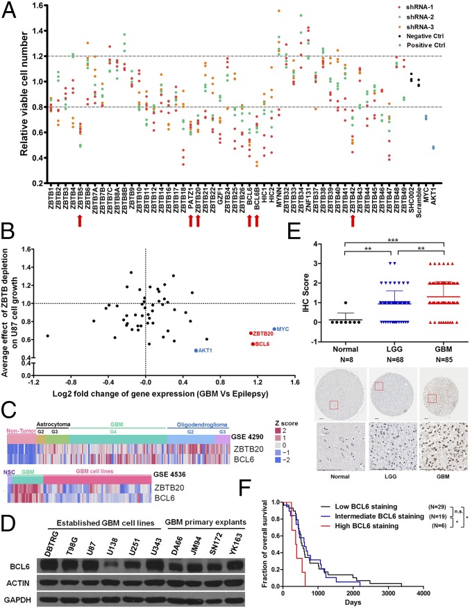
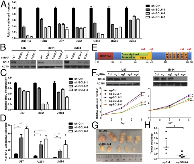
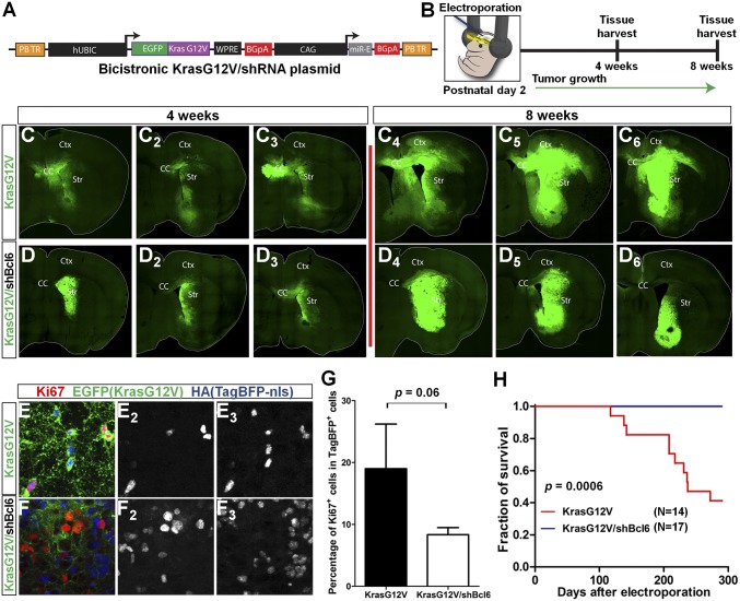
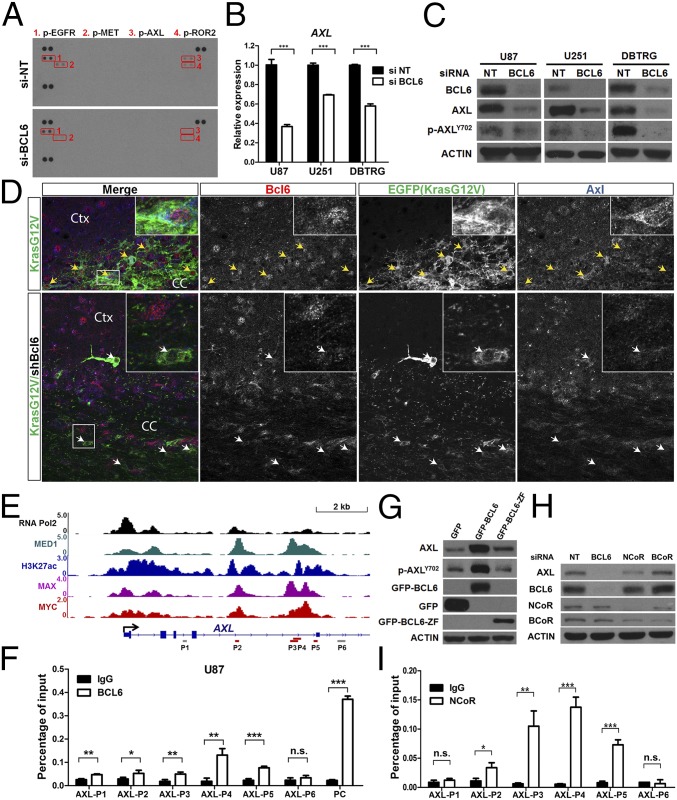
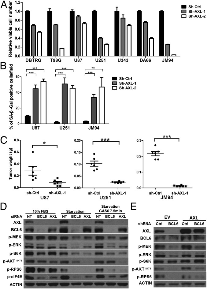
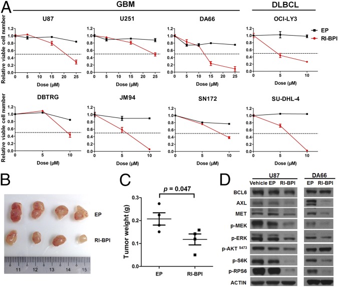
ZBTB transcription factors orchestrate gene transcription during tissue development. However, their roles in glioblastoma (GBM) remain unexplored. Here, through a functional screening of ZBTB genes, we identify that BCL6 is required for GBM cell viability and that BCL6 overexpression is associated with worse prognosis. In a somatic transgenic mouse model, depletion of Bcl6 inhibits the progression of KrasG12V-driven high-grade glioma. Transcriptome analysis demonstrates the involvement of BCL6 in tumor protein p53 (TP53), erythroblastic leukemia viral oncogene homolog (ErbB), and MAPK signaling pathways. Indeed, BCL6 represses the expression of wild-type p53 and its target genes in GBM cells. Knockdown of BCL6 augments the activation of TP53 pathway in response to radiation. Importantly, we discover that receptor tyrosine kinase AXL is a transcriptional target of BCL6 in GBM and mediates partially the regulatory effects of BCL6 on both MEK-ERK (mitogen-activated protein/extracellular signal-regulated kinase kinase-extracellular signal-regulated kinase) and S6K-RPS6 (ribosomal protein S6 kinase-ribosomal protein S6) axes. Similar to BCL6 silencing, depletion of AXL profoundly attenuates GBM proliferation both in vitro and in vivo. Moreover, targeted inhibition of BCL6/nuclear receptor corepressor 1 (NCoR) complex by peptidomimetic inhibitor not only significantly decreases AXL expression and the activity of MEK-ERK and S6K-RPS6 cascades but also displays a potent antiproliferative effect against GBM cells. Together, these findings uncover a glioma-promoting role of BCL6 and provide the rationale of targeting BCL6 as a potential therapeutic approach.

Keywords: AXL; BCL6; NCoR; ZBTB; glioblastoma multiforme.


Conflict of interest statement

The authors declare no conflict of interest.

Figures

Fig. 1.
Fig. 1.
Identification of BCL6 as a progrowth factor in glioma. (A) Relative viable number of U87 GBM cells after infection with lentiviral particles encoding indicated shRNAs. SHC002 and pLKO-scramble were used as negative controls (Negative Ctrl), and shRNAs targeting MYC and AKT1 were used as positive controls (Positive Ctrl). Candidate genes (marked by red arrows) were selected only if all independent shRNAs and all repeats showed consistent increase or decrease (>20%) in relative viable cell number compared with negative controls. (B) Plot showing the correlation between average effect of ZBTB depletion on U87 cell growth and differential gene expression (GBM vs. epilepsy; GSE4290). BCL6 and ZBTB20 were highlighted in red as candidates for growth-promoting genes. MYC and AKT1 were marked in blue as positive controls for oncogenes. (C) Heat map showing the expression of BCL6 and ZBTB20 in nontumor brain tissues (epilepsy), GBM cell lines, and primary GBM samples. G2, G3, and G4 denote World Health Organization grade II, III, and IV, respectively. NSC, neural stem cells. (D) Western blot result showing the endogenous BCL6 expression among 6 established GBM cell lines and 4 primary GBM explants. (E) IHC results and representative staining images showing BCL6 expression in normal brain, LGG (lower-grade glioma), and GBM samples. (Scale bars, 100 μm). IHC scores were compared using a nonparametric statistical test. (F) Survival curves for patients with GBM with differential BCL6 expression (Low, IHC score 0 or 1; Intermediate, IHC score 2; High, IHC score 3). Log-rank test was applied.
Fig. 2.
Fig. 2.
BCL6 expression is a functional requisite for GBM cell growth. (A) Effect of shRNA-mediated depletion of BCL6 on GBM cell viability. In each cell line, sh-BCL6 groups were significantly different from the control (sh-Ctrl) group. (B) Western blot results showing the efficiency of BCL6 knockdown. (C and D) Effect of shRNA-mediated depletion of BCL6 on BrdU incorporation (C) and senescence-associated β-galactosidase (SA-β-Gal) activity (D). (E) Schematic representation of potential BCL6 editing sites by four individual sgRNAs. (F) Western blot results showing the efficiency of each sgRNA against BCL6 and the effect of BCL6 silencing by CRISPR/Cas9 system on cell viability. (G and H) Effect of BCL6 depletion by CRISPR/Cas9 system on tumor growth. U87 cells stably expressing either sg-BCL6-4 or sg-Ctrl were s.c. injected into NOD scid gamma mice. Tumors were harvested 30 d after implantation and weighed.
Fig. 3.
Fig. 3.
Bcl6 silencing inhibits glioma progression in mice. (A) Schematic of bicistronic KrasG12V/shBcl6.275 miR-E knockdown construct. (B) Postnatal day 2 electroporation of wild-type CD-1 mice targeting left lateral ventricle of the brain with pBase, TagBFP-HA-nuclear localization sequence, and EGFP-KrasG12V/shBcl6 (or EGFP-KrasG12V) expressing plasmids. (C and D) Stitched images of tumors at 4- and 8-wk times in KrasG12V and KrasG12V/shBcl6 mice (n = 3 per time). CC, Ctx, and Str denote corpus callosum, cerebral cortex, and striatum, respectively. (E and F) Coronal section of 8-wk tumors from KrasG12V (E) and KrasG12V/shBcl6 (F) mice stained with Ki67, EGFP, and HA (TagBFP). (E2 and E3) Colocalization of Ki67 (E2) and TagBFP signals (E3). (F2 and F3) Ki67+ cells (F2) largely did not colocalize with TagBFP+ cells (F3). (G) Quantification of percentage of Ki67+ cells among the TagBFP-expressing cells at the ventral margin of tumors from indicated groups. Data represent means ± SEM (n = 3). (H) Kaplan-Meier survival curves for KrasG12V (n = 14) and KrasG12V/shBcl6 (n = 17) mice. Log-rank test was applied.
Fig. 4.
Fig. 4.
Identification of AXL as a target of BCL6. (A) Phospho-RTK array result showing the down-regulation of phosphorylated AXL in U87 cells with depletion of BCL6. Red boxes indicate the chemiluminescent signals in duplicate for p-EGFR (1), p-MET (2), p-AXL (3), and p-ROR2 (4). (B and C) qPCR (B) and Western blot (C) results showing the down-regulation of AXL after BCL6 depletion in GBM cell lines. RNA and protein samples were harvested 36 and 72 h posttransfection, respectively. Data represent means ± SD (n = 3). (D) Immunofluorescence staining showing the correlated expression between Bcl6 and Axl in EGFP+ tumor cells (indicated by arrows) at the margin of tumors in KrasG12V (Upper) and KrasG12V/shBcl6 (Lower). (E) The ChIP-seq signals of RNA Pol2, MED1, H3K27ac, MAX, and MYC in U87 AXL locus were visualized using Integrative Genomics Viewer. Y axis represents the value of reads per million. Gray and red bars indicate the template regions amplified by respective ChIP-qPCR primers. (F) ChIP-qPCR results showing the enrichment of BCL6 binding in AXL locus. Cells were fixed 2 h after the addition of fresh medium. The 5′ UTR region of BCL6 was applied as a positive control (PC). (G) Effect of either GFP-BCL6 or GFP-BCL6-ZF on AXL expression. (H) Effect of BCL6, NCoR, and BCoR silencing on AXL expression. (I) ChIP-qPCR results showing the enrichment of NCoR binding in AXL locus.
Fig. 5.
Fig. 5.
AXL contributes partially to the GBM-promoting effects of BCL6. (A–C) Effect of AXL depletion by shRNAs on cell viability (A), cellular senescence (B), and xenograft growth (C) in NOD scid gamma mice. (A) In all cell lines, sh-AXL groups were significantly different from the corresponding sh-Ctrl group. (D) Effect of siRNA-mediated silencing of BCL6 and AXL on downstream targets in U87 cells under complete medium (10% FBS), serum starvation (48 h), or GAS6 stimulation (100 ng/mL). (E) Effect of ectopic AXL expression on BCL6-depleted cells. U251 GBM cells expressing either ectopic AXL or empty vector (EV) were infected with indicated shRNAs and serum starved for 48 h before harvest.
Fig. 6.
Fig. 6.
Targeting BCL6 in glioma. (A) Effect of RI-BPI on GBM cell viability. GBM cells were treated with indicated concentration of either RI-BPI or empty peptide (EP) for 72 h before analysis. OCI-LY3 and SU-DHL-4 cells were used as positive controls. (B and C) Effect of RI-BPI treatment on JM94 cell growth in vivo. Peptides were administrated intraperitoneally (50 mg⋅kg−1⋅day−1 for 4 d). (D) Western blot results showing the effect of RI-BPI (20 µM, 24 h) on BCL6 downstream targets.

References

    1. Siggs OM, Beutler B. The BTB-ZF transcription factors. Cell Cycle. 2012;11:3358–3369. - PMC - PubMed
    1. Basso K, Dalla-Favera R. BCL6: Master regulator of the germinal center reaction and key oncogene in B cell lymphomagenesis. Adv Immunol. 2010;105:193–210. - PubMed
    1. Cattoretti G, et al. Deregulated BCL6 expression recapitulates the pathogenesis of human diffuse large B cell lymphomas in mice. Cancer Cell. 2005;7:445–455. - PubMed
    1. Phan RT, Dalla-Favera R. The BCL6 proto-oncogene suppresses p53 expression in germinal-centre B cells. Nature. 2004;432:635–639. - PubMed
    1. Duy C, et al. BCL6 is critical for the development of a diverse primary B cell repertoire. J Exp Med. 2010;207:1209–1221. - PMC - PubMed

Publication types

MeSH terms