. 2017 May 26;91(12):e00260-17.
doi: 10.1128/JVI.00260-17. Print 2017 Jun 15.

Minute Virus of Canines NP1 Protein Governs the Expression of a Subset of Essential Nonstructural Proteins via Its Role in RNA Processing

Affiliations

Minute Virus of Canines NP1 Protein Governs the Expression of a Subset of Essential Nonstructural Proteins via Its Role in RNA Processing

Olufemi O Fasina et al. J Virol. .

Abstract

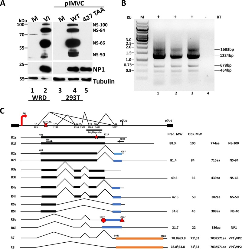
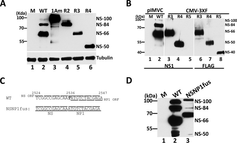
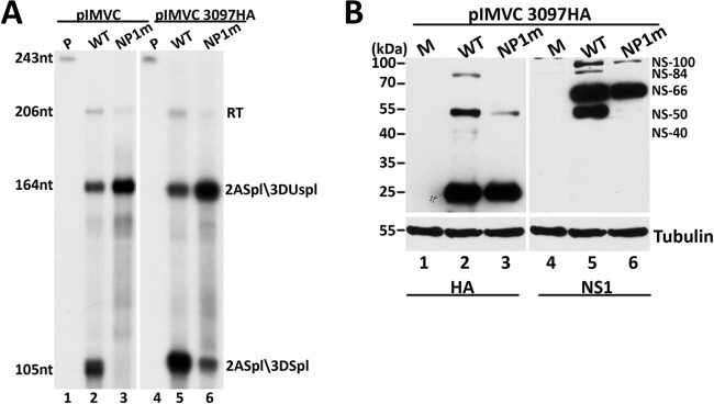
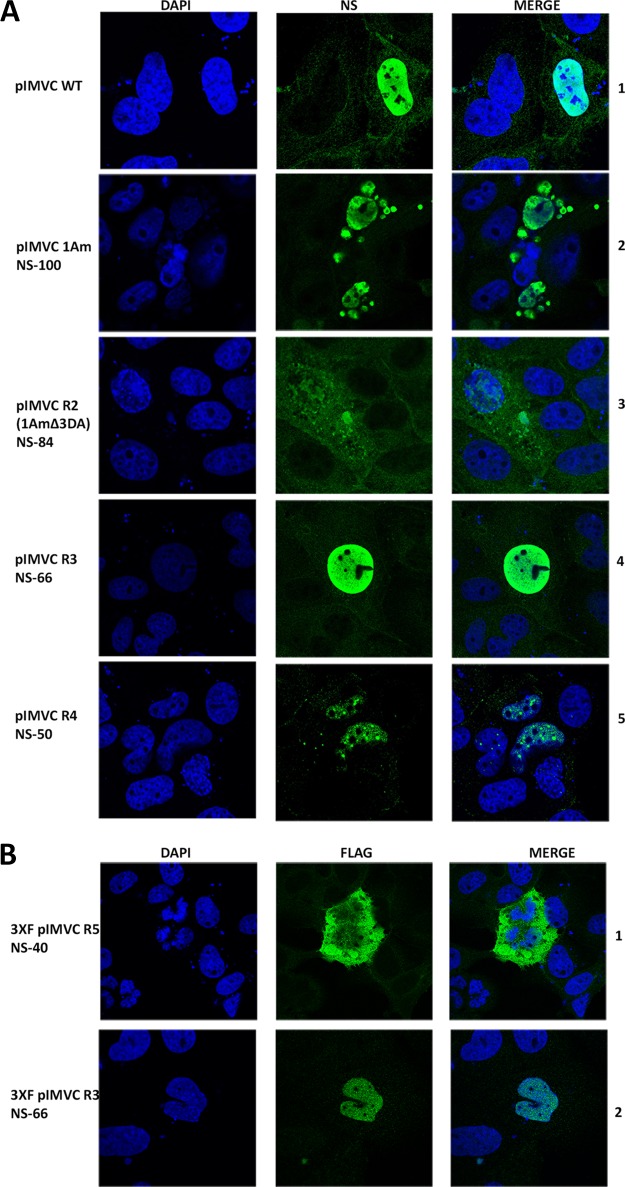
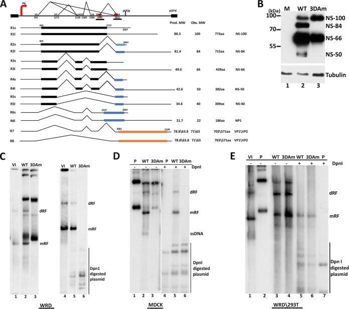
Parvoviruses use a variety of means to control the expression of their compact genomes. The bocaparvovirus minute virus of canines (MVC) encodes a small, genus-specific protein, NP1, which governs access to the viral capsid gene via its role in alternative polyadenylation and alternative splicing of the single MVC pre-mRNA. In addition to NP1, MVC encodes five additional nonstructural proteins (NS) that share an initiation codon at the left end of the genome and which are individually encoded by alternative multiply spliced mRNAs. We found that three of these proteins were encoded by mRNAs that excise the NP1-regulated MVC intron immediately upstream of the internal polyadenylation site, (pA)p, and that generation of these proteins was thus regulated by NP1. Splicing of their progenitor mRNAs joined the amino termini of these proteins to the NP1 open reading frame, and splice site mutations that prevented their expression inhibited virus replication in a host cell-dependent manner. Thus, in addition to controlling capsid gene access, NP1 also controls the expression of three of the five identified NS proteins via its role in governing MVC pre-mRNA splicing.IMPORTANCE The Parvovirinae are small nonenveloped icosahedral viruses that are important pathogens in many animal species, including humans. Minute virus of canine (MVC) is an autonomous parvovirus in the genus Bocaparvovirus It has a single promoter that generates a single pre-mRNA. NP1, a small genus-specific MVC protein, participates in the processing of this pre-mRNA and so controls capsid gene access via its role in alternative internal polyadenylation and splicing. We show that NP1 also controls the expression of three of the five identified NS proteins via its role in governing MVC pre-mRNA splicing. These NS proteins together are required for virus replication in a host cell-dependent manner.

Keywords: gene expression; parvovirus.


Figures

FIG 1
FIG 1
MVC encodes multiple NS isoforms during infection of WRD cells and transfection of 293T cells. (A) (Left) Immunoblots, using antibodies directed against the NS ORF (epitope depicted as a star in panel C) or NP1 (epitope depicted as a circle in panel C), of cell lysates of either mock-infected cells (M; lane 1) or WRD cells infected with MVC at a multiplicity of infection (MOI) of 7 harvested 48 h postinfection (lane 2). (Right) Immunoblots, using antibodies directed against the NS ORF (star), NP1 (circle), or tubulin, of 293T cell lysates harvested 48 h posttransfection with either pIMVC WT (lane 4) or pIMVC 427 TAA (terminating the NS ORF as described in the text) (lane 5) or mock transfection (lane 3). The bands correlating with NS isoforms (NS-100, NS-84, NS-66, and NS-50) and NP1 are indicated on the right, and molecular mass markers are on the left. (B) Reverse transcriptase PCR of total 293T cell RNA extracted 48 h posttransfection with pIMVC WT, primed with oligo(dT), and reverse transcribed with (lanes 1 to 3; each lane represents an individual complete experiment) and without (lane 4) RT. Amplicons (1,683 bp, 1,224 bp, 678 bp, and 464 bp) generated with MVC NS ORF-specific forward (nt 868) and NP1 ORF-specific reverse (nt 3097) primers, depicted by horizontal arrows in the transcription profile schematic in panel C, are shown on the right. The size markers (lane M) are depicted on the left. (C) Transcription profile of MVC showing the alternative RNA-processing events that generate MVC nonstructural protein (NS isoforms and NP1)-encoding transcripts (R1 to R6) and structural protein (VP1 and VP2)-encoding transcripts (R7 and R8). The NS- and NP1-encoding transcripts are illustrated as alternatively polyadenylated transcripts using the proximal (pA)p (R1 to R6s) or distal (pA)d (R1 to R6l) polyadenylation site. The P6 promoter, splice donors (D) and acceptors (A), and the proximal (pA)p and distal (pA)d polyadenylation sites are shown, along with relevant nucleotide landmarks within the MVC genome (GenBank accession number FJ214110.1). The position of the NS1 termination mutant (pIMVC 427 TAA) is shown with an ×, while the positions of the 5′ (nt 868) and 3′ (nt 3097) primers used in the RT-PCR analysis shown in panel B are depicted with horizontal arrows. The NS ORF (nt 403 to 2727) and NP1 ORF (nt 2537 to 3097) are indicated. The epitopes detected by the antibodies used to detect the NS proteins (NS ORF amino acids 687 to 700: PKKQRKTEHKVLID) and NP1 (NP1 amino acids 1 to 13: MSTRHMSKRSKARSR) are indicated by the star and circle, respectively. The triangle indicates the HA epitope at the NP1 ORF carboxyl termini in pIMVC 3097HA WT and pIMVC 3097HA NP1m constructs used for the experiments shown in Fig. 3. The predicted (pred. MW) and observed (obs. MW) molecular weights of the NS isoforms (NS-100, NS-84, NS-50, and NS-40), NP1 proteins, and capsid proteins (VP1 and VP2) are indicated with the corresponding number of amino acid residues (774 aa, 715 aa, 439 aa, 382 aa, 309 aa,186 aa, 703 aa, and 571 aa) encoded by their mRNAs.
FIG 2
FIG 2
MVC NS-84, NS-50, and NS-40 mRNAs are alternatively spliced, using the 3A acceptor, into the NP1 ORF. (A) Lysates of 293T cells taken 48 h following transfection of pIMVC WT (lane 2), pIMVC 1Am (lane 3), R2 cDNA (lane 4), R3 cDNA (lane 5), R4 cDNA (lane 6), and R5 cDNA (lane 7) were analyzed by immunoblotting with antibodies directed against NS (star in Fig. 1C) and tubulin. Molecular mass markers are indicated on the left. (B) Lysates of 293T cells taken 48 h following transfection with pIMVC WT (lane 2) or CMV-3XF constructs expressing R3 cDNA (lanes 3 and 6), R4 cDNA (lanes 4 and 7), and R5 cDNA (lanes 5 and 8) isoforms were analyzed by immunoblotting using either anti-NS antibody (epitope depicted by a star in Fig. 1C) (lanes 1 to 5) or anti-FLAG (lanes 6 to 8). (C) Schematic of wild-type MVC nt 2524 to 2574 and the pIMVC NSNP1fus mutant. The T nucleotide (nt 2536) deleted in the NS1 ORF to generate the NSNP1fus mutant fusing the NS and NP1 ORFs is italicized and boldface. Reading frames are indicated by lines above and below the sequences. (D) Lysates of 293T cells taken 48 h posttransfection with pIMVC WT (lane 2) and the pIMVC NS1NP1fus mutant (lane 3) were immunoblotted using antibodies to the NS ORF (star in Fig. 1C). The bands correlating with each NS1 isoform are indicated on the right.
FIG 3
FIG 3
MVC NP1 is required for the expression of NS-84, NS-50, and NS-40. (A) RNase protection of 20 μg of RNA extracted from 293T cells 48 h following transfection with pIMVC WT (lane 2), pIMVC NP1m (lane 3), pIMVC 3097HA WT (lane 5), and pIMVC NP1m 3097HA (lane 6), using the 2A/3D probe that spans nt 2344 to 2550 (shown in Fig. 1C). The sizes of the probe (243 nt) and protected RNAs (206 nt, 164 nt, and 105 nt) are shown on the left. Bands reflecting RNA species unspliced through this region (RT) or spliced at the second intron acceptor but not at the third intron donor (2Aspl/3DUnspl) and RNAs spliced at the second intron acceptor and also the third intron donor (2Aspl/3Dspl) are indicated on the right. (B) Samples taken from 293T cells transfected with pIMVC 3097HA WT (lanes 2 and 4) and pIMVC NP1m 3097HA (lanes 3 and 6) were subjected to immunoblot analysis using antibody directed against HA (lanes 1 to 3) or antibody directed against NS (epitope depicted as a star in Fig. 1C) (lanes 4 to 6). Tubulin was monitored as a loading control. The bands that correspond to each NS isoform are indicated on the right.
FIG 4
FIG 4
MVC NS isoforms exhibit differential localization and distinct morphology in transiently transfected WRD cells. (A) WRD cells were transfected with pIMVC WT or pIMVC constructs expressing cDNAs individually encoding each NS1 isoform (pIMVC 1Am-NS-100, pIMVC R2 1AmΔ3DA-NS-84, pIMVC R3-NS-66, and pIMVC R4-NS-50), as indicated on the left, and stained 48 h later with anti-NS antibody (epitope depicted as a star in Fig. 1C). Nuclei were visualized with DAPI staining. Representative images are shown. (B) Immunofluorescence of WRD cells 48 h posttransfection with CMV-3XF pIMVC R5 encoding the NS-40 isoform and CMV-3XF pIMVC R3 encoding the NS-66 isoform, using anti-FLAG antibodies. Nuclei were counterstained with DAPI. Representative images are shown.
FIG 5
FIG 5
MVC NS-84, NS-50, and NS-40 are required for viral genome replication in a host cell-dependent manner. (A) Schematic representation of the MVC transcription profile as described for Fig. 1C. The locations of the third-intron donor (3D) and acceptor (3A) mutations in pIMVC 3DAm are designated by × and are also underlined for clarity. (B) Lysates taken from the 293T cells 48 h following transfection with pIMVC WT (lane 2) and pIMVC 3DAm (lane 3) were subjected to immunoblotting using antibodies directed against NS (epitope designated by the star in Fig. 1C) and tubulin. The MVC NS isoforms are shown on the right. (C and D) Southern blots of total DNA extracts from canine WRD (C) or MDCK (D) cells taken 72 h posttransfection with pIMVC WT (lanes 2 and 5) or pIMVC 3DAm (lanes 3 and 6), or infected with MVC (VI) at an MOI of 7 (panel C, lanes 1 and 4), or input plasmid (panel D, lanes 1 and 4), transferred from 1% agarose gels. The DNAs shown in lanes 4 to 6 were treated with DpnI to differentiate transfected input plasmid from monomer (mRF) and dimer (dRF) replicative intermediates, indicated on the left. (E) Southern blots of total DNA extracts from human 293T cells taken 48 h posttransfection of pIMVC WT (lanes 3 and 5) or pIMVC 3DAm (lanes 4 and 6). Lysates from MVC-infected WRD cells (lane 1) and input plasmid (lane 2 and 7) were included as controls. Parallel DNA samples were treated with DpnI (lanes 5 to 7) to differentiate transfected input plasmid from monomer (mRF) and dimer (dRF) replicative intermediates, shown on the left.

Similar articles

Cited by

References

    1. Qiu J, Pintel D. 2008. Processing of adeno-associated virus RNA. Front Biosci 13:3101–3115. doi:10.2741/2912. - DOI - PubMed
    1. Cotmore SF, Tattersall P. 2014. Parvoviruses: small does not mean simple. Annu Rev Virol 1:517–537. doi:10.1146/annurev-virology-031413-085444. - DOI - PubMed
    1. Kailasan S, Agbandje-McKenna M, Parrish CR. 2015. Parvovirus family conundrum: what makes a killer? Annu Rev Virol 2:425–450. doi:10.1146/annurev-virology-100114-055150. - DOI - PubMed
    1. Cotmore S, Agbandje-McKenna M, Chiorini J, Mukha D, Pintel D, Qiu J, Soderlund-Venermo M, Tattersall P, Tijssen P, Gatherer D, Davison A. 2014. The family Parvoviridae. Arch Virol 159:1239–1247. doi:10.1007/s00705-013-1914-1. - DOI - PMC - PubMed
    1. Decaro N, Carmichael LE, Buonavoglia C. 2012. Viral reproductive pathogens of dogs and cats. Vet Clin North Am Small Anim Pract 42:583–598. doi:10.1016/j.cvsm.2012.01.006. - DOI - PMC - PubMed

MeSH terms