. 2017 May 26;91(12):e02384-16.
doi: 10.1128/JVI.02384-16. Print 2017 Jun 15.

Mutual Interplay between the Human Cytomegalovirus Terminase Subunits pUL51, pUL56, and pUL89 Promotes Terminase Complex Formation

Affiliations

Mutual Interplay between the Human Cytomegalovirus Terminase Subunits pUL51, pUL56, and pUL89 Promotes Terminase Complex Formation

Sebastian Neuber et al. J Virol. .

Abstract

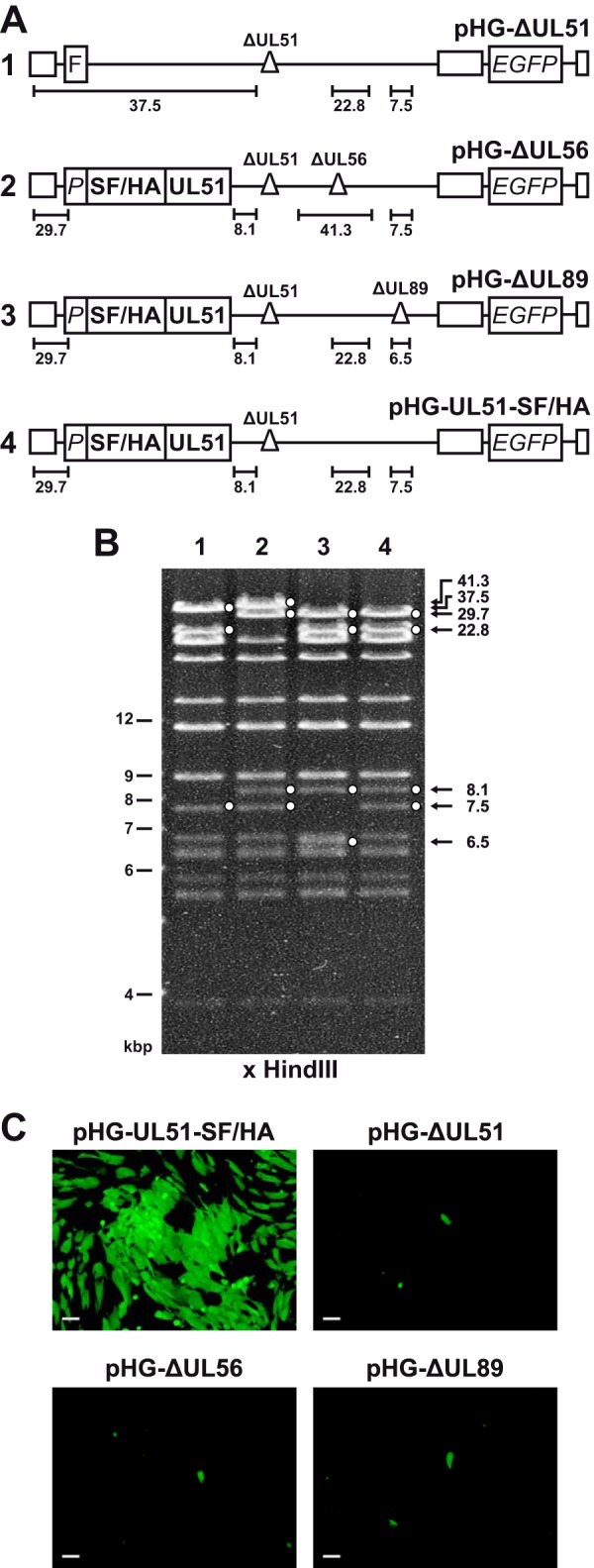
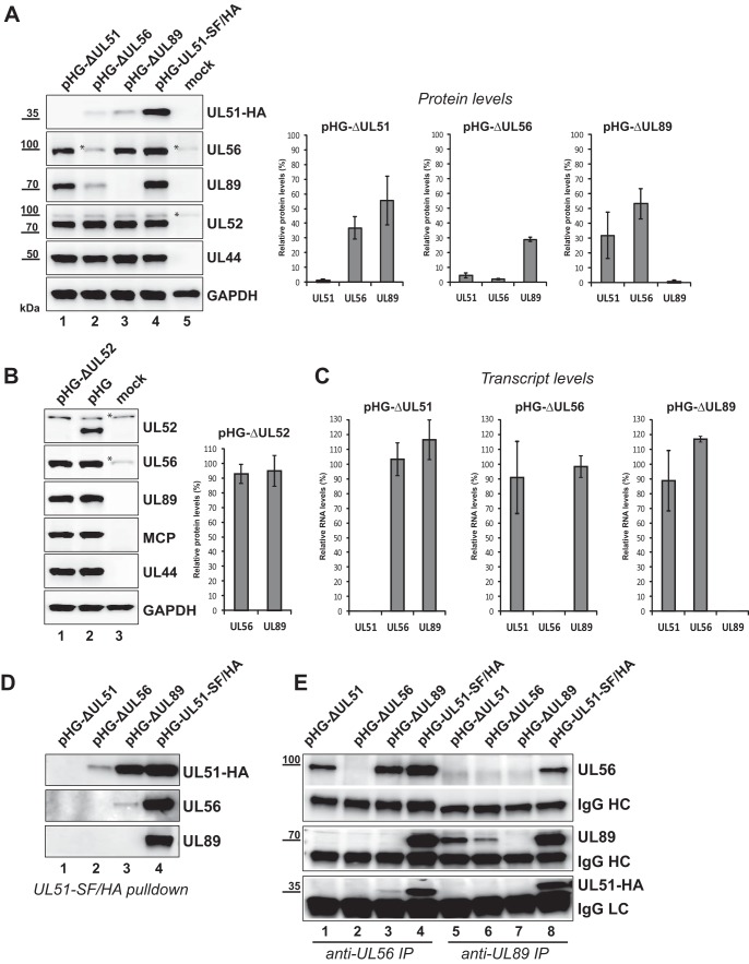
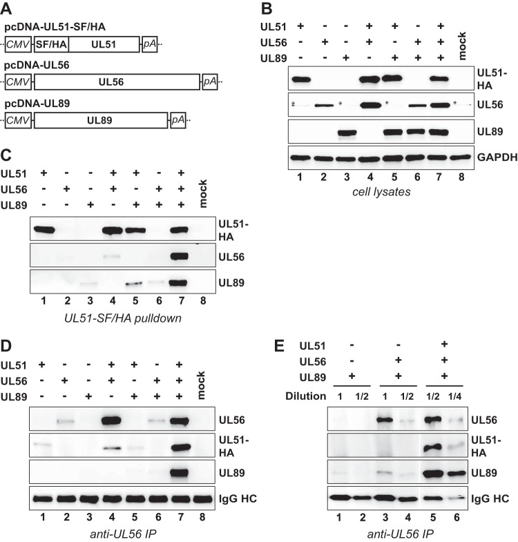
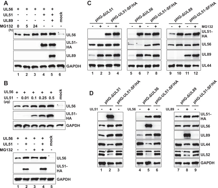
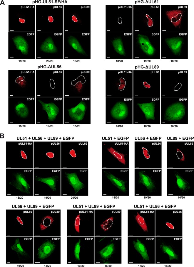
Human cytomegalovirus (HCMV) genome encapsidation requires several essential viral proteins, among them pUL56, pUL89, and the recently described pUL51, which constitute the viral terminase. To gain insight into terminase complex assembly, we investigated interactions between the individual subunits. For analysis in the viral context, HCMV bacterial artificial chromosomes carrying deletions in the open reading frames encoding the terminase proteins were used. These experiments were complemented by transient-transfection assays with plasmids expressing the terminase components. We found that if one terminase protein was missing, the levels of the other terminase proteins were markedly diminished, which could be overcome by proteasome inhibition or providing the missing subunit in trans These data imply that sequestration of the individual subunits within the terminase complex protects them from proteasomal turnover. The finding that efficient interactions among the terminase proteins occurred only when all three were present together is reminiscent of a folding-upon-binding principle leading to cooperative stability. Furthermore, whereas pUL56 was translocated into the nucleus on its own, correct nuclear localization of pUL51 and pUL89 again required all three terminase constituents. Altogether, these features point to a model of the HCMV terminase as a multiprotein complex in which the three players regulate each other concerning stability, subcellular localization, and assembly into the functional tripartite holoenzyme.IMPORTANCE HCMV is a major risk factor in immunocompromised individuals, and congenital CMV infection is the leading viral cause for long-term sequelae, including deafness and mental retardation. The current treatment of CMV disease is based on drugs sharing the same mechanism, namely, inhibiting viral DNA replication, and often results in adverse side effects and the appearance of resistant virus strains. Recently, the HCMV terminase has emerged as an auspicious target for novel antiviral drugs. A new drug candidate inhibiting the HCMV terminase, Letermovir, displayed excellent potency in clinical trials; however, its precise mode of action is not understood yet. Here, we describe the mutual dependence of the HCMV terminase constituents for their assembly into a functional terminase complex. Besides providing new basic insights into terminase formation, these results will be valuable when studying the mechanism of action for drugs targeting the HCMV terminase and developing additional substances interfering with viral genome encapsidation.

Keywords: complex assembly; cytomegalovirus; nuclear import; protein interactions; protein stability; terminase.


Figures

FIG 1
FIG 1
HCMV BAC genomes used in this study. (A) Schematic representation of the generated BACs containing the following elements: FLP recombination target (FRT [F]) site replacing the nonessential ORFs UL1 to UL10; UL51 promoter region (P); sequences encoding Strep-tag II, FLAG, and HA epitope tags (SF/HA); deletions (Δ) disrupting the indicated ORFs; ORF encoding the enhanced green fluorescent protein (EGFP). The terminal and internal repeat regions are indicated by open boxes. HindIII DNA fragments characteristic of the respective viral genomes are indicated by black lines. The illustration is not drawn to scale. (B) Restriction analysis of the recombinant HCMV genomes. BAC DNA was cut with HindIII, separated by agarose gel electrophoresis, and stained with ethidium bromide. Relevant bands characterizing the HCMV genomes are indicated by solid white circles. The lanes are numbered according to constructs 1 to 4 in the schematic drawings shown in panel A. (C) Ability of recombinant HCMV BAC genomes to generate infectious progeny virus. RPE-1 cells were adenofected with the indicated BACs. On day 4 posttransfection, cells were harvested and added to a monolayer of HFF cells, and 10 days later, viral spread was monitored by UV light microscopy. Bars, 100 μm.
FIG 2
FIG 2
Reduced amounts of the terminase subunits in cells transfected with HCMV BAC genomes in which either UL51, UL56, UL89, or UL52 is disrupted. (A, left) RPE-1 cells were adenofected with the indicated BACs or were mock transfected. On day 4 posttransfection, cells were harvested, whole-cell lysates were prepared, and immunoblot analysis was performed with the antibodies indicated. The HCMV late protein pUL52 and the early protein pUL44 served as controls for both transfection efficiency and viral gene expression, and GAPDH was used as a loading control. The asterisks denote unspecific reactivity with a cellular protein. (Right) Immunoblot signals of three independent experiments (means ± standard deviations [SD] [error bars]) were quantified with ImageJ software using membranes exposed for a few seconds only. For normalization, UL51, UL56, and UL89 protein levels of cells transfected with the parental BAC pHG-UL51-SF/HA were set at 100%. (B) RPE-1 cells were adenofected with the BAC genome pHG-ΔUL52 carrying a deletion within the UL52 ORF or with the parental BAC pHG and analyzed by immunoblotting as in panel A with the antibodies indicated (HCMV major capsid [late] protein [MCP]). Quantification of signals was done as described above for panel A. (C) RPE-1 cells adenofected with the indicated HCMV BACs were used for preparation of total RNA on day 4 posttransfection. UL51, UL56, UL89, and UL52 transcript levels were determined by quantitative RT-PCR, relative RNA levels were calculated using UL52 as the internal control and normalized to the values for cells transfected with the parental BAC pHG-UL51-SF/HA. Data are representative of two independent experiments. (D and E) Interactions between the terminase subunits in RPE-1 cells adenofected with the indicated BACs. pUL51-SF/HA was pulled down using Strep-Tactin Sepharose (D), or pUL56 and pUL89 were immunoprecipitated (IP) with specific antibodies from cell lysates prepared on day 4 posttransfection (E). Eluted proteins were analyzed by immunoblotting using antibodies directed against the HA tag (for pUL51), pUL56, or pUL89. IgG heavy chains (HC) and light chains (LC) served as controls.
FIG 3
FIG 3
Efficient interactions require all three terminase proteins. (A) Schematic representations of the expression plasmids used in this study. The major immediate early promoter of HCMV (CMV), sequences for Strep-tag II, FLAG, and HA epitope tags (SF/HA) and polyadenylation signal sequence (pA) are indicated. (B) RPE-1 cells were transfected with the expression plasmids depicted in panel A, either alone or in the indicated combinations, or were mock transfected. On day 2 posttransfection, whole-cell lysates were prepared and analyzed by immunoblotting using antibodies directed against the HA tag (for pUL51), against pUL56, pUL89, or GAPDH (which served as a loading control). The asterisks denote unspecific reactivity with a cellular protein. (C) The epitope-tagged pUL51 was pulled down using Strep-Tactin beads, and pUL56 or pUL89 bound to it was assessed by immunoblotting. (D) Following immunoprecipitation (IP) of pUL56, coprecipitated pUL51 or pUL89 was detected by immunoblotting. IgG HC, immunoglobulin heavy chain. (E) The samples displayed in panel D, lanes 6 and 7, were diluted to adjust immunoprecipitated pUL56 to comparable levels, and interacting pUL51 and pUL89 was analyzed by immunoblotting.
FIG 4
FIG 4
Mutual protection of the terminase subunits from proteasomal turnover. (A) RPE-1 cells were transfected with equal amounts of pcDNA-UL56 and treated with MG132 (5 μM) for the time periods indicated before harvesting (lanes 1 to 3), or cotransfected with pcDNA-UL56 and pcDNA-UL51-SF/HA (lane 4), or cotransfected with all three expression constructs (lane 5), or mock transfected (lane 6). Total cell lysates were prepared on day 2 posttransfection and analyzed by immunoblotting. (B, top) Stabilization of pUL56 levels by coexpression of pUL51. RPE-1 cells were cotransfected with a constant amount of pcDNA-UL56 and increasing amounts of pcDNA-UL51-SF/HA or were mock transfected and then analyzed by immunoblotting. (Bottom) Stabilization of pUL56 in the presence of both pUL51 and MG132. RPE-1 cells were transfected with the indicated expression plasmids and cultivated with MG132 (+) or without MG132 (−) for 24 h before harvesting. (C and D) Rescue of the terminase protein levels in BAC-transfected cells through proteasome inhibition (C) or by expressing the missing terminase constituent (D). (C) Adenofected RPE-1 cells were treated with MG132 (5 μM) for 24 h before harvesting (+) or were treated with solvent (dimethyl sulfoxide [DMSO]) only (−). Expression of the indicated proteins was examined by immunoblotting on day 4 posttransfection. (D) RPE-1 cells were transfected with the parental pHG-UL51-SF/HA BAC or the deletion BAC genomes indicated, with (+) or without (−) the pcDNA constructs encoding either the UL51, UL56, or UL89 protein. Analysis was done as described above for panel C. Please note that due to the high levels of pUL51, pUL56, and pUL89 in cells that received the expression plasmids (lanes 2, 5, and 8), the corresponding proteins expressed by the parental BAC pHG-UL51-SF/HA (lanes 3, 6, and 9) were detected only after overexposure of the blots (not shown).
FIG 5
FIG 5
Dependence of the subcellular localization of pUL51 and pUL89 on the presence of the other terminase subunits. (A) RPE-1 cells adenofected with the indicated BAC genomes were probed 4 days later with antibodies directed against the HA tag (for pUL51), pUL56, or pUL89 and were analyzed by confocal laser scanning microscopy. (B) HeLa cells were transfected with expression plasmids encoding pUL51, pUL56, or pUL89, either alone or in the given combinations, together with pEGFP-C1 to mark the transfected cells. After 2 days, cells were analyzed as described above for panel A. In panels A and B, contours of the nuclei are marked by white dashed lines. The numbers below each panel represent the proportion of cells exhibiting the localization pattern shown in the respective micrograph (e.g., 19 cells exhibiting the localization pattern/20 total cells). Images displaying the same antibody staining were taken with identical microscope settings. Bars, 10 μm.

References

    1. Casjens SR. 2011. The DNA-packaging nanomotor of tailed bacteriophages. Nat Rev Microbiol 9:647–657. doi: 10.1038/nrmicro2632. - DOI - PubMed
    1. Feiss M, Rao VB. 2012. The bacteriophage DNA packaging machine. Adv Exp Med Biol 726:489–509. doi: 10.1007/978-1-4614-0980-9_22. - DOI - PubMed
    1. Dittmer A, Drach JC, Townsend LB, Fischer A, Bogner E. 2005. Interaction of the putative human cytomegalovirus portal protein pUL104 with the large terminase subunit pUL56 and its inhibition by benzimidazole-d-ribonucleosides. J Virol 79:14660–14667. doi: 10.1128/JVI.79.23.14660-14667.2005. - DOI - PMC - PubMed
    1. Bogner E, Radsak K, Stinski MF. 1998. The gene product of human cytomegalovirus open reading frame UL56 binds the pac motif and has specific nuclease activity. J Virol 72:2259–2264. - PMC - PubMed
    1. Scheffczik H, Savva CG, Holzenburg A, Kolesnikova L, Bogner E. 2002. The terminase subunits pUL56 and pUL89 of human cytomegalovirus are DNA-metabolizing proteins with toroidal structure. Nucleic Acids Res 30:1695–1703. doi: 10.1093/nar/30.7.1695. - DOI - PMC - PubMed

Publication types

MeSH terms

LinkOut - more resources