. 2017 May 26;91(12):e00271-17.
doi: 10.1128/JVI.00271-17. Print 2017 Jun 15.

Herpes Simplex Virus 1 UL34 Protein Regulates the Global Architecture of the Endoplasmic Reticulum in Infected Cells

Affiliations

Herpes Simplex Virus 1 UL34 Protein Regulates the Global Architecture of the Endoplasmic Reticulum in Infected Cells

Fumio Maeda et al. J Virol. .

Abstract

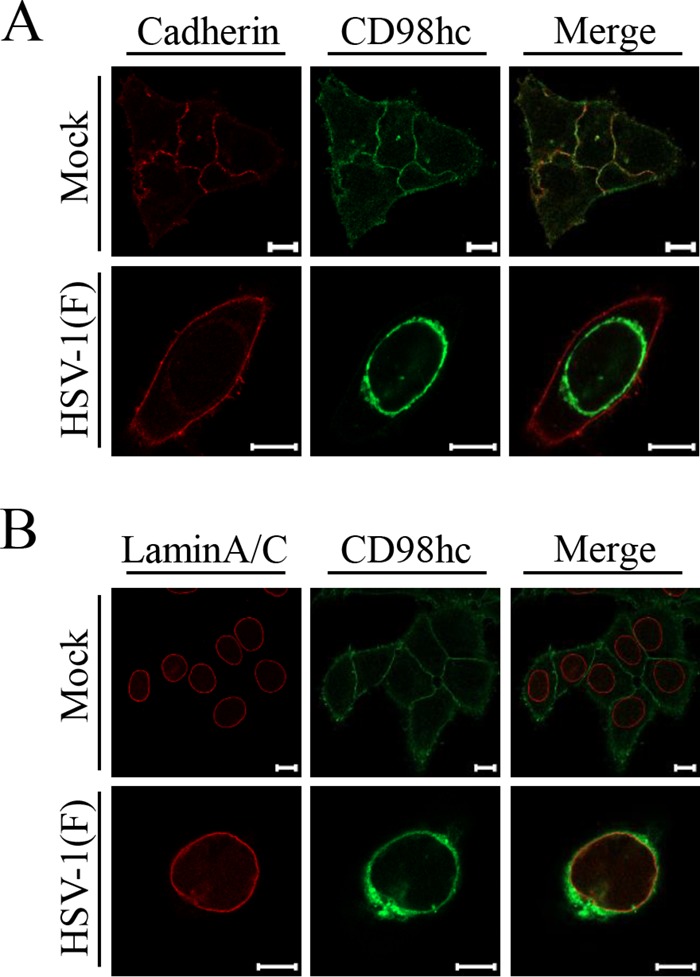
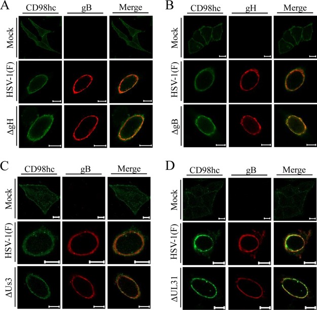
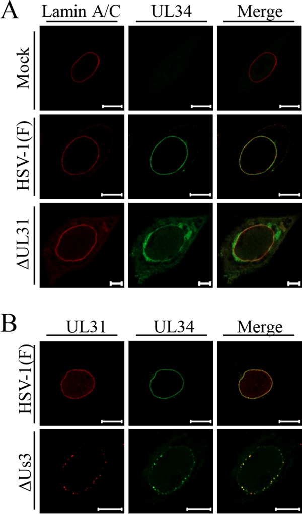
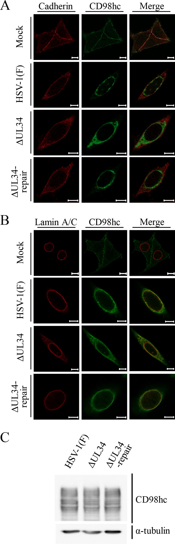
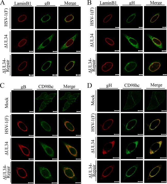
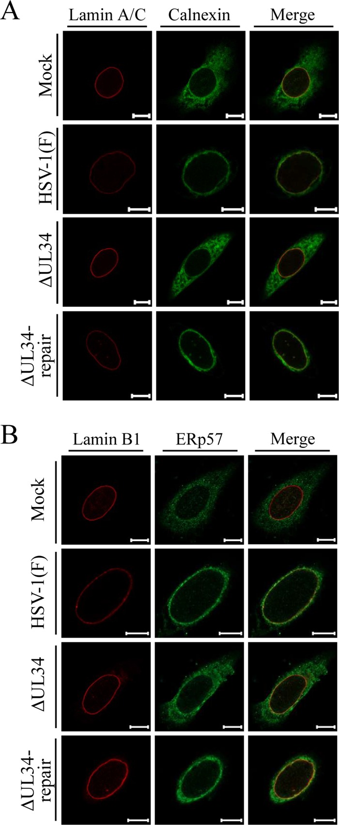
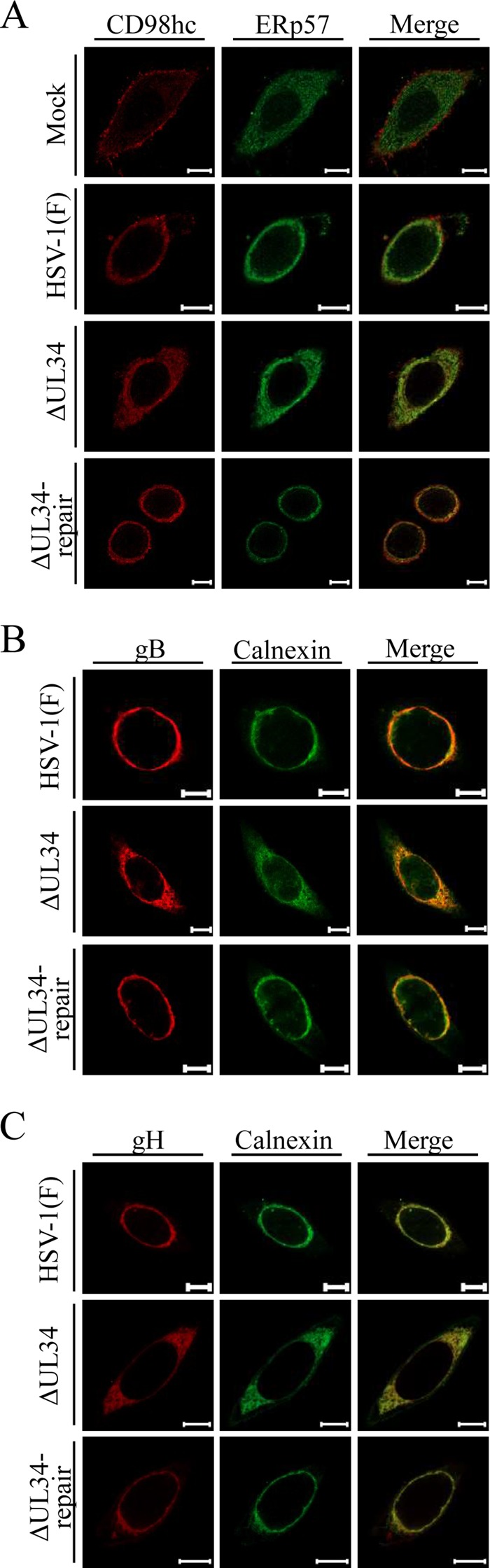
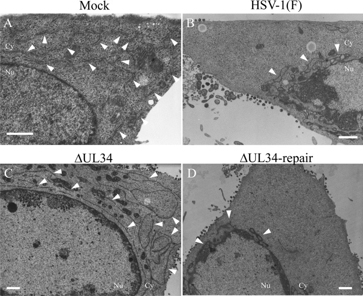
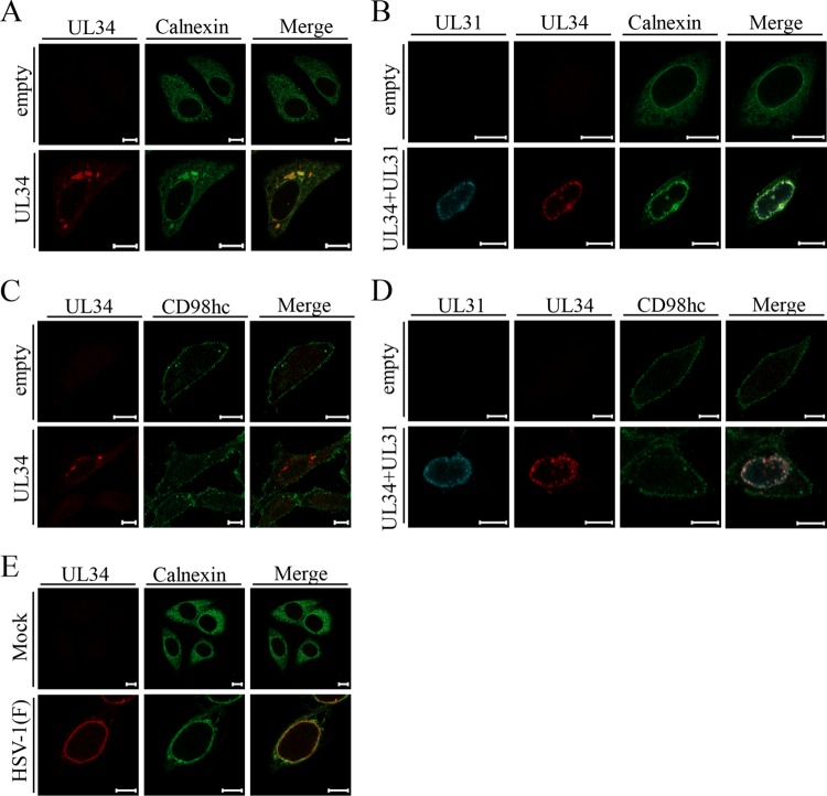
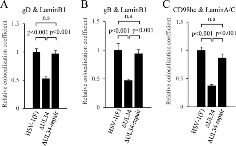
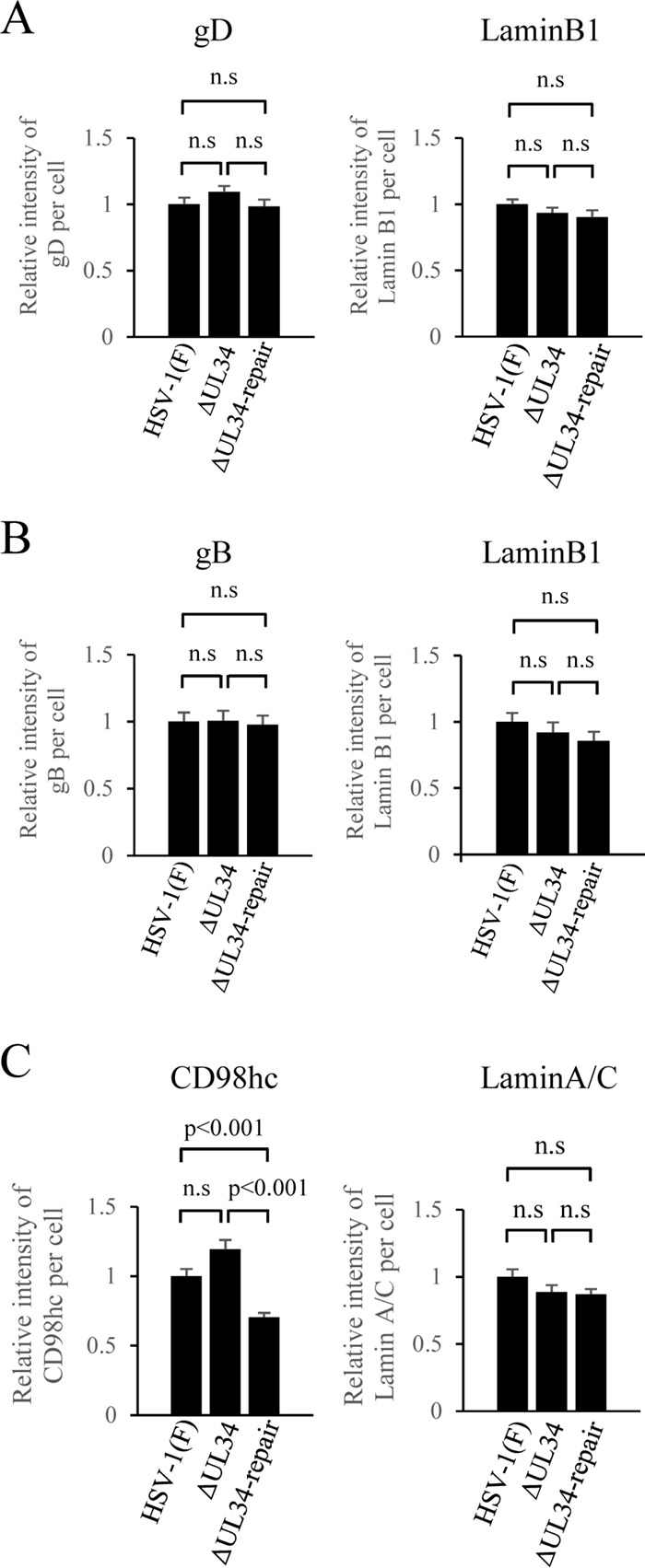
Upon herpes simplex virus 1 (HSV-1) infection, the CD98 heavy chain (CD98hc) is redistributed around the nuclear membrane (NM), where it promotes viral de-envelopment during the nuclear egress of nucleocapsids. In this study, we attempted to identify the factor(s) involved in CD98hc accumulation and demonstrated the following: (i) the null mutation of HSV-1 UL34 caused specific dispersion throughout the cytoplasm of CD98hc and the HSV-1 de-envelopment regulators, glycoproteins B and H (gB and gH); (ii) as observed with CD98hc, gB, and gH, wild-type HSV-1 infection caused redistribution of the endoplasmic reticulum (ER) markers calnexin and ERp57 around the NM, whereas the UL34-null mutation caused cytoplasmic dispersion of these markers; (iii) the ER markers colocalized efficiently with CD98hc, gB, and gH in the presence and absence of UL34 in HSV-1-infected cells; (iv) at the ultrastructural level, wild-type HSV-1 infection caused ER compression around the NM, whereas the UL34-null mutation caused cytoplasmic dispersion of the ER; and (v) the UL34-null mutation significantly decreased the colocalization efficiency of lamin protein markers of the NM with CD98hc and gB. Collectively, these results indicate that HSV-1 infection causes redistribution of the ER around the NM, with resulting accumulation of ER-associated CD98hc, gB, and gH around the NM and that UL34 is required for ER redistribution, as well as for efficient recruitment to the NM of the ER-associated de-envelopment factors. Our study suggests that HSV-1 induces remodeling of the global ER architecture for recruitment of regulators mediating viral nuclear egress to the NM.IMPORTANCE The ER is an important cellular organelle that exists as a complex network extending throughout the cytoplasm. Although viruses often remodel the ER to facilitate viral replication, information on the effects of herpesvirus infections on ER morphological integrity is limited. Here, we showed that HSV-1 infection led to compression of the global ER architecture around the NM, resulting in accumulation of ER-associated regulators associated with nuclear egress of HSV-1 nucleocapsids. We also identified HSV-1 UL34 as a viral factor that mediated ER remodeling. Furthermore, we demonstrated that UL34 was required for efficient targeting of these regulators to the NM. To our knowledge, this is the first report showing that a herpesvirus remodels ER global architecture. Our study also provides insight into the mechanism by which the regulators for HSV-1 nuclear egress are recruited to the NM, where this viral event occurs.

Keywords: ER; herpes simplex virus; organelle structure.


Figures

FIG 1
FIG 1
Schematic diagrams of the genome structure of wild-type HSV-1(F) and the relevant domains of the recombinant viruses generated in this study. 1, wild-type HSV-1(F) genome; 2, domain of the UL33 to UL35 genes; 3, domain of the UL34 gene; 4 and 5, recombinant viruses with mutations in the UL34 gene.
FIG 2
FIG 2
Characterization of UL34-Vero cells and recombinant viruses generated in this study. (A) (Left) Vero and UL34-Vero cells mock infected or infected with wild-type HSV-1(F) or YK722(ΔUL34) at an MOI of 5 for 18 h were analyzed by immunoblotting with the indicated antibodies. (Right) HEp-2 cells mock infected or infected with wild-type HSV-1(F), YK722(ΔUL34), or YK723(ΔUL34-repair) at an MOI of 5 for 24 h were analyzed by immunoblotting with the indicated antibodies. (B) HEp-2 cells were infected with wild-type HSV-1(F), YK722(ΔUL34), or YK723(ΔUL34-repair) at an MOI of 5 (left) or 0.01 (right). Total virus from the cell culture supernatants and infected cells was harvested and assayed on UL34-Vero cells. (C) HEp-2 cells were mock infected or infected with wild-type HSV-1(F), YK722(ΔUL34), or YK723(ΔUL34-repair) at an MOI of 5 for 24 h and then fixed, permeabilized, stained with the indicated antibodies, and examined by confocal microscopy. Scale bars, 10 μm.
FIG 3
FIG 3
Localization of CD98hc in HSV-1-infected cells. HEp-2 cells were mock infected or infected with wild-type HSV-1(F) at an MOI of 5 for 24 h and then fixed, permeabilized, stained with anti-CD98hc antibody in combination with anti-cadherin (A) or anti-lamin A/C (B), and examined by confocal microscopy. Scale bars, 10 μm.
FIG 4
FIG 4
Effects of mutations in gH, gB, or UL31 on localization of CD98hc in HSV-1-infected cells. HEp-2 cells were mock infected or infected with wild-type HSV-1(F) (A to D), YK713(ΔgH) (A), YK701(ΔgB) (B), R7040(ΔUs3) (C), or YK720(ΔUL31) (D) at an MOI of 5 for 24 h and then fixed, permeabilized, stained with the indicated antibodies, and examined by confocal microscopy. Scale bars, 10 μm.
FIG 5
FIG 5
Effects of mutations in UL31 or Us3 on localization of UL34 and/or UL31 in HSV-1-infected cells. HEp-2 cells were mock infected (A) or infected with wild-type HSV-1(F) (A and B), YK720(ΔUL31) (A), or R7040(ΔUs3) (B) at an MOI of 5 for 24 h and then fixed, permeabilized, stained with the indicated antibodies, and examined by confocal microscopy. Scale bars, 10 μm.
FIG 6
FIG 6
Effects of β1 integrin knockdown on localization of CD98hc in HSV-1 infected cells. (A) Expression of β1 integrin in sh-Luc-HEp-2 and sh-β1 integrin-HEp-2 cells was analyzed by immunoblotting with the indicated antibodies. (B) sh-Luc-HEp-2 and sh-β1 integrin-HEp-2 cells were mock infected or infected with wild-type HSV-1(F) at an MOI of 5 for 24 h and then fixed, permeabilized, stained with the indicated antibodies, and examined by confocal microscopy. Scale bars, 10 μm.
FIG 7
FIG 7
Effects of mutation in UL34 on localization and accumulation of CD98hc in HSV-1-infected cells. (A and B) HEp-2 cells were mock infected or infected with wild-type HSV-1(F), YK722(ΔUL34), or YK723(ΔUL34-repair) at an MOI of 5 for 24 h. The cells were fixed, permeabilized, stained with anti-CD98hc antibody in combination with anti-cadherin (A) or anti-lamin A/C (B) antibody, and examined by confocal microscopy. Scale bars, 10 μm. (C) The cells were also analyzed by immunoblotting with the indicated antibodies.
FIG 8
FIG 8
Effects of mutation in UL34 on localization of gB (A) and gH (B) and on colocalization of CD98hc with gB (C) or gH (D) in HSV-1-infected cells. HEp-2 cells were mock infected or infected with wild-type HSV-1(F), YK722(ΔUL34), or YK723(ΔUL34-repair) at an MOI of 5 for 24 h and then fixed, permeabilized, stained with the indicated antibodies, and examined by confocal microscopy. Scale bars, 10 μm.
FIG 9
FIG 9
Effect of mutation in UL34 on localization of the ER markers calnexin (A) and ERp57 (B) in HSV-1-infected cells. HEp-2 cells were mock infected or infected with wild-type HSV-1(F), YK722(ΔUL34), or YK723(ΔUL34-repair) at an MOI of 5 for 24 h and then fixed, permeabilized, stained with the indicated antibodies, and examined by confocal microscopy. Scale bars, 10 μm.
FIG 10
FIG 10
Effect of mutation in UL34 on colocalization of the ER markers ERp57 (A) and calnexin (B and C) with each of the de-envelopment factors CD98hc (A), gB (B), and gH (C) in HSV-1-infected cells. HEp-2 cells were mock infected or infected with wild-type HSV-1(F), YK722(ΔUL34), or YK723(ΔUL34-repair) at an MOI of 5 for 24 h and then fixed, permeabilized, stained with the indicated antibodies, and examined by confocal microscopy. Scale bars, 10 μm.
FIG 11
FIG 11
Effect of mutation in UL34 on the distribution of the ER in HSV-1-infected cells at the ultrastructural level. HEp-2 cells were mock infected (A) or infected with wild-type HSV-1(F) (B), YK722(ΔUL34) (C), or YK723(ΔUL34-repair) (D) at an MOI of 30 for 24 h; embedded; sectioned; stained; and examined by transmission electron microscopy. The arrowheads indicate the ER. Nu, nucleus; Cy, cytoplasm. Scale bars, 1 μm.
FIG 12
FIG 12
Localization of calnexin and CD98hc in cells ectopically expressing UL34 and/or UL31 or in HSV-1-infected cells. (A to D) HEp-2 cells were transfected with the empty plasmid alone (A to D), the UL34 expression plasmid alone (A and C), or the UL34 and UL31 expression plasmids (B and D) for 48 h and then fixed, permeabilized, stained with anti-calnexin (A and B) or anti-CD98hc (C and D) antibody in combination with the indicated antibodies, and examined by confocal microscopy. (E) HEp-2 cells were mock infected or infected with wild-type HSV-1(F) at an MOI of 5 for 24 h and then fixed, permeabilized, stained with anti-UL34 antibody and anti-calnexin antibody, and examined by confocal microscopy. Scale bars, 10 μm.
FIG 13
FIG 13
Effects of mutation in UL34 on colocalization of the NM markers with gD, gB, or CD98hc in HSV-1-infected cells. HEp-2 cells were infected with wild-type HSV-1(F), YK722(ΔUL34), or YK723(ΔUL34-repair) at an MOI of 5 for 24 h and then fixed; permeabilized; stained with anti-lamin B1 (A and B) or anti-lamin A/C (C) in combination with anti-gD (A), anti-gB (B), or anti-CD98hc antibody (C); and examined by confocal microscopy. Colocalization between gD and lamin B1 (A), gB and lamin B1 (B), or CD98hc and lamin A/C (C) was quantified using Manders' colocalization coefficient. Each value is the mean and standard error (n = 40) and is expressed relative to the mean value of wild-type HSV-1(F)-infected HEp-2 cells, which was normalized to 1. Statistical analysis was performed by one-way analysis of variance with the Tukey test; n.s., not significant.
FIG 14
FIG 14
Effect of mutation in UL34 on the total intensity of fluorescence of gD (A), gB (B), or CD98hc (C) in HSV-1-infected cells detected by confocal microscopy. Total intensities per cell in the infected HEp-2 cells analyzed in Fig. 13 were obtained using ZEN software (Zeiss). Each value is the mean and standard error (n = 40) and is expressed relative to the mean value of wild-type HSV-1(F)-infected HEp-2 cells, which was normalized to 1. Statistical analysis was performed by one-way analysis of variance with the Tukey test; n.s., not significant.

References

    1. Roizman B, Knipe DM, Whitley RJ. 2013. Herpes simplex viruses, p 1823–1897. In Knipe DM, Howley PM, Cohen JI, Griffin DE, Lamb RA, Martin MA, Racaniello VR, Roizman B (ed), Fields virology, 6th ed Lippincott-Williams &Wilkins, Philadelphia, PA.
    1. Johnson DC, Baines JD. 2011. Herpesviruses remodel host membranes for virus egress. Nat Rev Microbiol 9:382–394. doi: 10.1038/nrmicro2559. - DOI - PubMed
    1. Mettenleiter TC, Muller F, Granzow H, Klupp BG. 2013. The way out: what we know and do not know about herpesvirus nuclear egress. Cell Microbiol 15:170–178. doi: 10.1111/cmi.12044. - DOI - PubMed
    1. Reynolds AE, Ryckman BJ, Baines JD, Zhou Y, Liang L, Roller RJ. 2001. U(L)31 and U(L)34 proteins of herpes simplex virus type 1 form a complex that accumulates at the nuclear rim and is required for envelopment of nucleocapsids. J Virol 75:8803–8817. doi: 10.1128/JVI.75.18.8803-8817.2001. - DOI - PMC - PubMed
    1. Roller RJ, Zhou Y, Schnetzer R, Ferguson J, DeSalvo D. 2000. Herpes simplex virus type 1 U(L)34 gene product is required for viral envelopment. J Virol 74:117–129. doi: 10.1128/JVI.74.1.117-129.2000. - DOI - PMC - PubMed

MeSH terms

LinkOut - more resources