

Artonin E induces p53-independent G1 cell cycle arrest and apoptosis through ROS-mediated mitochondrial pathway and livin suppression in MCF-7 cells
- PMID: 28356713
- PMCID: PMC5367776
- DOI: 10.2147/DDDT.S124324
Artonin E induces p53-independent G1 cell cycle arrest and apoptosis through ROS-mediated mitochondrial pathway and livin suppression in MCF-7 cells
Abstract
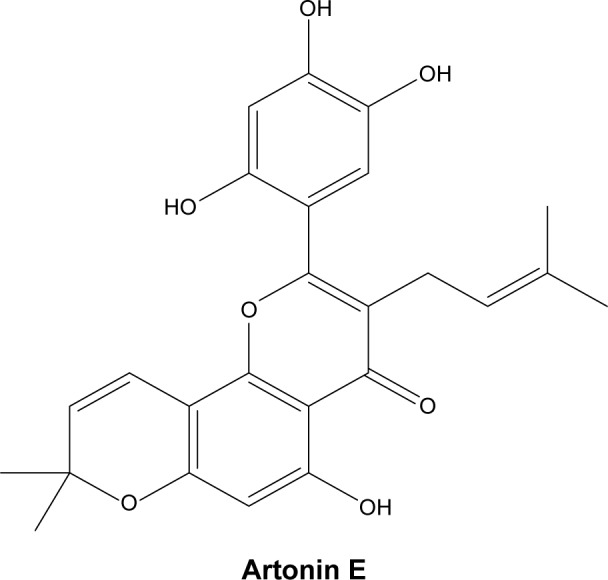
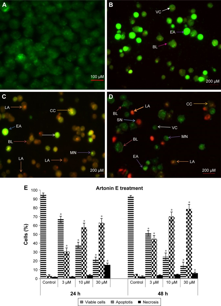
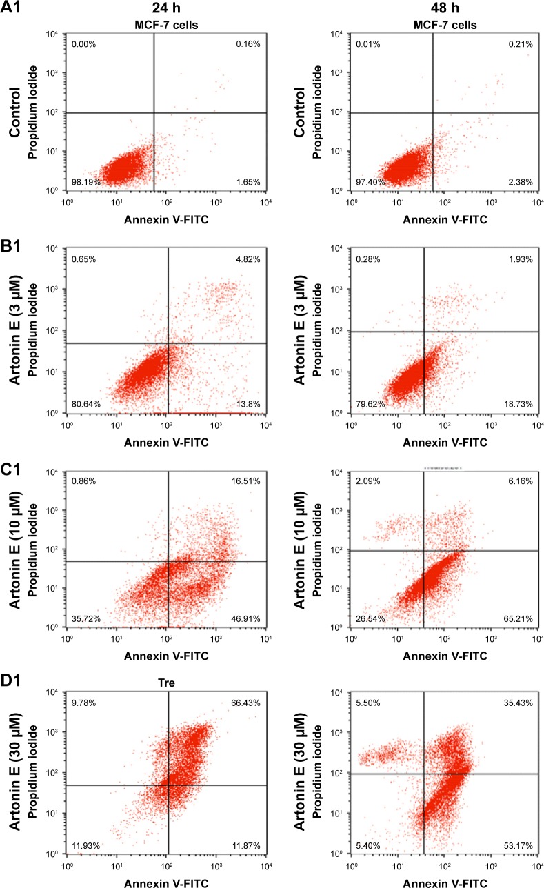
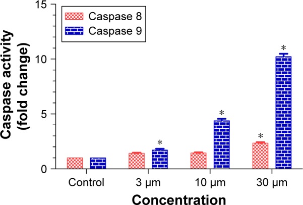
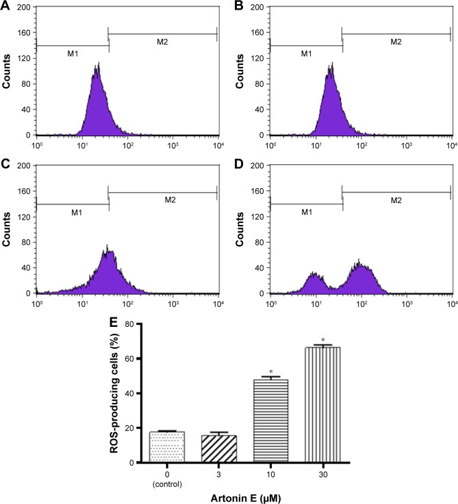
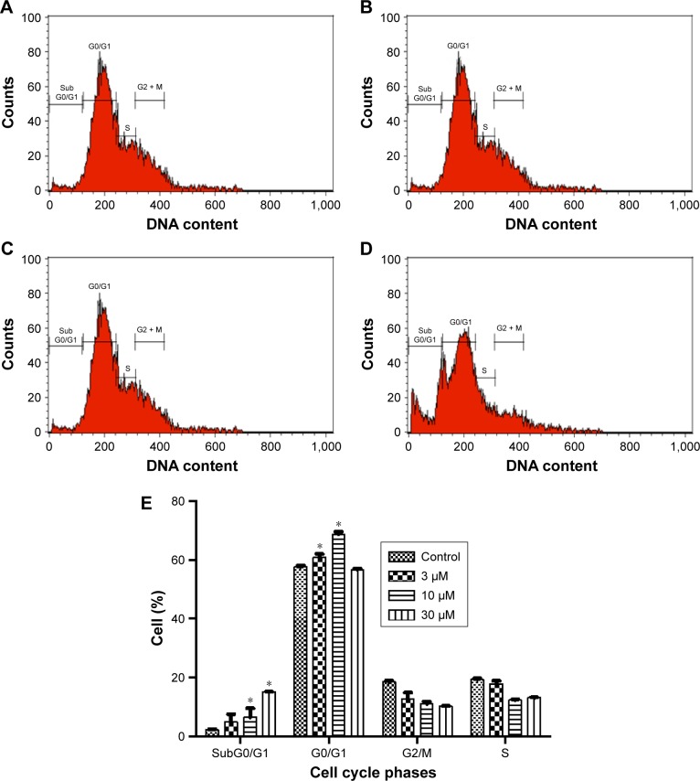
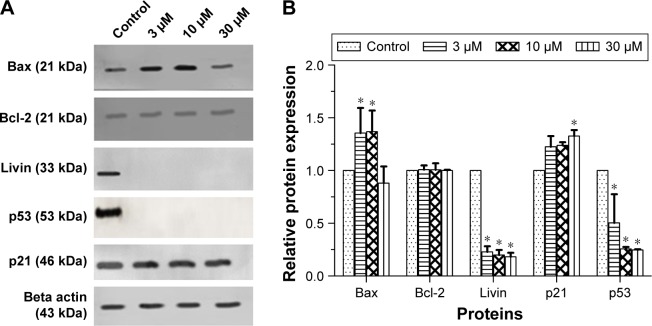
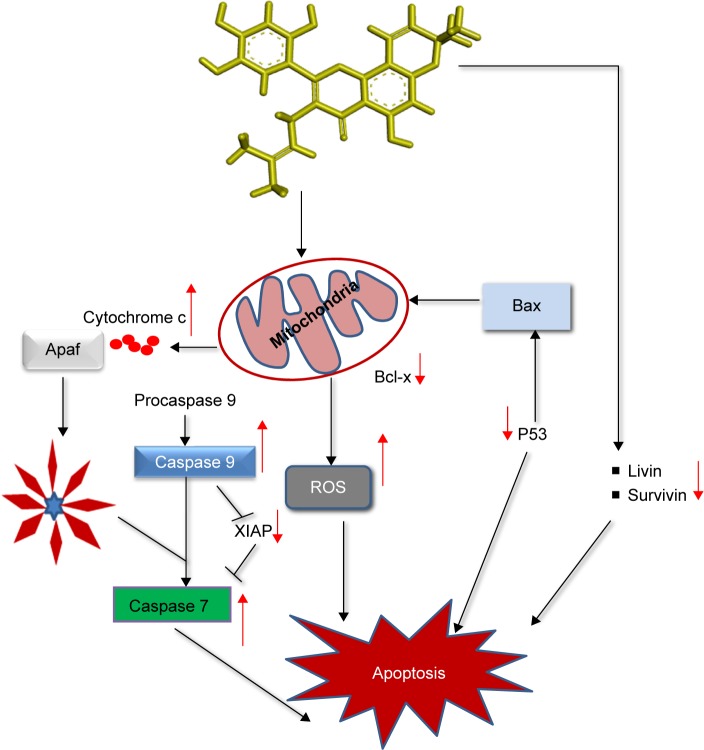
Artonin E is a prenylated flavonoid compound isolated from the stem bark of Artocarpus elasticus. This phytochemical has been previously reported to be drug-like with full compliance to Lipinski's rule of five and good physicochemical properties when compared with 95% of orally available drugs. It has also been shown to possess unique medicinal properties that can be utilized in view of alleviating most human disease conditions. In this study, we investigated the cytotoxic mechanism of Artonin E in MCF-7 breast cancer cells, which has so far not been reported. In this context, Artonin E significantly suppressed the breast cancer cell's viability while inducing apoptosis in a dose-dependent manner. This apoptosis induction was caspase dependent, and it is mediated mainly through the intrinsic pathway with the elevation of total reactive oxygen species. Gene and protein expression studies revealed significant upregulation of cytochrome c, Bax, caspases 7 and 9, and p21 in Artonin E-treated MCF-7 cells, while MAPK and cyclin D were downregulated. Livin, a member of the inhibitors of apoptosis, whose upregulation has been noted to precede chemotherapeutic resistance and apoptosis evasion was remarkably repressed. In all, Artonin E stood high as a potential agent in the treatment of breast cancer.
Keywords: Artonin E; apoptosis; breast cancer; cell cycle; livin.
Conflict of interest statement
Disclosure The authors report no conflicts of interest in this work.
Figures
Similar articles
-
The molecular mechanism of the anticancer effect of Artonin E in MDA-MB 231 triple negative breast cancer cells.PLoS One. 2017 Aug 3;12(8):e0182357. doi: 10.1371/journal.pone.0182357. eCollection 2017. PLoS One. 2017. PMID: 28771532 Free PMC article.
-
Artonin E Induces Apoptosis via Mitochondrial Dysregulation in SKOV-3 Ovarian Cancer Cells.PLoS One. 2016 Mar 28;11(3):e0151466. doi: 10.1371/journal.pone.0151466. eCollection 2016. PLoS One. 2016. PMID: 27019365 Free PMC article.
-
Induction of cell cycle arrest and apoptosis by betulinic acid-rich fraction from Dillenia suffruticosa root in MCF-7 cells involved p53/p21 and mitochondrial signalling pathway.J Ethnopharmacol. 2015 May 26;166:270-8. doi: 10.1016/j.jep.2015.03.039. Epub 2015 Mar 19. J Ethnopharmacol. 2015. PMID: 25797115
-
Fucoidan induces G1 phase arrest and apoptosis through caspases-dependent pathway and ROS induction in human breast cancer MCF-7 cells.J Huazhong Univ Sci Technolog Med Sci. 2013 Oct;33(5):717-724. doi: 10.1007/s11596-013-1186-8. Epub 2013 Oct 20. J Huazhong Univ Sci Technolog Med Sci. 2013. PMID: 24142726
-
Bufalin induces G0/G1 phase arrest through inhibiting the levels of cyclin D, cyclin E, CDK2 and CDK4, and triggers apoptosis via mitochondrial signaling pathway in T24 human bladder cancer cells.Mutat Res. 2012 Apr 1;732(1-2):26-33. doi: 10.1016/j.mrfmmm.2011.09.010. Epub 2012 Jan 20. Mutat Res. 2012. PMID: 22285700
Cited by
-
Phytochemistry and pharmacology of natural prenylated flavonoids.Arch Pharm Res. 2023 Apr;46(4):207-272. doi: 10.1007/s12272-023-01443-4. Epub 2023 Apr 14. Arch Pharm Res. 2023. PMID: 37055613 Free PMC article. Review.
-
Effects of Artonin E on Cell Growth Inhibition and Apoptosis Induction in Colon Cancer LoVo and HCT116 Cells.Molecules. 2022 Mar 24;27(7):2095. doi: 10.3390/molecules27072095. Molecules. 2022. PMID: 35408492 Free PMC article.
-
Effect of lycopene as an adjuvant therapy with 5-florouracil in human colon cancer.Saudi J Biol Sci. 2022 Sep;29(9):103392. doi: 10.1016/j.sjbs.2022.103392. Epub 2022 Jul 25. Saudi J Biol Sci. 2022. PMID: 35957702 Free PMC article.
-
p53 Gene (NY-CO-13) Levels in Patients with Chronic Myeloid Leukemia: The Role of Imatinib and Nilotinib.Diseases. 2018 Jan 25;6(1):13. doi: 10.3390/diseases6010013. Diseases. 2018. PMID: 29370077 Free PMC article.
-
Differential dose-response effect of cyclosporine A in regulating apoptosis and autophagy markers in MCF-7 cells.Inflammopharmacology. 2023 Aug;31(4):2049-2060. doi: 10.1007/s10787-023-01247-4. Epub 2023 May 19. Inflammopharmacology. 2023. PMID: 37204695
References
-
- Siegel R, Naishadham D, Jemal A. Cancer statistics. CA Cancer J Clin. 2013;63(1):11–30. - PubMed
-
- Perou CM, Sørlie T, Eisen MB, et al. Molecular portraits of human breast tumors. Nature. 2000;406(6797):747–752. - PubMed
-
- Ou JM, Ye B, Qiu MK, et al. Knockdown of livin inhibits growth and invasion of gastric cancer cells through blockade of the MAPK pathway in vitro and in vivo. Int J Oncol. 2014;44(1):276–284. - PubMed
-
- Li F, Yin X, Luo X, et al. Livin promotes progression of breast cancer through induction of epithelial-mesenchymal transition and activation of AKT signaling. Cell Signal. 2013;25(6):1413–1422. - PubMed
MeSH terms
Substances
LinkOut - more resources
Full Text Sources
Other Literature Sources
Research Materials
Miscellaneous
