. 2017 Mar 20:12:2189-2204.
doi: 10.2147/IJN.S106548. eCollection 2017.

Nanoliposomal artemisinin for the treatment of murine visceral leishmaniasis

Affiliations

Nanoliposomal artemisinin for the treatment of murine visceral leishmaniasis

Muzamil Y Want et al. Int J Nanomedicine. .

Abstract

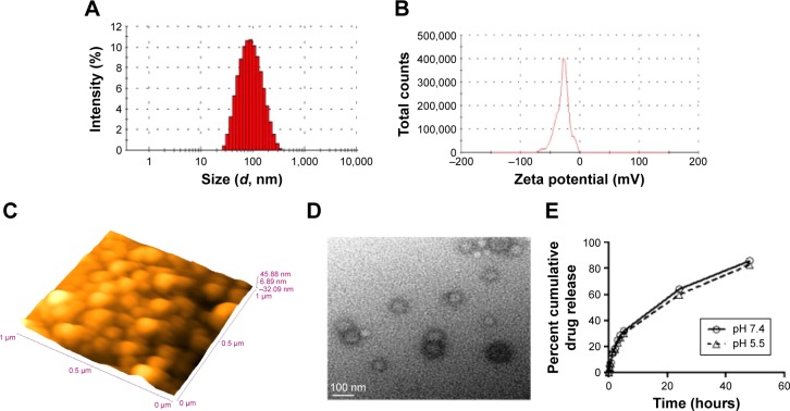
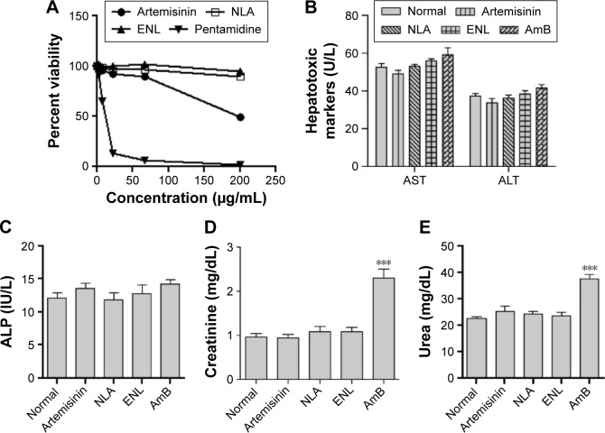
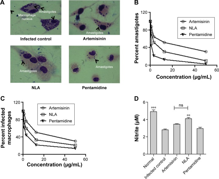
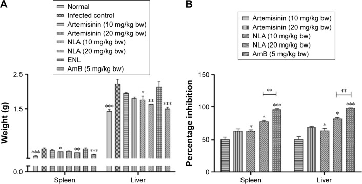
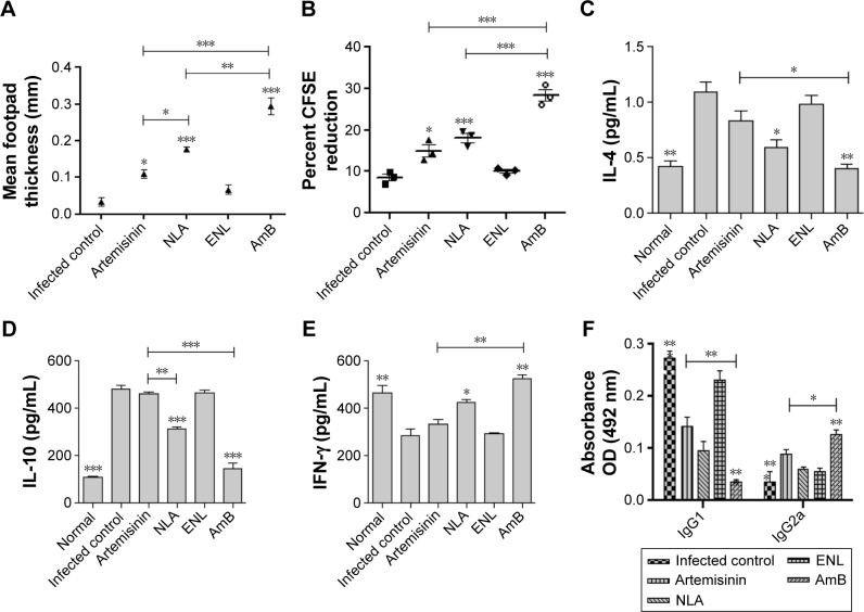
Visceral leishmaniasis (VL) is a fatal, vector-borne disease caused by the intracellular protozoa of the genus Leishmania. Most of the therapeutics for VL are toxic, expensive, or ineffective. Sesquiterpenes are a new class of drugs with proven antimicrobial and antiviral activities. Artemisinin is a sesquiterpene lactone with potent antileishmanial activity, but with limited access to infected cells, being a highly lipophilic molecule. Association of artemisinin with liposome is a desirable strategy to circumvent the problem of poor accessibility, thereby improving its efficacy, as demonstrated in a murine model of experimental VL. Nanoliposomal artemisinin (NLA) was prepared by thin-film hydration method and optimized using Box-Behnken design with a mean particle diameter of 83±16 nm, polydispersity index of 0.2±0.03, zeta potential of -27.4±5.7 mV, and drug loading of 33.2%±2.1%. Morphological study of these nanoliposomes by microscopy showed a smooth and spherical surface. The mechanism of release of artemisinin from the liposomes followed the Higuchi model in vitro. NLA was free from concomitant signs of toxicity, both ex vivo in murine macrophages and in vivo in healthy BALB/c mice. NLA significantly denigrated the intracellular infection of Leishmania donovani amastigotes and the number of infected macrophages ex vivo with an IC50 of 6.0±1.4 µg/mL and 5.1±0.9 µg/mL, respectively. Following treatment in a murine model of VL, NLA demonstrated superior efficacy compared to artemisinin with a percentage inhibition of 82.4%±3.8% in the liver and 77.6%±5.5% in spleen at the highest dose of 20 mg/kg body weight with modulation of cell-mediated immunity towards protective Th1 type. This study is the first report on the use of a liposomal drug delivery system for artemisinin as a promising alternative intervention against VL.

Keywords: Box–Behnken; Leishmania; artemisinin; drug delivery; nanoliposomes; visceral leishmaniasis.


Conflict of interest statement

Disclosure The authors report no conflicts of interest in this work.

Figures

Figure 1
Figure 1
Characterization of NLA. Notes: (A) Size distribution and (B) zeta potential by dynamic light scattering technique, (C) morphology by atomic force microscopy at a scale bar of 1 µm and (D) transmission electron microscopy at a scale bar of 100 nm, and (E) cumulative drug release for 48 hours at pH 5.5 and pH 7.4. All experiments were done in triplicate. Abbreviation: NLA, nanoliposomal artemisinin.
Figure 2
Figure 2
Toxicity of NLA. Notes: (A) Ex vivo on murine macrophages by MTT assay after 72 hours incubation at a concentration range of 0.82–200 µg/mL. Serum levels of (B) AST and ALT, (C) ALP, (D) creatinine, and (E) urea in healthy animals after 24 hours of administration of artemisinin, NLA, ENL, and AmB at the highest dose. Data are represented as mean ± SD and are from one of the two independent experiments. ***P<0.0001. Abbreviations: ALP, alkaline phosphatase; ALT, alanine transaminase; AmB, amphotericin B; AST, aspartate aminotransaminase; ENL, empty nanoliposome; NLA, nanoliposomal artemisinin.
Figure 3
Figure 3
In vitro antileishmanial activity. Notes: (A) Giemsa-stained murine macrophages infected with Leishmania donovani parasites at 48 hours in untreated and treated conditions. (B) Percent L. donovani amastigote infection and (C) percent macrophages infected with L. donovani amastigotes at 48 hours after incubation with different concentrations of artemisinin, NLA, and pentamidine (0–50 µg/mL). (D) Nitric oxide levels in the culture supernatant of uninfected, infected, and treated samples at 48 hours by Griess assay. **P<0.001, ***P<0.0001 Abbreviations: NLA, nanoliposomal artemisinin; ns, not significant.
Figure 4
Figure 4
Antileishmanial activity of nanoliposomes in experimental VL. Notes: (A) Spleen and liver weight in normal, infected, and treated animals 1 week post-treatment. (B) Percentage inhibition of parasites in the spleen and liver 1 week post-treatment. Data are represented as mean ± SD from one of the two independent experiments having five animals per group. *P<0.05, **P<0.001, ***P<0.0001. Abbreviations: AmB, amphotericin B; ENL, empty nanoliposome; NLA, nanoliposomal artemisinin; VL, visceral leishmaniasis.
Figure 5
Figure 5
Evaluation of cellular immunity. Notes: (A) Delayed type hypersensitivity response to freeze thawed leishmanial antigen (800 µg/mL) after 24 hours in age-matched, infected control and animals treated with artemisinin, NLA, ENL, and AmB at the highest dose. (B) Proliferative response by CFSE after 72 hours of in vitro stimulation of total lymphocytes with SLA (10 µg/mL) at the highest dose of artemisinin, NLA, ENL, and AmB. (C) Induction of IL-4, (D) IFN-γ, and (E) IL-10 in the culture supernatant of splenocytes at 72 hours by cytokine bead array at the highest dose of artemisinin, NLA, ENL, and AmB. (F) Parasite-specific antibodies IgG1 and IgG2a 1 week posttreatment with artemisinin, NLA, ENL, and AmB at the highest dose measured by ELISA. Data are represented as mean ± SE from one of the two independent groups having five animals per group. *P<0.05, **P<0.001, ***P<0.0001. Abbreviations: AmB, amphotericin B; CFSE, carboxy fluorescein succinidimyl ester; ELISA, enzyme-linked immunosorbent assay; ENL, empty nanoliposome; FT, freeze thawed; IFN-γ, interferon-γ; IgG, immunoglobulin G; IL, interleukin; NLA, nanoliposomal artemisinin; SLA, soluble leishmanial antigen.

References

    1. Alvar J, Velez ID, Bern C, et al. WHO Leishmaniasis Control Team Leishmaniasis worldwide and global estimates of its incidence. PLoS One. 2012;7(5):e35671. - PMC - PubMed
    1. Herwaldt BL. Leishmaniasis. Lancet. 1999;354(9185):1191–1199. - PubMed
    1. Cragg GM, Newman DJ. Natural products: a continuing source of novel drug leads. Biochim Biophys Acta. 2013;1830(6):3670–3695. - PMC - PubMed
    1. Singh N, Mishra BB, Bajpai S, Singh RK, Tiwari VK. Natural product based leads to fight against leishmaniasis. Bioorg Med Chem. 2014;22(1):18–45. - PubMed
    1. da Costa-Silva TA, Grecco SS, de Sousa FS, et al. Immunomodulatory and antileishmanial activity of phenylpropanoid dimers isolated from Nectandra leucantha. J Nat Prod. 2015;78(4):653–657. - PMC - PubMed

MeSH terms

LinkOut - more resources