. 2017:2017:4089214.
doi: 10.1155/2017/4089214. Epub 2017 Mar 5.

Neuroprotective Effect and Mechanism of Thiazolidinedione on Dopaminergic Neurons In Vivo and In Vitro in Parkinson's Disease

Affiliations

Neuroprotective Effect and Mechanism of Thiazolidinedione on Dopaminergic Neurons In Vivo and In Vitro in Parkinson's Disease

Yanqin Wang et al. PPAR Res. 2017.

Abstract

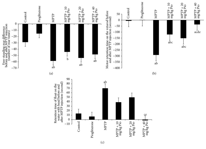
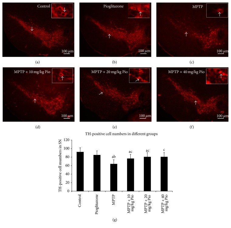
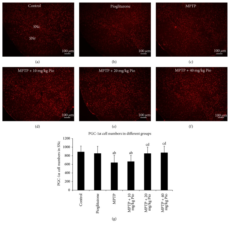
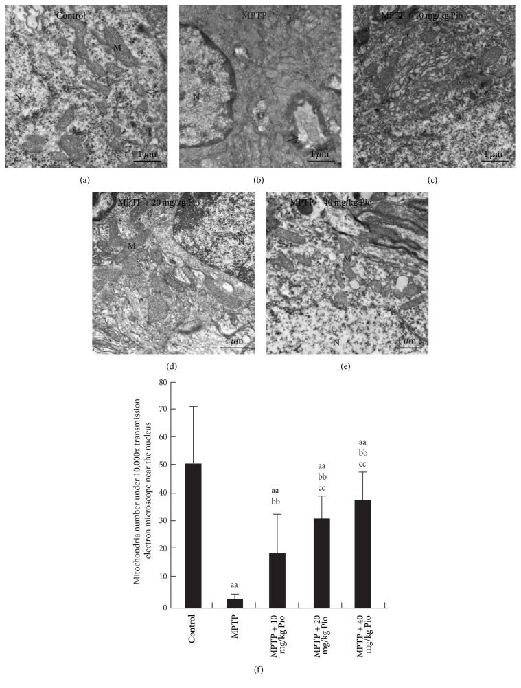
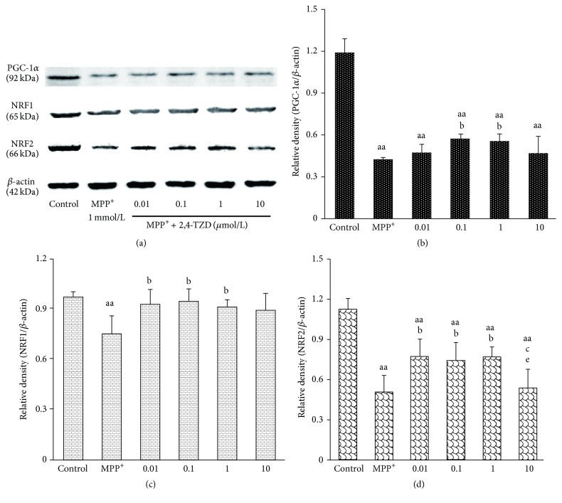
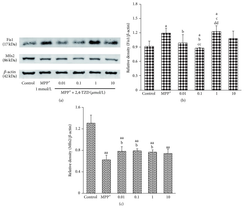
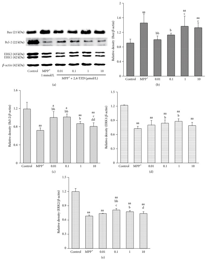
The aim of the present study was to gain insight into the neuroprotection effects and mechanism of thiazolidinedione pioglitazone in both in vitro and in vivo MPP+/MPTP induced PD models. In vivo experimental results showed that oral treatment of pioglitazone resulted in significant improvements in behavior symptoms damaged by MPTP and increase in the survival of TH positive neurons in the pioglitazone intervention groups. In addition, oral treatment of pioglitazone increased the expression of peroxisome proliferator-activated receptor-γ coactivator of 1α (PGC-1α) and increased the number of mitochondria, along with an observed improvement in mitochondrial ultrastructure. From in vitro studies, 2,4-thiazolidinedione resulted in increased levels of molecules regulated function of mitochondria, including PGC-1α, nuclear respiratory factor 1 (NRF1), NRF2, and mitochondria fusion 2 (Mfn2), and inhibited mitochondria fission 1 (Fis1). We show that protein levels of Bcl-2 and ERK were reduced in the MPP+-treated group compared with the control group. This effect was observed to be reversed upon treatment with 2,4-thiazolidinedione, as Bcl-2 and ERK expression levels were increased. We also observed that levels of the apoptotic protein Bax showed opposite changes compared to Bcl-2 and ERK levels. The results from this study confirm that pioglitazone/2,4-thiazolidinedione is able to activate PGC-1α and prevent damage of dopaminergic neurons and restore mitochondria ultrastructure through the regulation of mitochondria function.


Conflict of interest statement

The authors declare that they have no competing interests.

Figures

Figure 1
Figure 1
Effect of pioglitazone on behavior in MPTP-treated C57BL/6 mice. “a” indicates P < 0.05, compared with control group; “b” indicates P < 0.05, compared with pioglitazone group; “c” indicates P < 0.05, compared with MPTP group; “e” indicates P < 0.05, compared with MPTP + 20 mg/kg Pio group.
Figure 2
Figure 2
TH-positive neurons in different groups. “a” indicates P < 0.05, compared with control group; “b” indicates P < 0.05, compared with pioglitazone group; “c” indicates P < 0.05, compared with MPTP group. The bars in (a)–(f) indicate 100 μm; the arrows in each picture show the same neurons.
Figure 3
Figure 3
Effect of pioglitazone on PGC-1α expression in MPTP C57BL/6 mice. “a” indicates P < 0.05, compared with control group; “b” indicates P < 0.05, compared with pioglitazone group; “c” indicates P < 0.05, compared with MPTP group; “d” indicates P < 0.05, compared with MPTP + 10 mg/kg Pio group. The bars in (a)–(f) indicate 100 μm.
Figure 4
Figure 4
Effect of pioglitazone on the mitochondrial ultrastructure in SNc of MPTP C57BL/6 mice. “aa” indicates P < 0.01, compared with control group; “bb” indicates P < 0.01, compared with MPP+ group; “cc” indicates P < 0.01, compared with 0.01 μmol/L 2,4-TZD group. N indicates nucleus; M indicates mitochondria. Arrow points to the demyelination in (b). The bars in (a)–(e) indicate 1 μm.
Figure 5
Figure 5
Effect of thiazolidinedione on PGC-1α, NRF1, and NRF2 protein levels. (a) The expression of PGC-1α, NRF1, and NRF2 in different groups; β-actin was used as the loading control. ((b)–(d)) PGC-1α, NRF1, and NRF2 protein levels. “aa” indicates P < 0.01, compared with control group; “b” indicates P < 0.05, compared with MPP+ group; “c” indicates P < 0.05, compared with 0.01 μmol/L 2,4-TZD group; “e” indicates P < 0.05, compared with 1 μmol/L 2,4-TZD group.
Figure 6
Figure 6
Effect of thiazolidinedione on mitochondria fission and fusion. (a) The expression of Fis1 and Mfn2 in different groups; β-actin was used as the loading control. (b) and (c) express the protein level of Fis1 and Mfn2. “a” indicates P < 0.05 and “aa” indicates P < 0.01, compared with control group; “b” indicates P < 0.05, compared with MPP+ group; “c” indicates P < 0.05 and “cc” indicates P < 0.01, compared with 0.01 μmol/L 2,4-TZD group; “dd” indicates P < 0.05, compared with 0.1 μmol/L 2,4-TZD group.
Figure 7
Figure 7
Effect of thiazolidinedione on apoptosis and levels of antiapoptosis proteins. (a) The expression of Bax, Bcl-2, and ERK in different groups; β-actin was used as the loading control. (b)–(e) express the protein levels of Bax, Bcl-2, and ERK. “a” indicates P < 0.05 and “aa” indicates P < 0.01, compared with control group; “b” indicates P < 0.05 and “bb” indicates P < 0.01, compared with MPP+ group; “c” indicates P < 0.05 and “cc” indicates P < 0.01, compared with 0.01 μmol/L 2,4-TZD group; “d” indicates P < 0.05 and “dd” indicates P < 0.01, compared with 0.1 μmol/L 2,4-TZD group.

References

    1. Shin J.-H., Ko H. S., Kang H., et al. PARIS (ZNF746) repression of PGC-1α contributes to neurodegeneration in parkinson's disease. Cell. 2011;144(5):689–702. doi: 10.1016/j.cell.2011.02.010. - DOI - PMC - PubMed
    1. Dextera D. T., Jenner P. Parkinson disease: from pathology to molecular disease mechanisms. Free Radical Biology & Medicine. 2013;62:132–144. doi: 10.1016/j.freeradbiomed.2013.01.018. - DOI - PubMed
    1. Schaeffer E., Pilotto A., Berg D. Pharmacological strategies for the management of levodopa-induced dyskinesia in patients with parkinson's disease. CNS Drugs. 2014;28(12):1155–1184. doi: 10.1007/s40263-014-0205-z. - DOI - PubMed
    1. Freitas M. E., Fox S. H. Nondopaminergic treatments for Parkinson's disease: current and future prospects. Neurodegenerative Disease Management. 2016;6(3):249–268. doi: 10.2217/nmt-2016-0005. - DOI - PMC - PubMed
    1. Fischer R., Maier O. Interrelation of oxidative stress and inflammation in neurodegenerative disease: role of TNF. Oxidative Medicine and Cellular Longevity. 2015;2015:18. doi: 10.1155/2015/610813.610813 - DOI - PMC - PubMed

LinkOut - more resources