. 2017 Feb;13(2):857-866.
doi: 10.3892/ol.2016.5471. Epub 2016 Dec 7.

Influence of gap junction intercellular communication composed of connexin 43 on the antineoplastic effect of adriamycin in breast cancer cells

Affiliations

Influence of gap junction intercellular communication composed of connexin 43 on the antineoplastic effect of adriamycin in breast cancer cells

Guojun Jiang et al. Oncol Lett. 2017 Feb.

Abstract

Gap junctions (GJs) serve the principal role in the antineoplastic (cytotoxicity and induced apoptosis) effect of chemical drugs. The aim of the present study was to determine the effect of GJ intercellular communication (GJIC) composed of connexin 43 (Cx43) on adriamycin cytotoxicity in breast cancer cells. Four cell lines (Hs578T, MCF-7, MDA-MB-231 and SK-BR-3) with different degree of malignancy were used in the study. The results of western blotting and immunofluorescence revealed that, in Hs578T and MCF-7 cells, which have a low degree of malignancy, the expression levels of Cx43 and GJIC were higher than those in MDA-MB-231 and SK-BR-3 cells (which have a high degree of malignancy). In Hs578T and MCF-7 cells, where GJ could be formed, the function of GJ was modulated by a pharmacological potentiators [retinoid acid (RA)]/inhibitors [oleamide and 18-α-glycyrrhetinic acid (18-α-GA)] and small interfering RNA (siRNA). In high-density cells (where GJ was formed), enhancement of GJ function by RA increased the cytotoxicity of adriamycin, while inhibition of GJ function by oleamide/18-α-GA and siRNA decreased the cytotoxicity caused by adriamycin. Notably, the modulation of GJ did not affect the survival of cells treated with adriamycin when cells were in low density (no GJ was formed). The present study illustrated the association between GJIC and the antitumor effect of adriamycin in breast cancer cells. The cytotoxicity of adriamycin on breast cancer cells was increased when the function of gap junctions was enhanced.

Keywords: adriamycin; breast cancer; connexin 43; gap junction.


Figures

Figure 1.
Figure 1.
Expression of Cx43 in breast cancer cells (Hs578T, MCF-7, MDA-MB-231 and SK-BR-3). (A) Western blot analyses of total Cx43 protein in Hs578T, MCF-7, MDA-MB-231 and SK-BR-3 cells. (B) Fluorescence images show the expression of Cx43 on the membrane of breast cancer cells by immunofluorescence assay. (C) Fluorescence images show the degree of dye coupling by the ‘parachute’ dye-coupling assay. Magnifications, ×400. Cx43, connexin 43.
Figure 2.
Figure 2.
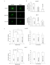
Effect of RA on the cytotoxicity of ADM. (A) Fluorescence images show the degree of dye coupling by the ‘parachute’ dye-coupling assay (magnification, ×400). RA (10 µM) increased gap junction intercellular communication in the two cell types analyzed. (B) Histograms show the quantitation of dye coupling. N=4; Bars are means ± SD. **P<0.01 vs. the control group. (C) Surviving fraction of Hs578T and MCF-7 cells expressing Cx43 incubated with 6 µM ADM for 24 h, with or without 10 µM RA, at high- and low-cell density. N=4; Bars are means ± SD. **P<0.01, vs. the ADM group. Cx43, connexin 43; RA, retinoic acid; ADM, adriamycin; SD, standard deviation.
Figure 3.
Figure 3.
Effect of oleamide or 18-α-GA on the cytotoxicity of ADM. (A) Fluorescence images show the degree of dye coupling by the ‘parachute’ dye-coupling assay (magnification, ×400). Oleamide (25 µM) or 10 µM 18-α-GA decreased gap junction intercellular communication in the two cell types evaluated. (B) Histograms show the quantitation of dye coupling. N=4; Bars are means ± SD. **P<0.01 vs. the control group. (C) Surviving fraction of Hs578T and MCF-7 cells expressing connexin 43 upon incubation with 6 µM ADM for 24 h, with or without either 25 µM oleamide or 10 µM 18-α-GA, at high- and low-cell density. N=4; Bars are means ± SD. **P<0.01 vs. the ADM group. ADM, adriamycin; SD, standard deviation; 18-α-GA, 18-α-glycyrrhetinic acid.
Figure 4.
Figure 4.
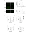
Effect of siRNA-mediated knockdown of Cx43 expression and gap junction intercellular communication. (A and B) Western blot analyses of siRNA-mediated knockdown of Cx43 expression in (A) Hs578T and (B) MCF-7 cells. (C) Fluorescence images show the expression of Cx43 on the membrane of Hs578T and MCF-7 cells by immunofluorescence assay (magnification, ×400). Cells in which Cx43 expression was knocked down by siRNA exhibited lower levels of Cx43 than control cells (not treated with siRNA) and cells expressing Cx43 or cells transfected with negative control siRNA. Cx43, connexin 43; siRNA, small interfering RNA.
Figure 5.
Figure 5.
Effect of siRNA-mediated knockdown of Cx43 expression on ADM cytotoxicity. (A) Fluorescence images show the degree of dye coupling by the ‘parachute’ dye-coupling assay (magnification, ×400). Cells in which Cx43 expression was knocked down by siRNA exhibited a lower extent of dye transfer than control cells (not treated with siRNA) and cells expressing Cx43 or cells transfected with negative control siRNA. (B) Histograms show the quantitation of dye coupling. N=4; Bars are means ± SD. **P<0.01 vs. the control group. (C) Survival fraction of Hs578T and MCF-7 cells incubated with 6 µM ADM and then transfected with negative control siRNA or Cx43 siRNA at high- or low-cell density. Bars are means ± SD. N=3. **P<0.01 vs. the control group. ##P<0.01 vs. the ADM group. ADM, adriamycin; SD, standard deviation; siRNA, small interfering RNA; Cx43, connexin 43.

Similar articles

Cited by

References

    1. Siegel R, Ward E, Brawley O, Jemal A. Cancer statistics, 2011: The impact of eliminating socioeconomic and racial disparities on premature cancer deaths. CA Cancer J Clin. 2011;61:212–236. doi: 10.3322/caac.20121. - DOI - PubMed
    1. Bernzweig J, Heiniger B, Prasain K, Lu J, Hua DH, Nguyen TA. Anti-breast cancer agents, quinolines, targeting gap junction. Med Chem. 2011;7:448–453. doi: 10.2174/157340611796799131. - DOI - PMC - PubMed
    1. Zhang S, Chen Y, Guo W, Yuan L, Zhang D, Xu Y, Nemeth E, Ganz T, Liu S. Disordered hepcidin-ferroportin signaling promotes breast cancer growth. Cell Signal. 2014;26:2539–2550. doi: 10.1016/j.cellsig.2014.07.029. - DOI - PubMed
    1. Li WJ, Zhong SL, Wu YJ, Xu WD, Xu JJ, Tang JH, Zhao JH. Systematic expression analysis of genes related to multidrug-resistance in isogenic docetaxel- and adriamycin-resistant breast cancer cell lines. Mol Biol Rep. 2013;40:6143–6150. doi: 10.1007/s11033-013-2725-x. - DOI - PubMed
    1. Cao B, Li M, Zha W, Zhao Q, Gu R, Liu L, Shi J, Zhou J, Zhou F, Wu X, et al. Metabolomic approach to evaluating adriamycin pharmacodynamics and resistance in breast cancer cells. Metabolomics. 2013;9:960–973. doi: 10.1007/s11306-013-0517-x. - DOI - PMC - PubMed