. 2017 Feb;13(2):955-960.
doi: 10.3892/ol.2016.5521. Epub 2016 Dec 20.

Interleukin-1β activates focal adhesion kinase and Src to induce matrix metalloproteinase-9 production and invasion of MCF-7 breast cancer cells

Affiliations

Interleukin-1β activates focal adhesion kinase and Src to induce matrix metalloproteinase-9 production and invasion of MCF-7 breast cancer cells

Naing Naing Mon et al. Oncol Lett. 2017 Feb.

Abstract

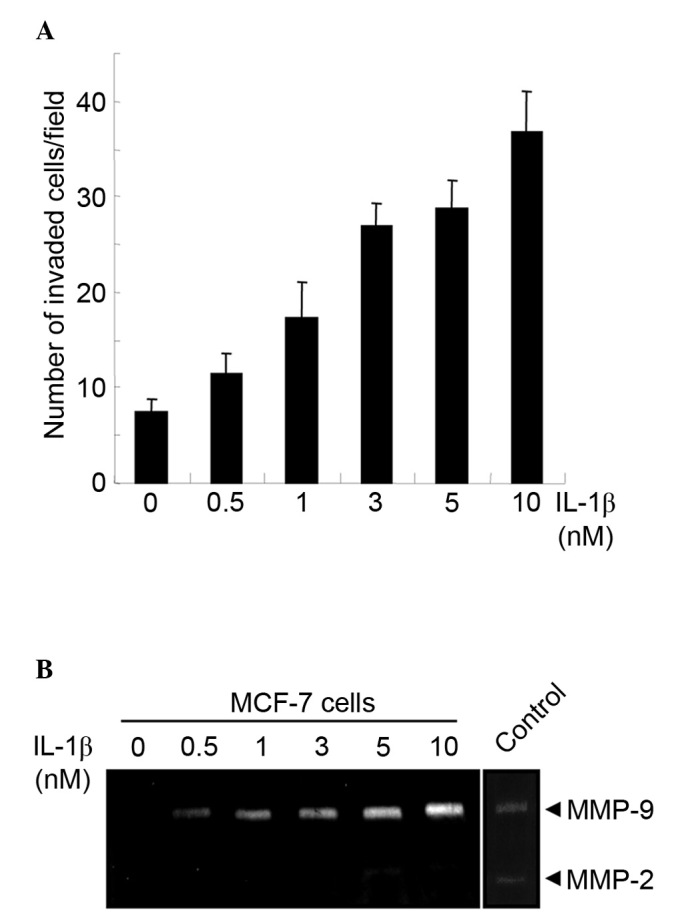
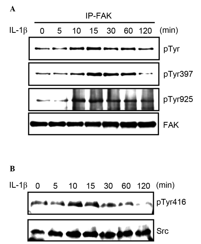
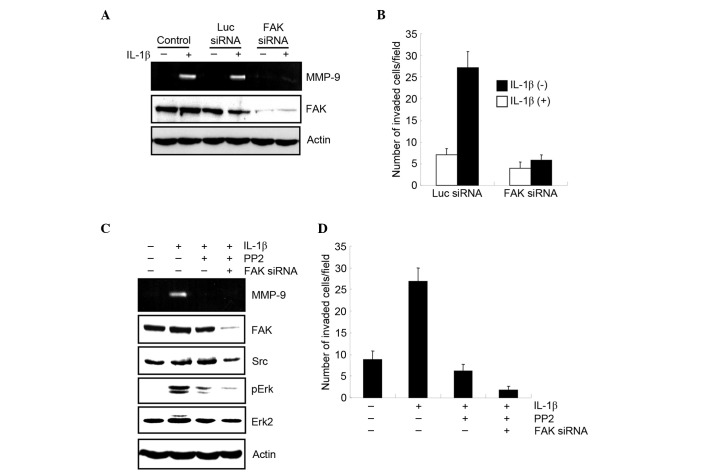
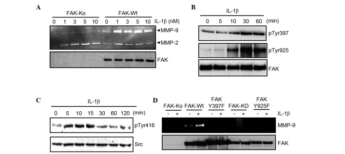
Interleukin-1β (IL-1b) is a pleiotropic cytokine that is important in tumor progression and invasion. Matrix metalloproteinase-9 (MMP-9), which is a secreted matrix-degrading enzyme, is one of the key regulators of tumor invasion and metastasis. The current report indicated that IL-1b promotes MMP-9 production and cell invasion in non-metastatic MCF-7 breast cancer cells. IL-1b activated focal adhesion kinase (FAK) and proto-oncogene tyrosine-protein kinase Src (Src). Moreover, inhibiting the Src/FAK pathway reduced the IL-1b-induced production of MMP-9 and cell invasion. To investigate the functional role of FAK in MMP-9 production cell lines expressing mutant FAK in FAK knock-out mouse fibroblasts were generated. In wild-type FAK-expressing cells, MMP-9 production was induced by IL-1b stimulation. By contrast, IL-1b-induced MMP-9 production was abrogated in FAK knock-out, FAK Y397F, FAK Y925F, and kinase dead mutant-expressing cells. Therefore the results of the current study indicate that FAK and Src kinases are activated by IL-1b and play a critical role in MMP-9 production and tumor cell invasion.

Keywords: FAK; IL-1β; MMP-9; Src; breast cancer cell; invasion.


Figures

Figure 1.
Figure 1.
IL-1β induces MMP-9 production and promotes MCF-7 cell invasion. (A) MCF-7 cells were serum-starved for 6 h and pretreated with the indicated concentrations of human IL-1β for 12 h. Cells were resuspended in serum-free DMEM and seeded onto a Matrigel-coated Boyden chamber with or without IL-1β. Following incubation for 7 h, MCF-7 cells that invaded the lower surface of the filter were fixed, stained and quantified by counting three randomly selected fields under the microscope. Data represent the mean ± SD of three independent experiments. (B) MCF-7 cells were serum-starved for 6 h and incubated with the indicated concentrations of human IL-1β for 16 h. Conditioned media were collected and subjected to gelatin zymography.
Figure 2.
Figure 2.
IL-1β activates FAK and Src in MCF-7 cells. (A) MCF-7 cells were serum-starved and stimulated with 3 nM IL-1β. FAK was immunoprecipitated at the indicated time points and tyrosine phosphorylation was examined by using an anti-phospho-tyrosine antibody. Phospho-FAK-specific antibodies were used to determine the phosphorylation of Tyr397 and Tyr925. FAK expression acted as a reference (B) MCF-7 cells were serum-starved and stimulated with 3 nM IL-1β for the indicated times, and the phosphorylation of Tyr416 was determined by a phospho-Src-specific antibody. Src expression acted as a reference.
Figure 3.
Figure 3.
FAK and Src are required for IL-1β-induced MMP-9 production and invasion in MCF-7 cells. (A) MCF-7 cells were transfected with either luciferase or FAK siRNA. 30 h following transfection, cells were serum-starved for 6 h and incubated with or without 3 nM 1L-1β for 16 h. Conditioned media were collected and subjected to zymography (upper panel). Expression of FAK was determined by western blot analysis (lower panel). Actin expression was used as a reference. (B) Cells were transfected with either luciferase or FAK siRNA and stimulated with IL-1β. Cell invasion was determined by using a modified Boyden chamber. Data represent the mean ± SD from three independent experiments. (C) Following treatment with either luciferase or FAK siRNA for 30 h, cells were serum-starved for 6 h, stimulated by 3 nM IL-1β for 16 h, and were treated with or without PP2 for 1 h. Conditioned media were collected and subjected to zymography. Cell lysates were subjected to western blot analysis by the indicated antibodies and actin expression was used as a reference. (D) Cells were treated with FAK siRNA, PP2, or both, stimulated with IL-1β and subjected to an invasion assay. Data represent the mean ± SD from three independent experiments.
Figure 4.
Figure 4.
FAK is required for IL-1β induced MMP-9 production in mouse fibroblast cells. (A) FAK-Ko and FAK-Wt cells were serum-starved and stimulated with the indicated doses of IL-1β for 16 h. Conditioned media were collected and subjected to gelatin zymography (upper panel). FAK expression in cell lysates is presented in the lower panel, and acted as a reference (B) FAK-Wt cells were serum-starved and stimulated with 3 nM of murine IL-1β for the indicated times. Tyr397 and Tyr925 phosphorylation of FAK was assessed by western blot analysis. FAK expression was used as a reference. (C) FAK-Wt cells were serum-starved and treated with 3 nM murine IL-1β for the indicated times, and Tyr416 phosphorylation of Src was assessed by western blot analysis. Src expression was used as a reference. (D) Cells expressing either wild-type or mutant FAK were serum-starved and stimulated with or without 3 nM of IL-1β for 16 h. Conditioned media were collected and subjected to gelatin zymography (upper panel). Expression of FAK in cell lysates was assessed by western blot analysis (lower panel). FAK expression was used as a reference.

References

    1. Grivennikov SI, Greten FR, Karin M. Immunity, inflammation, and cancer. Cell. 2010;140:883–899. doi: 10.1016/j.cell.2010.01.025. - DOI - PMC - PubMed
    1. Hussain SP, Harris CC. Inflammation and cancer: An ancient link with novel potentials. Int J Cancer. 2007;121:2373–2380. doi: 10.1002/ijc.23173. - DOI - PubMed
    1. Hugo HJ, Saunders C, Ramsay RG, Thompson EW. New insights on COX-2 in chronic inflammation driving breast cancer growth and metastasis. J Mammary Gland Biol Neoplasia. 2015;20:109–119. doi: 10.1007/s10911-015-9333-4. - DOI - PubMed
    1. Harris RE, Casto BC, Harris ZM. Cyclooxygenase-2 and the inflammogenesis of breast cancer. World J Clin Oncol. 2014;5:677–692. doi: 10.5306/wjco.v5.i4.677. - DOI - PMC - PubMed
    1. Hanahan D, Weinberg RA. Hallmarks of cancer: The next generation. Cell. 2011;144:646–674. doi: 10.1016/j.cell.2011.02.013. - DOI - PubMed