. 2017 Feb;6(2):261-265.
doi: 10.3892/mco.2016.1107. Epub 2016 Dec 13.

Paclitaxel and platinum-based chemotherapy results in transient dyslipidemia in cancer patients

Affiliations

Paclitaxel and platinum-based chemotherapy results in transient dyslipidemia in cancer patients

Guoping Wang et al. Mol Clin Oncol. 2017 Feb.

Abstract

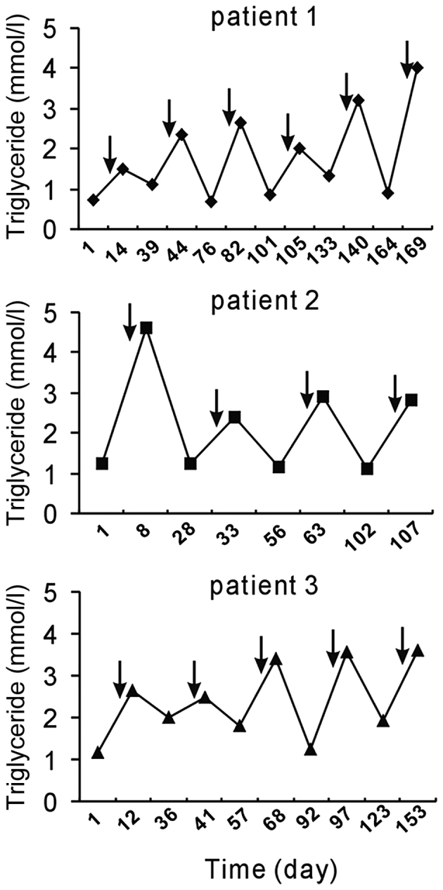
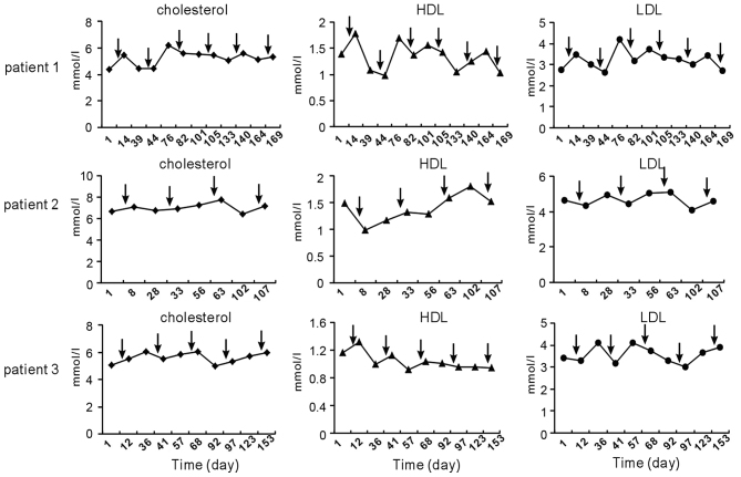
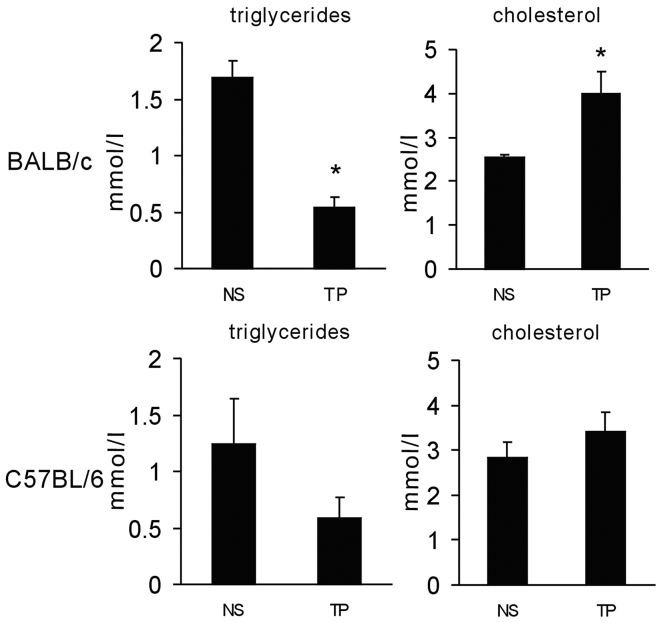
Paclitaxel and cisplatin (TP) are common chemotherapeutic agents extensively used for treating lung and esophageal cancers. The present study reported three patients with transient hypertriglyceridemia (HTG) following TP chemotherapy. Serum triglyceride (TG) levels returned to baseline at chemotherapy intermission. No patient had any history of HTG or any evidence of pancreatitis or other complications of HTG. No regular elevation of any other serum lipids, including cholesterol, high-density lipoprotein and low-density lipoprotein, was observed. However, treatment of mice with TP decreased TG and slightly increased cholesterol. The findings of the present study suggested that TP chemotherapy results in transient dyslipidemia, and physicians must monitor TG levels during chemotherapy to avoid TG-associated complications.

Keywords: chemotherapy; cisplatin; hypertriglyceridemia; paclitaxel.


Figures

Figure 1.
Figure 1.
Chemotherapy with TP induced a fluctuation in serum triglyceride levels. Arrows indicate the time-points of TP chemotherapy administration. TP chemotherapy induced transient hyperglyceridemia. Normal range of serum TG: 0.29–1.83 mmol/l. Patients: 1, lung squamous carcinoma; 2, esophageal squamous carcinoma; 3, synovial carcimoma with lung metastasis. TP, paclitaxel and cisplatin.
Figure 2.
Figure 2.
Serum cholesterol, HDL and LDL levels before and after TP chemotherapy in patients 1–3. Arrows indicate the time-points of TP chemotherapy. The fluctuation trend of cholesterol, HDL and LDL after chemotherapy was dissimilar to that of triglycerides. Normal range: cholesterol, 2.8–5.7 mmol/l; HDL >0.9 mmol/l; LDL <4.0 mmol/l. HDL, high-density lipoprotein; LDL, low-density lipoprotein; TP, paclitaxel and cisplatin.
Figure 3.
Figure 3.
Liver-associated parameters before and after TP chemotherapy. Arrows indicate time-points of TP chemotherapy. Normal range: albumin, 40.0–55.0 g/l; ALT <50 U/l; AST <40 U/l. ALT, alanine transaminase; AST, aspartate aminotransferase; TP, paclitaxel and cisplatin.
Figure 4.
Figure 4.
Serum levels of parameters associated with renal function before and after TP chemotherapy. Normal range: creatinine, 53.0–140.0 µmol/l; urea, 2.40–7.20 mmol/l. Arrows indicate time-points of TP chemotherapy. TP, paclitaxel and cisplatin.
Figure 5.
Figure 5.
Chemotherapy with TP disturbed serum lipids in experimental animals. BALB/c mice were treated intravenously with paclitaxel (10 mg/kg body weight) at day 1, and cisplatin (5 mg/kg body weight) at days 1, 2 and 3. C57BL/6 mice were administered intraperitoneally with the same dose of chemotherapeutical drugs. Serum was obtained on day 4 to evaluate the triglyceride and cholesterol levels. *P<0.05 vs. NS. TP, paclitaxel and cisplatin. Results were expressed as the mean ± standard deviation. NS, normal saline.

References

    1. Fearon KC, Glass DJ, Guttridge DC. Cancer cachexia: Mediators, signaling, and metabolic pathways. Cell Metab. 2012;16:153–166. doi: 10.1016/j.cmet.2012.06.011. - DOI - PubMed
    1. Landen CN, Jr, Birrer MJ, Sood AK. Early events in the pathogenesis of epithelial ovarian cancer. J Clin Oncol. 2008;26:995–1005. doi: 10.1200/JCO.2006.07.9970. - DOI - PubMed
    1. Nieman KM, Kenny HA, Penicka CV, Ladanyi A, Buell-Gutbrod R, Zillhardt MR, Romero IL, Carey MS, Mills GB, Hotamisligil GS, et al. Adipocytes promote ovarian cancer metastasis and provide energy for rapid tumor growth. Nat Med. 2011;17:1498–1503. doi: 10.1038/nm.2492. - DOI - PMC - PubMed
    1. Zhang Y, Daquinag AC, Amaya-Manzanares F, Sirin O, Tseng C, Kolonin MG. Stromal progenitor cells from endogenous adipose tissue contribute to pericytes and adipocytes that populate the tumor microenvironment. Cancer Res. 2012;72:5198–5208. doi: 10.1158/0008-5472.CAN-12-0294. - DOI - PubMed
    1. Dirat B, Bochet L, Dabek M, Daviaud D, Dauvillier S, Majed B, Wang YY, Meulle A, Salles B, Le Gonidec S, et al. Cancer-associated adipocytes exhibit an activated phenotype and contribute to breast cancer invasion. Cancer Res. 2011;71:2455–2465. doi: 10.1158/0008-5472.CAN-10-3323. - DOI - PubMed