. 2017:2017:2624170.
doi: 10.1155/2017/2624170. Epub 2017 Mar 5.

The Noncell Autonomous Requirement of Proboscipedia for Growth and Differentiation of the Distal Maxillary Palp during Metamorphosis of Drosophila melanogaster

Affiliations

The Noncell Autonomous Requirement of Proboscipedia for Growth and Differentiation of the Distal Maxillary Palp during Metamorphosis of Drosophila melanogaster

Anthony Percival-Smith et al. Genet Res Int. 2017.

Abstract

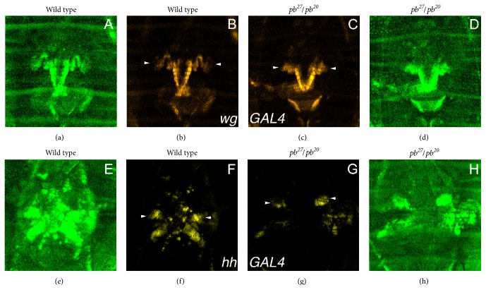
The Drosophila maxillary palpus that develops during metamorphosis is composed of two elements: the proximal maxillary socket and distal maxillary palp. The HOX protein, Proboscipedia (PB), was required for development of the proximal maxillary socket and distal maxillary palp. For growth and differentiation of the distal maxillary palp, PB was required in the cells of, or close to, the maxillary socket, as well as the cells of the distal maxillary palp. Therefore, PB is required in cells outside the distal maxillary palp for the expression, by some mechanism, of a growth factor or factors that promote the growth of the distal maxillary palp. Both wingless (wg) and hedgehog (hh) genes were expressed in cells outside the distal maxillary palp in the lancinia and maxillary socket, respectively. Both wg and hh were required for distal maxillary palp growth, and hh was required noncell autonomously for distal maxillary palp growth. However, expression of wg-GAL4 and hh-GAL4 during maxillary palp differentiation did not require PB, ruling out a direct role for PB in the regulation of transcription of these growth factors.


Conflict of interest statement

The authors have no competing interests.

Figures

Figure 1
Figure 1
The proboscipedia phenotype. In both panels the ventral side is on the left. Panel (A) is wild type mouthparts. The mp bracket indicates the distal maxillary palp and the ms bracket indicates the proximal maxillary socket. The insert at the bottom left is a close-up of the portion of the maxillary socket with three proximal palpus bristles indicated by the arrows. Panel (B) is a pb27/pb20 transformed mouthpart with the reduced distal maxillary palp (mp) and reduced proximal maxillary socket (ms) indicated with brackets. The arrowheads indicate the lancinia in both panels. The inserts on the top left of each panel show the long tricombs found on the maxillary socket of wild type (panel A) and the short tricombs found on the maxillary socket of pb mutants (panel B).
Figure 2
Figure 2
The expression of PB and pb-GAL4 during metamorphosis. Panel (A) is the expression of pb-GAL4 recorded using the UASlacZ reporter gene fixed just before head eversion. The arrows indicate the fused labial discs, asterisks indicate the maxillary palp primordia, and the arrowheads show the antennal primordia. Panel (B) is the expression of pb-GAL4 in developing mouthparts (approximately 18 h AHE) using UASEGFP as the reporter gene (green). The arrowheads indicate the distal maxillary palps. The tissue is stained with DAPI (blue). Panels (C) and (D) are the expression of PB (C) and the expression of PB (green) and SCR (red) (D) at approximately 36 h AHE. The arrowheads indicate the distal maxillary palp. Panel (E) is the time line of metamorphosis indicating the stages and major events observed. The start and stop point for labial fusion, antenna migration, and maxillary palp morphogenesis are estimates based on first evidence of movement. The letters indicate the relative time of the images shown in panels (F)–(M). Panels (F)–(M) are individual frames from live imaging shown in Supplemental Data Movie 1. The time the image was recorded is indicated (h: min: sec BHE or AHE) and the arrows indicate the developing labial segment and the arrowheads indicate the aristal primordia and antennal primordia expression. Panels (N)–(Q) are the first 7.5 minutes AHE of YFP expression driven by pb-GAL4 shown in Supplemental Data Movie 2. The arrowheads indicate the proboscis primordia and the arrows the maxillary palpus primordia. In panels (A)–(D) and (I)–(Q) the dorsal side of the head is at the top and the ventral is at the bottom.
Figure 3
Figure 3
The maxillary palpus phenotypes of three independent mosaic analyses. Panels (A) and (B) are scanning electron micrographs of the effects of clonal loss of PB function generated in flies with the genotype y w; P{hspFLP}/+; P{ry+, neor, FRT}82B pb27/P{ry+, neor, FRT}82B Sb63b M(3)95A2 P{y+, ry+}96E. In panel (A), a Sb+ pb27 clone in the distal maxillary palp is shown and the maxillary palp is reduced. In panel (B), Sb pb+ distal maxillary palps are shown; the right is wild type, and the left is reduced indicated by the arrow. In panels (A) and (B), the arrows indicate Sb bristles and the arrowheads Sb+ bristles. Panels (C) and (D) are bright field micrographs of clonal loss of PB function generated in the genotype y w; P{hspFLP}/+; P{ry+, neor, FRT}82B pb27 Scr2 P{w+, ry+}90E/P{ry+, neor, FRT}82B M(3)95A2 P{y+, ry+}96E P{exd+, w+}. Panel (C) is one of the 22 pb27/+ wild type distal maxillary palps with the y+ proximal palpus bristle indicated by the arrow and y+ maxillary socket tricombs indicated with the arrowhead. Panel (D) shows a y pb27Scr2 maxillary palpus. Panels (E)–(G) are bright field micrographs of distal maxillary palps from the clonal ectopic expression of PB in a pb27/pb20 mutant background generated in the genotype y w, P{w+, pba>y+>Tubα1}B; P{hspFLP}/+; pb27/pb20. Panel (E) is a rescued y and PB expressing (pba>Tubα1) maxillary palpus, panel (F) is a reduced y and PB expressing (pba>Tubα1) maxillary palpus, and panel (G) is a reduced y+ pb (pba>y+>Tubα1) maxillary palpus.
Figure 4
Figure 4
Expression of pb, hh, wg, and dpp-GAL4 drivers during maxillary palpus development. In panels (A)–(D), the driver is indicated on the bottom lefthand corner. The arrowheads indicate the developing maxillary palpus and the arrows in panel (C) indicate the developing lancinia. The tissue is stained with DAPI (blue), and the expression of the drivers was detected with a UASEGFP reporter (green). Panel (E) is the expression of hh-GAL4 in the wing marked by expression of TRCS292A  T453A from the UAStrcS292A T453A reporter. The dotted line indicates the anterior-posterior compartment boundary, and multiple short bristles are observed in the posterior compartment. Panels (F) and (G) are the expression of hh-GAL4 in the maxillary palpus marked by expression of TRCS292A  T453A from the UAStrcS292A T453A reporter. The box in (F) indicates the close-up shown in (G). The dotted line in (G) indicates the field of cells that have multiple short tricombs indicating expression of TRCS292A  T453A.
Figure 5
Figure 5
Genetic analysis of the requirement of hh. Panels (A) and (B) are scanning electron micrographs of the effects of clonal loss of HH function generated in flies with the genotype y w; P{hspFLP}/+; P{ry+, neor, FRT}82B e hh9/P{ry+, neor, FRT}82B Sb63b M(3)95A2 P{y+, ry+}96E. The arrow in panel (A) points to a hh clone in the distal maxillary palp that was marked with Sb+ M+ bristles and that did not affect growth; the growth of the other three distal maxillary palps in panels (A) and (B) were affected to varying degrees and lacked bristles. Panel (C) is a hh9 genetic mosaic in a pb mutant background generated in the genotype y w; P{hspFLP}/+; P{ry+, neor, FRT}82B pb27 Scr2 e hh9/P{ry+, neor, FRT}82B pb20 Sb63b M(3)95A2 P{y+, ry+}96E. The arrows indicate reduced maxillary palpus and the arrowheads indicate the loss of the maxillary palpus in panels (B) and (C). In panel (C) the left vestigial maxillary palpus was missing and remaining vestigial palpus on the right was Sb M (hh9/+).
Figure 6
Figure 6
Expression of wg-GAL4 and hh-GAL4 in wild type and pb27/pb20 mutants. Panels (A)–(D) are wg-GAL4; panels (E)–(H) are hh-GAL4. Panels (A), (D), (E), and (H) show expression of UbiGFP (green). Panels (B), (C), (F), and (G) show expression of YFP (yellow) from a UASYFP reporter gene. Panels (A), (B), (E), and (F) are wild type and panels (C), (D), (G), and (H) are pb27/pb20 mutants. The arrowheads indicate expression of YFP in the maxillary palps of wild type and pb mutants at 16 h AHE.
Figure 7
Figure 7
Clonal analysis of the requirement of PB in hh-GAL4 expression. All clones were generated in the genotype y w; P{UASRFP}/P{hspFLP}; P{UAStrcS292A T453A}, FRT82B pb27hh-GAL4/FRT82B P{UbiGFP}. Panels (A)–(D) are a pupal wing and panels (E)–(H) are a pupal maxillary palp. Panels (A) and (E) are GFP expression; panels (B) and (F) are GFP expression (green) and nuclei visualized with DAPI (red); panels (C) and (G) are RFP expression; and panels (D) and (H) are GFP (green) and RFP (blue) expression with the nuclei visualized with DAPI (red). The two arrowheads in panels (A)–(D) indicate clones of cells that are homozygous for UbiGFP and have lost RFP expression due to loss of hh-GAL4. In panels (E)–(H), the arrow indicates the developing distal maxillary palp, and the arrowhead indicates a pb27 mutant clone that shows strong expression of RFP indicating strong expression of hh-GAL4.

Similar articles

Cited by

References

    1. Snodgrass R. E. Principles of Insect Morphogenesis. New York, NY, USA: McGraw-Hill Book Co.; 1935.
    1. Grant P. R., Grant B. R. Unpredictable evolution in a 30-year study of Darwin's finches. Science. 2002;296(5568):707–711. doi: 10.1126/science.1070315. - DOI - PubMed
    1. Kaufman T. C. Cytogenetic analysis of chromosome 3 in Drosophila melanogaster: isolation and characterization of four new alleles of the proboscipedia (pb) locus. Genetics. 1978;90(3):579–596. - PMC - PubMed
    1. Struhl G. Genes controlling segmental specification in the Drosophila thorax. Proceedings of the National Academy of Sciences of the United States of America. 1982;79(23 I):7380–7384. doi: 10.1073/pnas.79.23.7380. - DOI - PMC - PubMed
    1. Merrill V. K. L., Turner F. R., Kaufman T. C. A genetic and developmental analysis of mutations in the Deformed locus in Drosophila melanogaster. Developmental Biology. 1987;122(2):379–395. doi: 10.1016/0012-1606(87)90303-4. - DOI - PubMed

LinkOut - more resources