. 2017 Mar 22;2(2):e00040-17.
doi: 10.1128/mSphere.00040-17. eCollection 2017 Mar-Apr.

A Novel Staphylococcus Podophage Encodes a Unique Lysin with Unusual Modular Design

Affiliations

A Novel Staphylococcus Podophage Encodes a Unique Lysin with Unusual Modular Design

Katie Cater et al. mSphere. .

Abstract

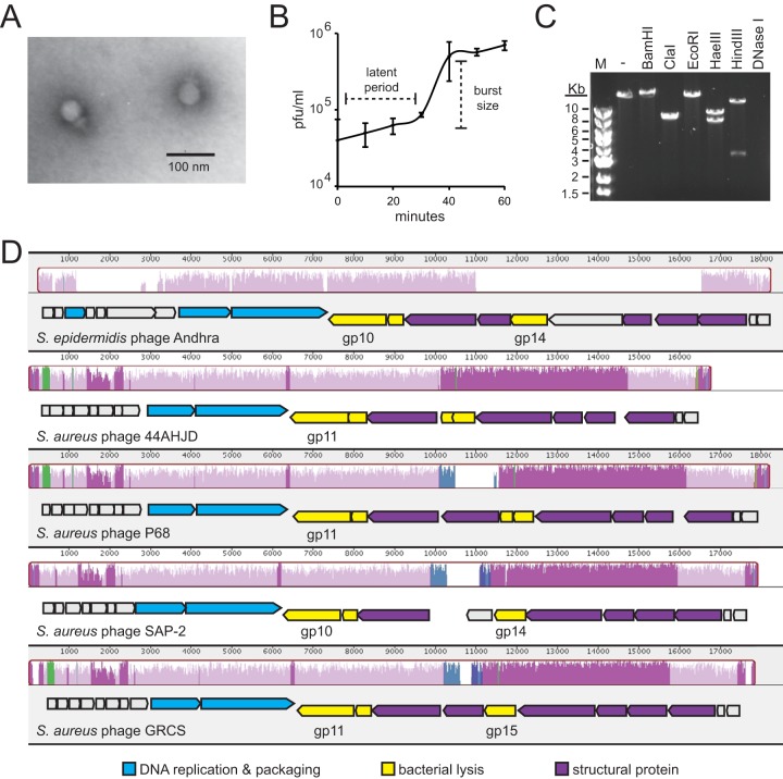
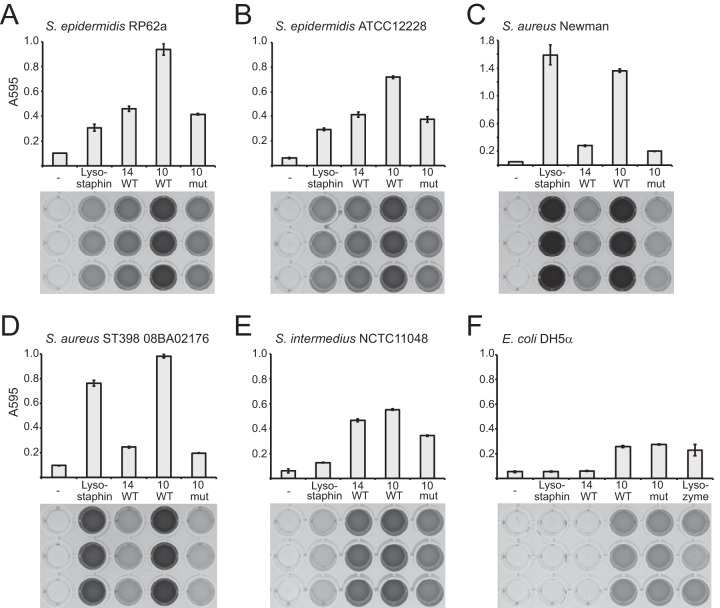
Drug-resistant staphylococci, particularly Staphylococcus aureus and Staphylococcus epidermidis, are leading causes of hospital-acquired infections. Bacteriophages and their peptidoglycan hydrolytic enzymes (lysins) are currently being explored as alternatives to conventional antibiotics; however, only a limited diversity of staphylococcal phages and their lysins has yet been characterized. Here, we describe a novel staphylococcal phage and its lysins. Bacteriophage Andhra is the first reported S. epidermidis phage belonging to the family Podoviridae. Andhra possesses an 18,546-nucleotide genome with 20 open reading frames. BLASTp searches revealed that gene product 10 (gp10) and gp14 harbor putative catalytic domains with predicted peptidase and amidase activities, characteristic functions of phage lysins. We purified these proteins and show that both Andhra_gp10 and Andhra_gp14 inhibit growth and degrade cell walls of diverse staphylococci, with Andhra_gp10 exhibiting more robust activity against the panel of cell wall substrates tested. Site-directed mutagenesis of its predicted catalytic residues abrogated the activity of Andhra_gp10, consistent with the presence of a catalytic CHAP domain on its C terminus. The active site location combined with the absence of an SH3b cell wall binding domain distinguishes Andhra_gp10 from the majority of staphylococcal lysins characterized to date. Importantly, close homologs of Andhra_gp10 are present in related staphylococcal podophages, and we propose that these constitute a new class of phage-encoded lysins. Altogether, our results reveal insights into the biology of a rare family of staphylococcal phages while adding to the arsenal of antimicrobials with potential for therapeutic use. IMPORTANCE The spread of antibiotic resistance among bacterial pathogens is inciting a global public health crisis. Drug-resistant Staphylococcus species, especially S. aureus and S. epidermidis, have emerged in both hospital and community settings, underscoring the urgent need for new strategies to combat staphylococcal infections. Bacterial viruses (phages) and the enzymes that they use to degrade bacterial cell walls (lysins) show promise as alternative antimicrobials; however, only a limited variety of staphylococcal phages and their lysins have yet been identified. Here, we report the discovery and characterization of a novel staphylococcal phage, Andhra. We show that Andhra encodes two lysins (Andhra_gp10 and Andhra_gp14) that inhibit growth and degrade the cell walls of diverse staphylococci, including S. aureus and S. epidermidis strains. Andhra and its unique lysins add to the arsenal of antimicrobials with potential for therapeutic use.

Keywords: Staphylococcus; antimicrobial agents; bacteriophage lysis; bacteriophage therapy; bacteriophages.


Figures

FIG 1
FIG 1
Phenotypic and genotypic features of bacteriophage Andhra. (A) Andhra was stained with uranyl acetate and imaged using transmission electron microscopy at ×200,000 magnification. The morphology is consistent with Podoviridae. (B) A one-step growth curve is shown. S. epidermidis RP62a was challenged with phage Andhra, and PFU were enumerated at 10-min intervals. An average of triplicate measurements is shown. The latent period is defined as the time between adsorption and the beginning of the first burst, and the burst size is defined as the ratio of final plaque count to the initial count. (C) Andhra genomic DNA was extracted, subjected to digestion by the indicated enzymes, and resolved on a 1% agarose gel. Lane M, 1-kb DNA marker; lane -, no enzyme added. (D) A whole-genome sequence alignment of S. epidermidis phage Andhra with indicated S. aureus podophages was generated using the Mauve software (http://darlinglab.org/mauve/mauve.html). Homologous genome regions are indicated with similarly colored histograms, and regions that lack detectible homology remain uncolored. Below the similarity histograms, open reading frames are indicated by block arrows pointing in the direction of transcription. Specific gene products that are discussed in the text are labeled underneath each open reading frame. BLASTp was used to assign putative functions for phage Andhra genes, and the GenBank annotations were used for gene function assignments for the other podophages. Gene products with related functions are similarly colored according to the key at the bottom.
FIG 2
FIG 2
Cell wall hydrolytic activities of Andhra lysins. A dye release assay was used to quantify the cell wall hydrolytic activity of Andhra_gp14 (14 WT), Andhra_gp10 (10 WT), Andhra_gp10C354A,H420A (10 mut), lysostaphin, or lysozyme. Cell walls from indicated bacterial strains were labeled with Remazol brilliant blue dye and used as the substrates. Enzymes (3 μg) or IMAC buffer (-) was incubated with an excess of labeled cell wall substrate at 37°C for 3 h. The dye released into the supernatant was quantified by measuring the optical density of the soluble fraction at 595 nm. The experiment was conducted in triplicate, and average measurements are shown (top). Images of 96-well plates containing soluble fractions corresponding to each measurement are also shown (bottom).

Similar articles

Cited by

References

    1. Rossolini GM, Arena F, Pecile P, Pollini S. 2014. Update on the antibiotic resistance crisis. Curr Opin Pharmacol 18:56–60. doi:10.1016/j.coph.2014.09.006. - DOI - PubMed
    1. Lowy FD. 1998. Staphylococcus aureus infections. N Engl J Med 339:520–532. doi:10.1056/NEJM199808203390806. - DOI - PubMed
    1. Kluytmans J, van Belkum A, Verbrugh H. 1997. Nasal carriage of Staphylococcus aureus: epidemiology, underlying mechanisms, and associated risks. Clin Microbiol Rev 10:505–520. - PMC - PubMed
    1. Peacock SJ, de Silva I, Lowy FD. 2001. What determines nasal carriage of Staphylococcus aureus? Trends Microbiol 9:605–610. doi:10.1016/S0966-842X(01)02254-5. - DOI - PubMed
    1. Otto M. 2009. Staphylococcus epidermidis—the “accidental” pathogen. Nat Rev Microbiol 7:555–567. doi:10.1038/nrmicro2182. - DOI - PMC - PubMed