. 2017 Mar 30:8:14741.
doi: 10.1038/ncomms14741.

FUS affects circular RNA expression in murine embryonic stem cell-derived motor neurons

Affiliations

FUS affects circular RNA expression in murine embryonic stem cell-derived motor neurons

Lorenzo Errichelli et al. Nat Commun. .

Abstract

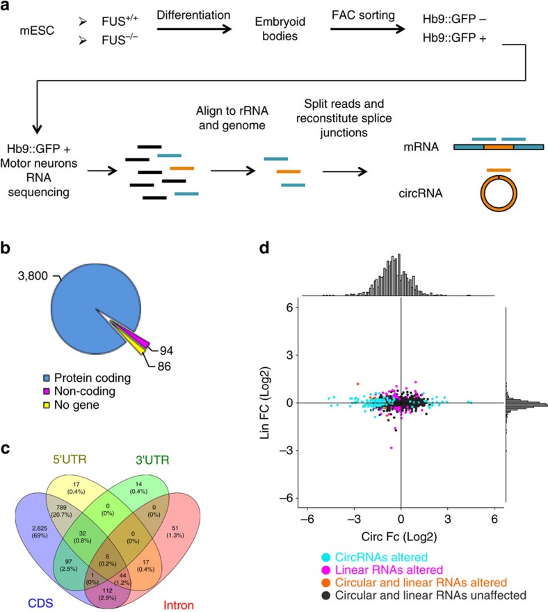
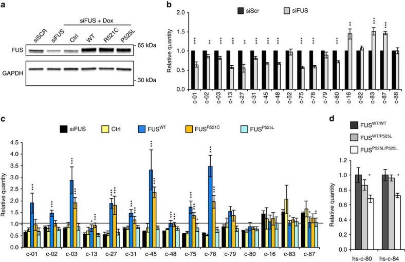
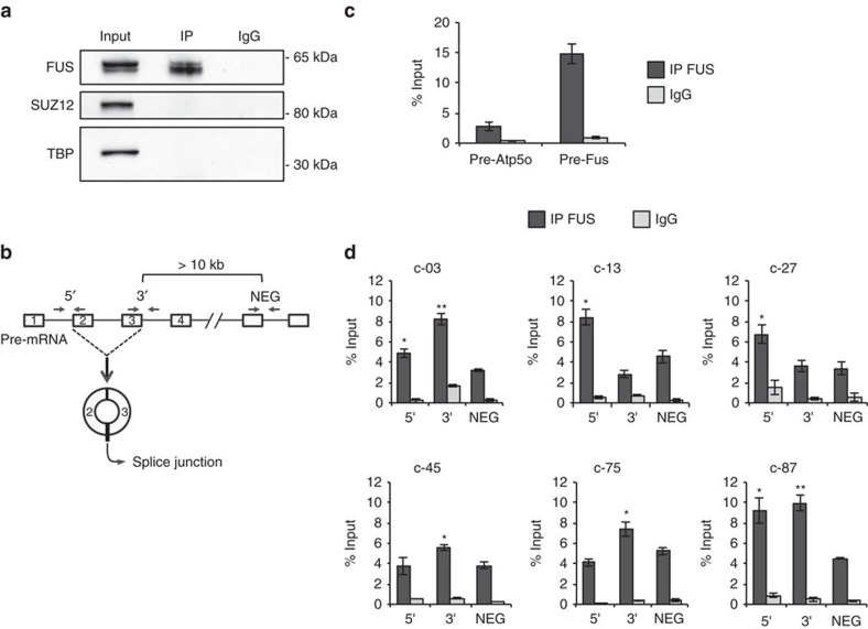
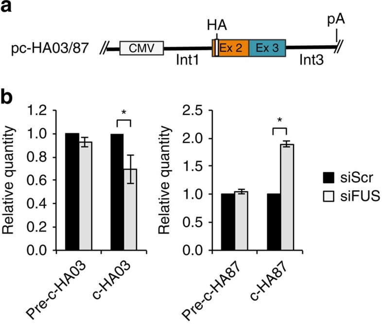
The RNA-binding protein FUS participates in several RNA biosynthetic processes and has been linked to the pathogenesis of amyotrophic lateral sclerosis (ALS) and frontotemporal dementia. Here we report that FUS controls back-splicing reactions leading to circular RNA (circRNA) production. We identified circRNAs expressed in in vitro-derived mouse motor neurons (MNs) and determined that the production of a considerable number of these circRNAs is regulated by FUS. Using RNAi and overexpression of wild-type and ALS-associated FUS mutants, we directly correlate the modulation of circRNA biogenesis with alteration of FUS nuclear levels and with putative toxic gain of function activities. We also demonstrate that FUS regulates circRNA biogenesis by binding the introns flanking the back-splicing junctions and that this control can be reproduced with artificial constructs. Most circRNAs are conserved in humans and specific ones are deregulated in human-induced pluripotent stem cell-derived MNs carrying the FUSP525L mutation associated with ALS.


Conflict of interest statement

The authors declare no competing financial interests.

Figures

Figure 1
Figure 1. mESC in vitro differentiation to spinal motor neurons and RNA-seq analysis.
(a) Schematic overview of the murine motor neuron differentiation protocol and of the RNA-seq analysis. The procedures are reported in Supplementary Fig. 1a and in Methods section. (b) Pie chart showing the number of circRNAs hosted by protein-coding, non-coding genes and intergenic regions (No gene). (c) Venn diagram showing circRNA localization within the body of protein-coding genes. Numbers refer to circRNA species for each category. (d) Scatter plot showing the correlation of log2 fold change of circRNAs (x axis) and their cognate linear RNAs (y axis) in FUS−/− conditions. The distributions of the fold change values are shown above and aside the scatter plot. Turquoise dots indicate when only circRNA species are altered in FUS−/−; magenta dots indicate when only linear RNAs are deregulated; circRNA and linear counterparts either deregulated or unaffected are indicated by orange and black dots, respectively.
Figure 2
Figure 2. CircRNA expression upon FUS depletion and in MN differentiation of mESCs.
(a) circRNA expression analysed by qRT–PCR in sorted GFP+-FUS+/+ and GFP+-FUS−/− cells. The 14 downregulated (left) and 5 upregulated (right) species are reported. CircRNA levels were normalized against Atp5o mRNA levels and expressed as relative quantity with respect to the GFP+-FUS+/+ sample set to a value of 1. (b) The histogram shows the expression level of circRNAs, measured by qRT–PCR, in FUS+/+ mESCs and GFP+-FUS+/+ and GFP-FUS+/+ cells. Their values were normalized against Atp5o mRNA levels and expressed as relative quantity with respect to GFP samples set to a value of 1. For a,b, error bars represent s.e.m. of three independent experiments. *P<0.05, **P<0.01 and ***P<0.001 correspond to two-tailed Student's t-test. (c) Left panel: location on the circRNA of the DIG-labelled probe used for northern blot analysis. Right panel: Northern blots on 10 μg of total RNA from FUS+/+ mESCs, GFP-FUS+/+, GFP+-FUS+/+ and GFP+-FUS−/− cells. The circular forms are indicated aside the gels; asterisks indicate two additional bands likely corresponding to the two alternative linear forms of the c-31 host mRNAs (Hdgfrp3-001, ENSMUST00000107305.7, 5,887 nt; Hdgfrp3-002, ENSMUST00000026094.5, 2,865 nt). Each blot is shown in parallel to the EtBr staining of the agarose gel, where the migration of the 18S and 28S rRNAs is indicated.
Figure 3
Figure 3. CircRNA expression upon ectopic expression of wild-type and mutant FUS.
(a) Western blot analysis on total protein extracts from differentiated N2a cells treated with control siRNAs (siScr) or with siRNAs against FUS (siFUS). In the same blot, shown also are proteins derived from differentiated N2a cells stably transfected with an Ctrl or with FUSWT (WT), FUSR521C (R521C) and FUSP525L (P525L) cDNA expression cassettes and treated with siFUS and Dox. GAPDH was used as a loading control. (b) Histograms show the levels of circRNAs in differentiated N2a cells treated with control siRNAs (siScr; black bars) or with siRNAs against FUS (siFUS; grey bars). CircRNA were quantified by qRT–PCR, normalized against Atp5o mRNA levels and expressed as relative quantity with respect to siScr samples set to a value of 1. (c) Histograms show the levels of circRNAs in the samples treated as in a; the values were normalized against Atp5o mRNA levels and siScr samples set to a value of 1. Statistical analysis was performed on Ctrl, FUSWT, FUSR521C and FUSP525L samples against siFUS samples. (d) Histograms show the level of hsc-80 and hsc-84 in FUSWT/WT, FUSWT/P525L and FUSP525L/P525L iPSC-derived MNs. CircRNAs were normalized against Atp5o mRNA levels and expressed as relative quantity with respect to FUS WT/WT samples set to a value of 1. For all the experiments shown in the figure, error bars represent s.e.m. of at least three independent experiments. *P<0.05, **P<0.01 and ***P<0.001 correspond to two-tailed Student's t-test.
Figure 4
Figure 4. FUS binds the introns flanking the circularized exons.
(a) Protein extracts from FUS CLIP experiments, performed in differentiated N2a cells, were analysed by western blot with FUS antibodies and with SUZ12 and TBP antibodies as negative controls. Input samples account to 4% of the extract; IP and IgG represent 1/5 of the sample used for subsequent RNA analysis. (b) Schematic representation of the primers used for amplifying: the intron–exon junction upstream to the first exon included in the circRNA (5′); the exon–intron junction downstream to the last exon included in the circRNA (3′); the exon–intron junction at a distance of at least 10 Kb away from circularizing exons (NEG). (c) Histogram show the levels of enrichment in IP FUS and IgG samples for the Atp5o pre-mRNA (negative control—pre-Atp5o) and for the region across exon 7 and intron 7 of the FUS pre-mRNA (positive control—pre-Fus). The values are measured as a percentage of the input. (d) Histograms show the levels of enrichment of 5′, 3′ and NEG regions in IP FUS and IgG samples for six circRNA primary transcripts. The values are measured as a percentage of the input. For c,d, error bars represent s.e.m. of at least three independent experiments. *P<0.05, **P<0.01 and ***P<0.001 corresponds to two-tailed Student's t-test.
Figure 5
Figure 5. Artificial constructs reproduce FUS-dependent back-splicing.
(a) Schematic representation of the pc-HA-c03 and pc-HA-c87 constructs. The second and third exons were cloned into pc-DNA3.1+ vector together with ∼1,500 nucleotides of the flanking introns. An HA tag was inserted in order to discriminate the ectopically expressed circRNAs from the endogenous ones. (b) Histograms show the circRNA levels derived from pc-HA03 and pc-HA87 together with the corresponding linear transcripts (pre-c-HA03 and pre-c-HA87) in N2a cells treated with control siRNAs (siScr; black bars) or with siRNAs against FUS (siFUS; grey bars). The values were normalized against Neomycin mRNA levels and expressed as relative quantity with respect to siScr samples set to a value of 1. For all the experiments shown in the figure, error bars represent s.e.m. of three independent experiments. *P<0.05 corresponds to two-tailed Student's t-test.

Comment in

References

    1. Sanger H. L., Klotz G., Riesner D., Gross H. J. & Kleinschmidt A. K. Viroids are single-stranded covalently closed circular RNA molecules existing as highly base-paired rod-like structures. Proc. Natl Acad. Sci. USA 73, 3852–3856 (1976). - PMC - PubMed
    1. Kjems J. & Garrett R. A. Novel splicing mechanism for the ribosomal RNA intron in the archaebacterium desulfurococcus mobilis. Cell 54, 693–703 (1988). - PubMed
    1. Capel B. et al.. Circular transcripts of the testis-determining gene Sry in adult mouse testis. Cell 73, 1019–1030 (1993). - PubMed
    1. Salzman J., Gawad C., Wang P. L., Lacayo N. & Brown P. O. Circular RNAs are the predominant transcript isoform from hundreds of human genes in diverse cell types. PLoS ONE 7, e30733 (2012). - PMC - PubMed
    1. Danan M., Schwartz S., Edelheit S. & Sorek R. Transcriptome-wide discovery of circular RNAs in Archaea. Nucleic Acids Res. 40, 3131–3142 (2012). - PMC - PubMed

Publication types