. 2017 Mar 30:7:45499.
doi: 10.1038/srep45499.

Tubular Cardiac Tissues Derived from Human Induced Pluripotent Stem Cells Generate Pulse Pressure In Vivo

Affiliations

Tubular Cardiac Tissues Derived from Human Induced Pluripotent Stem Cells Generate Pulse Pressure In Vivo

Hiroyoshi Seta et al. Sci Rep. .

Abstract

Human induced pluripotent stem (iPS) cell-derived cardiac cells provide the possibility to fabricate cardiac tissues for transplantation. However, it remains unclear human bioengineered cardiac tissues function as a functional pump in vivo. Human iPS cells induced to cardiomyocytes in suspension were cultured on temperature-responsive dishes to fabricate cardiac cell sheets. Two pairs of triple-layered sheets were transplanted to wrap around the inferior vena cava (IVC) of nude rats. At 4 weeks after transplantation, inner pressure changes in the IVC were synchronized with electrical activations of the graft. Under 80 pulses per minute electrical stimulation, the inner pressure changes at 8 weeks increased to 9.1 ± 3.2 mmHg, which were accompanied by increases in the baseline inner pressure of the IVC. Immunohistochemical analysis revealed that 0.5-mm-thick cardiac troponin T-positive cardiac tissues, which contained abundant human mitochondria, were clearly engrafted lamellar around the IVC and surrounded by von Willebrand factor-positive capillary vessels. The mRNA expression of several contractile proteins in cardiac tissues at 8 weeks in vivo was significantly upregulated compared with those at 4 weeks. We succeeded in generating pulse pressure by tubular human cardiac tissues in vivo. This technology might lead to the development of a bioengineered heart assist pump.


Conflict of interest statement

Tatsuya Shimizu is a stakeholder in CellSeed Inc. Tokyo Women’s Medical University received research funds from CellSeed Inc. Tatsuya Shimizu and Katsuhisa Matsuura are inventors of bioreactor systems. Tokyo Women’s Medical University, Tatsuya Shimizu and Katsuhisa Matsuura received a licence fee from ABLE Corporation. The other authors have no conflicts.

Figures
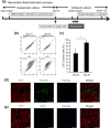
Figure 1
Figure 1. Preparation of human cardiac cell sheets.
(a) Schematic illustration of the culture process including cardiac differentiation, cardiomyocyte purification, and cell sheet fabrication. (b,c) Flow cytometric analysis of cells on days 21 and 27 of differentiation. (b) Representative images of flow cytometry analysis. (c) The percentage of cardiac troponin T (cTnT)-positive cells was calculated and shown in the graph (n = 5). *p < 0.05. (d,e) High content confocal image analysis of cells in cardiac cell sheets (day 27). Nuclei were stained with Hoechst 33258. (d) Vimentin (green) expression in cTnT-positive cells (red). Bars, 100 μm. (e) CD31 (green) expression in cTnT-positive cells (red). Bars, 200 μm. All data are represented as means ± SD.
Figure 2
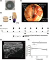
Figure 2. Transplantation of cardiac cell sheets around the inferior vena cava (IVC).
(a) Representative macroscopic image of a monolayer cell sheet. (b) Schematic illustration of transplantation of two pairs of triple-layered cardiac cell sheets around the IVC. (c) Macroscopic image of cardiac cell sheet transplantation around the IVC. The area surrounded by white dots indicates the transplanted cardiac cell sheets around the IVC. (d) Schematic illustration of the time course of transplantation and analysis. (e) Representative image of echographic analysis at 4 weeks after transplantation. The area surrounded by white dots indicates the transplanted tissues around the IVC. Ao: Aorta. Bars, 1 mm. (f) Echographic analysis of the transplanted tissue thickness 2–8 weeks after transplantation (n = 4). *p < 0.05. All data are represented as means ± SD.
Figure 3
Figure 3. Immunohistochemical analysis of transplanted tissues.
(a) Hematoxylin-eosin staining of transplanted tissues at 4 weeks. CM and Int. indicate cardiomyocyte and interstitial layer, respectively. Bars, 500 μm. (b–e) Immunofluorescence analysis of transplanted tissues around the IVC at 4 weeks. (b) Cardiac troponin T (cTnT)-positive cells (red). Nuclei were stained with 4′, 6-diamidino-2-phenylindole (DAPI). Bars, 500 μm. (c) Connexin 43 (green, arrow heads) expression in the graft area (red). Nuclei were stained with DAPI. Bars, 20 μm. (d) von Willebrand factor (vWF)-positive microvascular networks (green) in cTnT-positive cardiac tissues (red). Nuclei were stained with DAPI. Bars: 50 μm. (e) Human mitochondria (red) in transplanted cardiac tissues (green). Area between the white lines indicates the transplanted tissues. Nuclei were stained with DAPI. Bars, 100 μm.
Figure 4
Figure 4. Flow diagram of study population.
Figure 5
Figure 5. Physiological analysis of tubular cardiac tissues around the IVC.
At 4 and 8 weeks after transplantation, rats were intubated and attached to an artificial ventilator under anesthesia. The pressure catheter was inserted into the right femoral vein and the head of the catheter was positioned just into the transplanted cardiac tissue. Pressure measurements were performed under the apnea by turning off the artificial ventilator. (a,b) The inner pressure changes evaluation of the transplanted tissue spontaneous pulsation without clamping IVC lumen. (a) Schematic illustration of the experiment. (b) Pulsation-mediated inner pressure changes before clamping the proximal and distal region of transplanted site in each experiment. (4 weeks, 0–0.19 mmHg, n = 6; 8 weeks, 0.09–1.06 mmHg, n = 4, p = 0.08). Pressure measurements were performed under spontaneous beating. The data include pacing failure cases and pressure control failure case. Bars indicate the median value. (c-g) The inner pressure changes evaluation of the transplanted tissue pulsation with clamping IVC lumen and the electrical stimulation. (c) Schematic illustration of the experiment on the clamping site and the electrical stimulation. In order to change basal inner pressure of IVC, the proximal side of IVC was tightly clamped with an atraumatic bulldog clamp and the distal side of IVC was ligated with 6–0 silk with varying degree. (d) Representative images of the rat electrocardiogram (ECG) (upper), graft electrical potential (middle), and IVC inner pressure (lower) at 4 weeks. The data under the electrical stimulation at 0 (left), 60 (middle) and 120 (right) pulses per minute (ppm). (e,f) Relationship between graft pulsation-mediated inner pressure changes and basal inner pressure of the IVC after clamping. Pressure measurements were performed under electrical stimulation at 80 ppm. (e) 4 weeks (n = 4). (f) 8 weeks (n = 3). (g) Maximum graft pulsation-mediated inner pressure changes in each experiment were calculated and shown in the graph (4 weeks, n = 4; 8 weeks, n = 3, p = 0.44). All data are represented as means ± SEM.
Figure 6
Figure 6. Quantitative polymerase chain reaction analysis of cardiac gene expression in cardiac tissues.
Y-axis indicates relative gene expression compared with the cardiac cell sheet before transplantation. In vitro, cardiac cells from the same preparation of cell sheets were cultured for a further 4 weeks in vitro (n = 4). In vivo V, cardiac tissues around the IVC (n = 3). In vivo Ao, cardiac tissues around the abdominal aorta (n = 3). All data are represented as means ± SD. *p < 0.05; **p < 0.01.

References

    1. Nishida K. et al.. Corneal reconstruction with tissue-engineered cell sheets composed of autologous oral mucosal epithelium. N Engl J Med 351, 1187–1196, doi: 10.1056/NEJMoa040455 (2004). - DOI - PubMed
    1. Ohki T. et al.. Prevention of esophageal stricture after endoscopic submucosal dissection using tissue-engineered cell sheets. Gastroenterology 143, 582–588 e582, doi: 10.1053/j.gastro.2012.04.050 (2012). - DOI - PubMed
    1. Sawa Y. et al.. Tissue engineered myoblast sheets improved cardiac function sufficiently to discontinue LVAS in a patient with DCM: report of a case. Surg Today 42, 181–184, doi: 10.1007/s00595-011-0106-4 (2012). - DOI - PubMed
    1. Takahashi K. et al.. Induction of pluripotent stem cells from adult human fibroblasts by defined factors. Cell 131, 861–872, doi: 10.1016/j.cell.2007.11.019 (2007). - DOI - PubMed
    1. Matsuura K. et al.. Creation of human cardiac cell sheets using pluripotent stem cells. Biochem Biophys Res Commun 425, 321–327, doi: 10.1016/j.bbrc.2012.07.089 (2012). - DOI - PubMed

Publication types

MeSH terms

LinkOut - more resources