Review
. 2017 May 4;13(5):781-819.
doi: 10.1080/15548627.2017.1290751. Epub 2017 Feb 23.

New frontiers in the treatment of colorectal cancer: Autophagy and the unfolded protein response as promising targets

Affiliations
Review

New frontiers in the treatment of colorectal cancer: Autophagy and the unfolded protein response as promising targets

Pooneh Mokarram et al. Autophagy. .

Abstract

Colorectal cancer (CRC), despite numerous therapeutic and screening attempts, still remains a major life-threatening malignancy. CRC etiology entails both genetic and environmental factors. Macroautophagy/autophagy and the unfolded protein response (UPR) are fundamental mechanisms involved in the regulation of cellular responses to environmental and genetic stresses. Both pathways are interconnected and regulate cellular responses to apoptotic stimuli. In this review, we address the epidemiology and risk factors of CRC, including genetic mutations leading to the occurrence of the disease. Next, we discuss mutations of genes related to autophagy and the UPR in CRC. Then, we discuss how autophagy and the UPR are involved in the regulation of CRC and how they associate with obesity and inflammatory responses in CRC. Finally, we provide perspectives for the modulation of autophagy and the UPR as new therapeutic options for CRC treatment.

Keywords: Beclin 1; ER-stress; GRP78; autophagy; cancer therapy; colorectal cancer; unfolded protein response.


Figures

Figure 1.
Figure 1.
CRC distribution—relation to the genetic background. The graph shows percentages of sporadic, familial, and hereditary (familial adenomatous polyposis, FAP; Lynch syndrome) subtypes of CRC.
Figure 2.
Figure 2.
Schematic representation of factors that increase risk or prevent CRC. Both lifestyle and diet affect incidence of CRC as they can be both preventive and risk factors.
Figure 3.
Figure 3.
Dual role of autophagy in cancer chemotherapy. Autophagy may induce stress adaptation (A) in cancer cells allowing them to obtain a resistance phenotype against cancer chemotherapy agents, or it may induce cytotoxicity (B) resulting in autophagic cell death of cancer cells (adapted from ref. 49).
Figure 4.
Figure 4.
Dual role of autophagy during tumorigenesis: Autophagy may suppress tumorigenesis by eliminating damaged organelles in transformed cells and protect them against oxidative stress, resulting in subsequent genome stabilization and prevention of malignant transformation. Autophagy may also initiate an oncogene-induced senescence, thus preventing malignant transformation. It may prevent necrosis in apoptosis-deficient cells in tumors in response to metabolic stress. This reduces pro-tumorigenic inflammation and release of tumorigenic compounds from necrotic tumor cells. Tumor-supportive functions of autophagy are fulfilled mainly by stimulating tumor cell survival and protection against detachment-induced apoptosis (anoikis), which can facilitate chemoresistance and EMT induced-metastasis (adapted from. refs. 50,51).
Figure 5.
Figure 5.
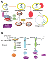
Autophagy and the UPR signaling pathways. (A) Depiction of autophagy pathways. Autophagy is a catabolic process that sequesters specific intracellular cargo by engulfing them within a cytosolic double-membraned vesicle, called an autophagosome. Extracellular stimuli or recognition of a cargo material induces the formation of the phagophore. ULK1 is an important upstream initiator that induces activation of nucleation complex, including PtdIns3K and BECN1, to engage phagophores for autophagy. LC3 is conjugated to the phagophores and controls their maturation and elongation. Upon vesicle completion, the autophagosome fuses with a lysosome, releasing its contents to be degraded by hydrolases. (B) Initiation of the UPR: The domain structures of ERN1, EIF2AK3, and ATF6 and their associations with HSPA5 are illustrated. ERN1, EIF2AK3, and ATF6 are docked and inactive in non-ER stress condition by binding to HSPA5. Upon ER stress, HSPA5 is released from the lumenal domain of ERN1, EIF2AK3 and ATF6 and this initiates the UPR.
Figure 6.
Figure 6.
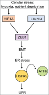
Hypoxia- and nutrient deprivation-induced stress cause EMT-mediated UPR. The relationship between EMT and ER stress. At the invasive front of CRCs, cellular stress conditions (hypoxia or changes in the microenvironment) induce EMT via activation of HIF1A or CTNNB1/β-catenin, which consequently leads to ZEB1 activation and EMT induction. EMT activates the UPR which induces the activation of UPR-related transcription factors (ATF6) (adapted from ref. 199).
Figure 7.
Figure 7.
The role of MTOR in cancer-associated signaling pathways that regulate autophagy in mammalian cells. The best known regulator of autophagy is MTOR (mechanistic target of rapamycin), a serine/threonine kinase conserved throughout eukaryotes. The activity of MTORC1 is inversely correlated with autophagy induction. The µTORC1 inhibitor rapamycin potently induces autophagy, even in the presence of abundant nutrients (adapted from ref. 50). The PI3K-AKT regulates autophagy. This regulation is mediated via the small RHO-GTPase RHEB.
Figure 8.
Figure 8.
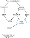
Regulation of tumor survival and angiogenesis via glucose and oxygen supply. Glucose deprivation increases PGE2 by upregulating PTGS2 and downregulating HPGD expression via PI3K-AKT and DDIT3-dependent mechanisms. Hypoxia increases PGE2 levels by upregulating PTGS2 expression via HIF1A). Elevated PGE2 increases survival of colon cancer cells exposed to both glucose deprivation and hypoxic conditions (adapted from ref. 218).
Figure 9.
Figure 9.
The role for the MAPK p38 signaling pathway in cellular response to 5-FU. There is a critical role for the MAPK p38-signaling pathway in the cellular response to 5-FU by controlling the balance between apoptosis and autophagy (adapted from ref. 240). This pathway is tightly controlled by the TP53-mediated regulation of autophagy.
Figure 10.
Figure 10.
Autophagy targeting strategies in in vitro and in vivo models of CRC. All chemical compounds, drugs, and inhibitors have been introduced in the section “Therapeutic targeting of the autophagy pathway.”
Figure 11.
Figure 11.
Endoplasmic reticulum (ER) stress induction by docosahexaenoic acid (DHA). DHA induces ER stress in colorectal cells. Diagram showing transcripts affected by DHA treatment in SW620 colon cancer cells by gene expression analysis in the main pathways of ER stress signaling. Three transmembrane proteins mediate the unfolded protein response (UPR) across the ER membrane after dissociation from HSPA5-ATF6, EIF2AK3 and ERN1 (adapted from ref. 278).
Figure 12.
Figure 12.
ER stress and UPR targeting strategies in in vitro and in vivo models for CRC therapy. All chemical compounds, drugs, and inhibitors have been introduced in the section “Therapeutic targeting of ER stress and the UPR.”
Figure 13.
Figure 13.
Lysosomal targeting strategies in in vitro and in vivo models for CRC therapy. All chemical compounds, drugs, and inhibitors have been introduced in the section “Cancer therapy using lysosomal targeting.”
Figure 14.
Figure 14.
MTOR targeting strategies in in vitro and in vivo models for CRC therapy. All chemical compounds, drugs, and inhibitors have been introduced in the section “Anticancer potential of MTOR inhibitors.”
Figure 15.
Figure 15.
Therapeutic targeting of epigenetic regulators in in vitro and in vivo models for CRC therapy. All chemical compounds, drugs, and inhibitors have been introduced in the section “Therapeutic targeting of epigenetic regulators.”
Figure 16.
Figure 16.
Miscellaneous drugs targeting autophagy in in vitro and in vivo models for CRC therapy. All chemical compounds, drugs, and inhibitors have been introduced in the section “Other pharmacological autophagy inducers.”
Figure 17.
Figure 17.
TP53 status of CRC and the respective targeting strategies in in vitro and in vivo models for CRC therapy. All chemical compounds, drugs, and inhibitors have been introduced in the section “TP53 status-dependent therapeutic strategies.”
Figure 18.
Figure 18.
Proteasome activity modulators in in vitro and in vivo models for CRC therapy. All chemical compounds, drugs, and inhibitors have been introduced in the section “Proteasome inhibitor-based antitumor strategies.”

References

    1. Jemal A, Bray F, Center MM, Ferlay J, Ward E, Forman D. Global cancer statistics. CA Cancer J Clin 2011; 61:69-90; PMID:21296855; http://dx.doi.org/10.3322/caac.20107 - DOI - PubMed
    1. Canada CCSNCIo Canadian Cancer Statistics 2008. Toronto: Canadian Cancer Society, 2008
    1. Center MM, Jemal A, Smith RA, Ward E. Worldwide variations in colorectal cancer. CA Cancer J Clin 2009; 59:366-78; PMID:19897840; http://dx.doi.org/10.3322/caac.20038 - DOI - PubMed
    1. Saudi Moh Health awareness center: “Cancer incidence in the Kingdom, Compared to the global incidence, Is still low. Saudi Arabia: Ministry of health, 2013
    1. Dolatkhah R, Somi MH, Bonyadi MJ, Asvadi Kermani I, Farassati F, Dastgiri S. Colorectal cancer in iran: Molecular epidemiology and screening strategies. J Cancer Epidemiol 2015; 2015:643020; PMID:25685149; http://dx.doi.org/10.1155/2015/643020 - DOI - PMC - PubMed

MeSH terms

LinkOut - more resources