. 2017 Mar 30;8(3):e2718.
doi: 10.1038/cddis.2017.147.

Novel prosurvival function of Yip1A in human cervical cancer cells: constitutive activation of the IRE1 and PERK pathways of the unfolded protein response

Affiliations

Novel prosurvival function of Yip1A in human cervical cancer cells: constitutive activation of the IRE1 and PERK pathways of the unfolded protein response

Yuki Taguchi et al. Cell Death Dis. .

Abstract

Cancer cells are under chronic endoplasmic reticulum (ER) stress due to hypoxia, low levels of nutrients, and a high metabolic demand for proliferation. To survive, they constitutively activate the unfolded protein response (UPR). The inositol-requiring protein 1 (IRE1) and protein kinase RNA-like ER kinase (PERK) signaling branches of the UPR have been shown to have cytoprotective roles in cancer cells. UPR-induced autophagy is another prosurvival strategy of cancer cells, possibly to remove misfolded proteins and supply nutrients. However, the mechanisms by which cancer cells exploit the UPR and autophagy machinery to promote survival and the molecules that are essential for these processes remain to be elucidated. Recently, a multipass membrane protein, Yip1A, was shown to function in the activation of IRE1 and in UPR-induced autophagy. In the present study, we explored the possible role of Yip1A in activation of the UPR by cancer cells for their survival, and found that depletion of Yip1A by RNA interference (RNAi) induced apoptotic cell death in HeLa and CaSki cervical cancer cells. Intriguingly, Yip1A was found to activate the IRE1 and PERK pathways of the UPR constitutively in HeLa and CaSki cells. Yip1A mediated the phosphorylation of IRE1 and also engaged in the transcription of PERK. The activation of these signaling pathways upregulated the expression of anti-apoptotic proteins and autophagy-related proteins. These events might enhance resistance to apoptosis and promote cytoprotective autophagy in HeLa and CaSki cells. The present study is the first to uncover a key prosurvival modulator, Yip1A, which coordinates IRE1 signaling with PERK signaling to support the survival of HeLa and CaSki cervical cancer cells.


Conflict of interest statement

The authors declare no conflict of interest.

Figures

Figure 1
Figure 1
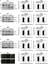
Depletion of Yip1A induces apoptotic cell death in HeLa and CaSki cervical cancer cells. (a) HeLa and CaSki cells were transfected with control scramble siRNA (left panel) or Yip1A siRNA (right panel). Representative confocal micrographs show morphological differences between control and Yip1A-knockdown cells at the indicated time points. Scale bars are 50 μm. (b) The viability of transfected cells was evaluated at the indicated time points using the MTT Cell Proliferation Assay. Data are means±S.D. from three independent experiments; *P<0.05, **P<0.01. (c) Representative flow cytometric data for control (left panels) and Yip1A-knockdown (right panels) cells at the indicated time points after siRNA transfection. The percentages of apoptotic cells (Annexin V+/PI + Annexin V+/PI+) are shown in the bar graphs. Data are means±S.D. from three independent experiments; *P<0.05. (d) Representative confocal micrographs of control (upper panels) and Yip1A-knockdown (lower panels) cells at the indicated time points after siRNA transfection. Scale bars are 10 μm. The percentages of TUNEL-positive cells are shown in the bar graphs. Data are means±S.D. from three independent experiments; *P<0.05, **P<0.01. (e) Western blotting shows relative levels of cleaved caspase 3 and cleaved PARP protein in control and Yip1A-knockdown cells at the indicated time points after siRNA transfection. GAPDH was used for normalization. Data are means±S.D. from three independent experiments; *P<0.05, **P<0.01
Figure 2
Figure 2
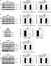
Depletion of Yip1A inhibits the activation of IRE1 and PERK. (a) HeLa cells were treated with control scramble siRNA or Yip1A siRNA and cell lysates were prepared at the indicated time points. Western blotting shows the relative levels of phosphorylated IRE1 (pIRE1) and IRE1 protein in control and Yip1A-knockdown cells at 24 h and 48 h after siRNA transfection. GAPDH was used for normalization. Data are means±S.D. from three independent experiments; **P<0.01. (b) Western blotting shows relative levels of phosphorylated PERK (pPERK) and PERK protein in control and Yip1A-knockdown cells at 24 h and 48 h after siRNA transfection. GAPDH was used for normalization. Data are means±S.D. from three independent experiments; *P<0.05, **P<0.01. (c) CaSki cells were treated with control scramble siRNA or Yip1A siRNA and the cell lysates were prepared at the indicated time points. Western blotting shows the relative levels of pIRE1 and IRE1 protein in control and Yip1A-knockdown cells at 48 h and 72 h after siRNA transfection. GAPDH was used for normalization. Data are means±S.D. from three independent experiments; **P<0.01. (d) Western blotting shows relative levels of pPERK and PERK protein in control and Yip1A-knockdown cells at 48 h and 72 h after siRNA transfection. GAPDH was used for normalization. Data are means±S.D. from three independent experiments; **P<0.01. (e) Representative confocal micrographs of control (upper panels) and Yip1A-knockdown (lower panels) HeLa cells at 24 h after siRNA transfection, showing the depletion of PERK by Yip1A knockdown. Fixed cells were double-stained for Yip1A (green) and PERK (red). Scale bars are 10 μm. (f) RT-PCR shows relative levels of PERK mRNA in control and Yip1A-knockdown cells. GAPDH was used for normalization. Data are means±S.D. from three independent experiments. **P<0.01
Figure 3
Figure 3
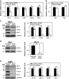
Depletion of Yip1A impairs signaling downstream of the IRE1 and PERK pathways. (a) HeLa cells were treated with control scramble siRNA or Yip1A siRNA and the cell lysates were prepared at the indicated time points. Western blotting shows the relative levels of phosphorylated IKKα/β (pIKKα/β) and phosphorylated NF-κB p65 (p-p65) protein in control and Yip1A-knockdown cells at 24 h and 48 h after siRNA transfection. GAPDH was used for normalization. Data are means±S.D. from three independent experiments; **P<0.01. (b) Western blotting shows the relative levels of phosphorylated eIF2α (peIF2α) and ATF4 protein in control and Yip1A-knockdown cells at 24 h and 48 h after siRNA transfection. GAPDH was used for normalization. Data are means±S.D. from three independent experiments; **P<0.01. (c) Western blotting shows relative levels of p-p65 and ATF4 protein in the nuclear fraction of control and Yip1A-knockdown HeLa cells at 48 h after siRNA transfection. Nup98 was used as a control for loading of the nuclear fraction. Data are means±S.D. from three independent experiments; **P<0.01. (d) CaSki cells were treated with control scramble siRNA or Yip1A siRNA and the cell lysates were prepared at the indicated time points. Western blotting shows the relative levels of pIKKα/β and phosphorylated JNK (pJNKp46 and pJNKp54) protein in control and Yip1A-knockdown cells at 48 h and 72 h after siRNA transfection. GAPDH was used for normalization. Data are means±S.D. from three independent experiments; **P<0.01. (e) Western blotting shows the relative levels of peIF2α and ATF4 protein in control and Yip1A-knockdown cells at 48 h and 72 h after siRNA transfection. GAPDH was used for normalization. Data are means±S.D. from three independent experiments. *P<0.05, **P<0.01
Figure 4
Figure 4
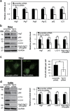
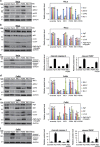
Depletion of Yip1A downregulates the expression of the anti-apoptotic proteins Bcl-2, cIAP2, and Mcl-1, and the phosphorylation of Bcl-2. (a) HeLa cells were transfected with control scramble siRNA or Yip1A siRNA. RT-PCR shows the relative mRNA levels of the indicated anti-apoptotic factors in control and Yip1A-knockdown cells at 48  after siRNA transfection. GAPDH was used for normalization. Data are means±S.D. from three independent experiments; **P<0.01. (b) RT-PCR shows the relative mRNA levels of the indicated proapoptotic factors in control and Yip1A-knockdown HeLa cells at 48 h after siRNA transfection. GAPDH was used for normalization. Data are means±S.D. from three independent experiments. (c) Western blotting shows the relative levels of anti-apoptotic Bcl-2, cIAP2, and Mcl-1 protein in control and Yip1A-knockdown cells at 48 h after siRNA transfection. GAPDH was used for normalization. Data are means±S.D. from three independent experiments; *P<0.05, **P<0.01. (d) Western blotting shows the relative levels of phosphorylated Bcl-2 (pBcl-2) protein in control and Yip1A-knockdown cells at 48 h after siRNA transfection. GAPDH was used for normalization. Data are means±S.D. from three independent experiments; **P<0.01. (e) CaSki cells were transfected with control scramble siRNA or Yip1A siRNA. Western blotting shows the relative levels of anti-apoptotic Bcl-2, pBcl-2, cIAP2, and Mcl-1 protein in control and Yip1A-knockdown cells at 72 h after siRNA transfection. GAPDH was used for normalization. Data are means±S.D. from three independent experiments; **P<0.01
Figure 5
Figure 5
Depletion of Yip1A suppresses autophagy. (a) HeLa cells were transfected with control scramble siRNA or Yip1A siRNA. RT-PCR shows the relative mRNA levels of the indicated autophagy-related factors in the control and Yip1A-knockdown cells at 48 h after siRNA transfection. GAPDH was used for normalization. Data are means±S.D. from three independent experiments; *P<0.05, **P<0.01. (b) Western blotting shows the relative levels of Atg7, p62, and LC3-II protein, and Atg5–Atg12 conjugate in the control and Yip1A-knockdown cells at 48 h after siRNA transfection. GAPDH was used for normalization. Data are means±S.D. from three independent experiments; **P<0.01. (c) HeLa cells were co-transfected with either control scramble siRNA or Yip1A siRNA and pEGFP-LC3 expression plasmid DNA. After 48 h, autophagosome formation was measured by visualizing LC3-positive dots with the use of a confocal microscope. Scale bars are 10 μm. Quantification of the number of LC3-positive dots per cell is shown in the bar graph (n=30 cells); **P<0.01. (d) CaSki cells were transfected with control scramble siRNA or Yip1A siRNA. Western blotting shows the relative levels of Atg7, p62, and LC3-II protein, and Atg5–Atg12 conjugate in the control and Yip1A-knockdown cells at 72 h after siRNA transfection. GAPDH was used for normalization. Data are means±S.D. from three independent experiments; **P<0.01
Figure 6
Figure 6
Both the IRE1 and PERK pathways engage in the upregulation of anti-apoptotic proteins and autophagy-related proteins in HeLa and CaSki cells. (a) HeLa cells were transfected with the indicated siRNAs, and cell lysates were prepared at 48 h. Western blotting shows the relative levels of Bcl-2, pBcl-2, cIAP2, and Mcl-1 protein in respective siRNA-transfected cells. GAPDH was used for normalization. Data are means±S.D. from three independent experiments. The significant difference from the control (scramble) was determined using Dunnett's post hoc test; **P<0.01. (b) Western blotting shows relative levels of Atg7, p62, and LC3-II protein, and Atg5–Atg12 conjugate in respective siRNA-transfected HeLa cells. GAPDH was used for normalization. Data are means±S.D. from three independent experiments. The significant difference from the control (scramble) was determined using Dunnett's post hoc test; **P<0.01. (c) Western blotting shows relative levels of cleaved caspase 3 and cleaved PARP protein in respective siRNA-transfected HeLa cells. GAPDH was used for normalization. Data are means±S.D. from three independent experiments. The significant difference from the control (scramble) was determined using Dunnett's post hoc test; **P<0.01. (d) CaSki cells were transfected with the indicated siRNAs, and the cell lysates were prepared at 72 h. Western blotting shows the relative levels of Bcl-2, pBcl-2, cIAP2, and Mcl-1 protein in respective siRNA-transfected cells. GAPDH was used for normalization. Data are means±S.D. from three independent experiments. The significant difference from the control (scramble) was determined using Dunnett's post hoc test; **P<0.01. (e) Western blotting shows relative levels of Atg7, p62, and LC3-II protein, and Atg5–Atg12 conjugate in respective siRNA-transfected CaSki cells. GAPDH was used for normalization. Data are means±S.D. from three independent experiments. The significant difference from the control (scramble) was determined using Dunnett's post hoc test; *P<0.05, **P<0.01. (f) Western blotting shows relative levels of cleaved caspase 3 and cleaved PARP protein in respective siRNA-transfected CaSki cells. GAPDH was used for normalization. Data are means±S.D. from three independent experiments. The significant difference from the control (scramble) was determined using Dunnett's post hoc test; **P<0.01
Figure 7
Figure 7
Schematic representation of how Yip1A operates as a prosurvival modulator that coordinately activates the IRE1 and PERK pathways of the UPR to support the survival of HeLa and CaSki cervical cancer cells. See text for details. Ub, ubiquitination

Similar articles

Cited by

References

    1. Ma Y, Hendershot LM. The role of the unfolded protein response in tumour development: friend or foe? Nat Rev Cancer 2004; 4: 966–977. - PubMed
    1. Moenner M, Pluquet O, Bouchecareilh M, Chevet E. Integrated endoplasmic reticulum stress responses in cancer. Cancer Res 2007; 67: 10631–10634. - PubMed
    1. Wang M, Kaufman RJ. The impact of the endoplasmic reticulum protein-folding environment on cancer development. Nat Rev Cancer 2014; 14: 581–597. - PubMed
    1. Nagelkerke A, Bussink J, FCGJ Sweep, Span PN. The unfolded protein response as a target for cancer therapy. Biochim Biophys Acta 2014; 1846: 277–284. - PubMed
    1. Walter P, Ron D. The unfolded protein response: from stress pathway to homeostatic regulation. Science 2011; 334: 1081–1086. - PubMed

MeSH terms