. 2017 Mar 30;12(3):e0174517.
doi: 10.1371/journal.pone.0174517. eCollection 2017.

Phenotypically silent Cre recombination within the postnatal ventricular conduction system

Affiliations

Phenotypically silent Cre recombination within the postnatal ventricular conduction system

Samadrita Bhattacharyya et al. PLoS One. .

Abstract

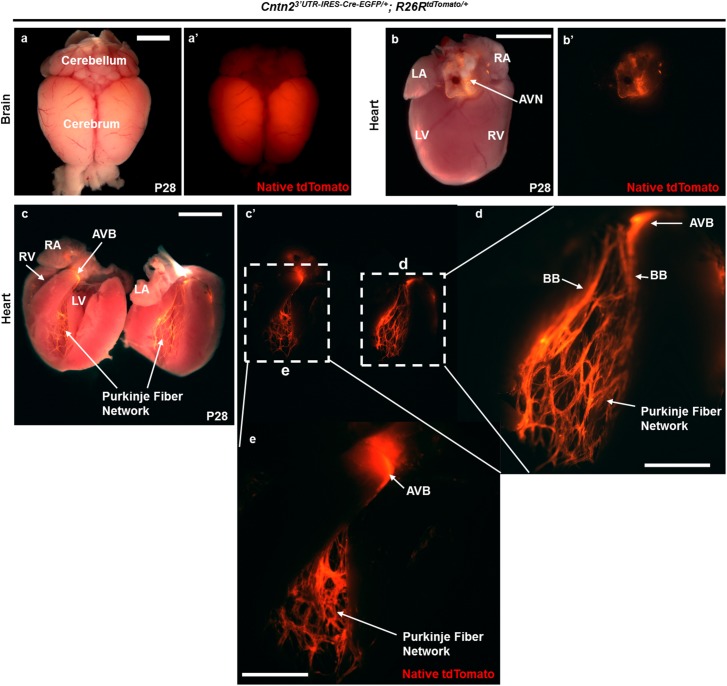
The cardiac conduction system (CCS) is composed of specialized cardiomyocytes that initiate and maintain cardiac rhythm. Any perturbation to the normal sequence of electrical events within the heart can result in cardiac arrhythmias. To understand how cardiac rhythm is established at the molecular level, several genetically modified mouse lines expressing Cre recombinase within specific CCS compartments have been created. In general, Cre driver lines have been generated either by homologous recombination of Cre into an endogenous locus or Cre expression driven by a randomly inserted transgene. However, haploinsufficiency of the endogenous gene compromises the former approach, while position effects negatively impact the latter. To address these limitations, we generated a Cre driver line for the ventricular conduction system (VCS) that preserves endogenous gene expression by targeting the Contactin2 (Cntn2) 3' untranslated region (3'UTR). Here we show that Cntn23'UTR-IRES-Cre-EGFP/+ mice recombine floxed alleles within the VCS and that Cre expression faithfully recapitulates the spatial distribution of Cntn2 within the heart. We further demonstrate that Cre expression initiates after birth with preservation of native Cntn2 protein. Finally, we show that Cntn23'UTR-IRES-Cre-EGFP/+ mice maintain normal cardiac mechanical and electrical function. Taken together, our results establish a novel VCS-specific Cre driver line without the adverse consequences of haploinsufficiency or position effects. We expect that our new mouse line will add to the accumulating toolkit of CCS-specific mouse reagents and aid characterization of the cell-autonomous molecular circuitry that drives VCS maintenance and function.


Conflict of interest statement

Competing Interests: The authors have declared that no competing interests exist.

Figures

Fig 1
Fig 1. Genomic architecture of the Cntn23’UTR-IRES-Cre-EGFP/+ allele.
a) Schematic of the endogenous Contactin-2 (Cntn2) locus comprising 23 exons as indicated by blue numbered blocks. Green block denotes the 3’UTR of the Cntn2 gene located in exon 23. The black right-handed arrow indicates the Cntn2 transcriptional start site driven by endogenous regulatory elements. The IRES-Cre-EGFP-FRT-neo-PGK-FRT KI cassette was targeted to the 3’ UTR of the Cntn2 locus to ensure unperturbed bi-cistronic expression of endogenous Cntn2 protein and a Cre-EGFP fusion protein under the control of the endogenous regulatory elements upon FLP recombination. b) PCR genotyping of mice before FLP-mediated recombination using the indicated primer sets to amplify the 5’ (i) and 3’ (ii) insertion sites. In this example, 4 out of 5 pups were positive by F1-R1 genotyping, and these 4 were subjected to the second round of genotyping using F2-R2 primer sets. C) PCR genotyping of mice after FLP-mediated recombination using the indicated primer sets to amplify across the deleted NeoR cassette (i) and the Cre coding sequence (ii). In this example, 4 out of 6 pups were positive by F3-R3 genotyping, and these 4 were subjected to the second round of genotyping using F4-R4 primer sets.
Fig 2
Fig 2. Cntn23’UTR-IRES-Cre-EGFP/+; R26RtdTomato/+ mice display robust reporter expression in the VCS.
(a-e) Cntn23’UTR-IRES-Cre-EGFP/+ mice were bred with R26RtdTomato/tdTomato reporter mice to characterize the Cntn23’UTR-IRES-Cre-EGFP/+ allele by monitoring tdTomato expression at P28 by whole mount fluorescence imaging (a and a’) in brain (dorsal view, used as a positive control) where endogenous Contactin 2 is known to be expressed abundantly and (b-e) in heart. We monitored tdTomato expression in at least 15 independent Cntn23’UTR-IRES-Cre-EGFP/+; R26RtdTomato/+ mice from multiple litters in comparison to 12 WT control littermates. (b and b’) Native tdTomato fluorescence was visualized at the Atrio-Ventricular Junction (AVJ) by whole-mount microscopy following Cre recombination of the R26R locus. (c-e) A P28 mouse heart was sectioned grossly in the four-chamber orientation to visualize native tdTomato expression in the VCS. (c and c’) Robust tdTomato fluorescence was observed in the Atrio-Ventricular Bundle (AVB), right and left Bundle Branches (BB), and right and left Purkinje Fiber (PF) network. Faint speckled fluorescent signal was also observed in the Right Atrium (RA) (not visible in the image). (a-c) Scale bar: 500 μM. (d and e) Higher magnification images of (d) right inlet in (c’) and (e) left inlet in (c’) to visualize the intricate structures of AVB and highly branched PF network in the adult mouse heart. Scale bar: 250 μm. LA, Left Atrium; LV, Left Ventricle; RV, Right Ventricle.
Fig 3
Fig 3. tdTomato co-localizes with VCS markers in Cntn23’UTR-IRES-Cre-EGFP/+; R26RtdTomato/+ mice.
(a-d) Cntn23’UTR-IRES-Cre-EGFP/+ mice were crossed with R26RtdTomato/tdTomato reporter mice, and P42 heart cryosections were imaged by confocal microscopy. (a, a’, a”, a”‘) High resolution confocal images of the AVN region demonstrates co-localization of tdTomato (a’, red), endogenous Cntn2 (a”, green), and endogenous Hcn4 (a”‘, magenta) with the merged image shown in (a). (b, b’, b”, b”‘) High specificity and complete overlap (b: merged signal) of native tdTomato (b’, red), endogenous Cntn2 (b”, green), and endogenous Hcn4 (b’”, magenta) expression upon Cre recombination in the AVB and BB. (c, c’, c”, c”‘) Confocal images of mouse heart sections showing that Cre recombined tdTomato cells of left PFs (c’, red) co-localize with endogenous Cntn2 (c”, green) and Hcn4 (c”‘, magenta). (d, d’, d”, d”‘) Images of serial Cntn23’UTR-IRES-Cre-EGFP/+; R26RtdTomato/+ mice heart sections verified co-expression of endogenous Cntn2 (d”, green) and Hcn4 (d”‘, magenta) in recombined tdTomato cells (d’, red) of right PFs. Blue signal indicates nuclear counterstaining with DAPI. The cardiac anatomical location for each confocal micrograph is shown in S2A Fig. Scale bar: 100 μm.
Fig 4
Fig 4. Cre-mediated recombination in Cntn23’UTR-IRES-Cre-EGFP/+; R26RtdTomato/+ mice is only evident after birth.
(a, a’) Whole mount fluorescent image of an E18.5 double heterozygous embryo with robust expression of the recombined reporter protein in the cerebrum. tdTomato expression in the brain was also seen at E16.5 (data not shown). (b, b’) Following microdissection of the heart at E18.5, native tdTomato signal was not observed. (c, c’) Double heterozygous P0 mouse heart after dissection revealed bright reporter protein expression in the AVB, right and left BB, and right and left PF network. P0 was the earliest point at which reporter expression was observed in the heart. (d,d’) Micro-dissected P7 double heterozygous hearts express tdTomato protein in the identical anatomical regions as described in (c,c’). The cardiac PF network appears fully developed by P7 based on fluorescent reporter expression. At least 8 hearts were dissected per group of animals at each time point. Scale bar: 500 μm.
Fig 5
Fig 5. Cardiac mechanical function is preserved in Cntn23’UTR-IRES-Cre-EGFP/+ mice.
(a-b) M-mode echocardiography was performed in conscious mice to analyze heart function parameters in P42 (a) wild-type (WT) and (b) Cntn23’UTR-IRES-Cre-EGFP/+ mice. Representative traces show normal heart function in both groups. (c) Heart Rate (HR) in Beats Per Minute (BPM), (d) Ejection Fraction (EF) as a percentage, (e) Fractional Shortening (FS) as a percentage, and (f) Cardiac Output (CO) in milliliters per minute were calculated for n = 9 animals in both groups. Black circles represent individual WT and Cntn23’UTR-IRES-Cre-EGFP/+ mice used in this study. Blue and red bars represent mean values of each parameter in WT and Cntn23’UTR-IRES-Cre-EGFP/+ mice, respectively. No statistically significant differences were observed between groups. ns, not significant.
Fig 6
Fig 6. Cardiac electrical function is preserved in Cntn23’UTR-IRES-Cre-EGFP/+ mice.
(a-j) Serial surface lead II ECGs were recorded on both groups of mice (n = 9 per group) under mild anesthesia at P7, P14, P21, P28, and P42. Exemplary ECG traces for P42 (a) WT and (b) Cntn23’UTR-IRES-Cre-EGFP/+ mice are shown. Scale size: 40 milliseconds (ms). (c) PR interval (in seconds), (d) QRS width (in seconds), (e) RR interval (in seconds), and (f) PR/RR ratio were measured at P7, P14, P21, and P42. At P28, (g) PR interval (in seconds), (h) QRS width (in seconds), (i) RR interval (in seconds), and (j) PR/RR ratio were measured pre- and post-isoproterenol (-/+ Iso) injection. Black circles represent individual WT and Cntn23’UTR-IRES-Cre-EGFP/+ mice used in this study. Blue and red bars represent mean values for each parameter in WT and Cntn23’UTR-IRES-Cre-EGFP/+ mice, respectively. No statistically significant differences were observed between groups. ns, not significant.

References

    1. Munshi NV. Gene regulatory networks in cardiac conduction system development. Circ Res. 2012;110(11):1525–37. 10.1161/CIRCRESAHA.111.260026 - DOI - PMC - PubMed
    1. van Weerd JH, Christoffels VM. The formation and function of the cardiac conduction system. Development. 2016;143(2):197–210. 10.1242/dev.124883 - DOI - PubMed
    1. Arnolds DE, Chu A, McNally EM, Nobrega MA, Moskowitz IP. The emerging genetic landscape underlying cardiac conduction system function. Birth Defects Res A Clin Mol Teratol. 2011;91(6):578–85. 10.1002/bdra.20800 - DOI - PMC - PubMed
    1. Park DS and Fishman GI. The cardiac conduction system. Circulation. 2011;123(9):904–15. - PMC - PubMed
    1. Rubart M, Zipes DP. Mechanisms of sudden cardiac death. The Journal of clinical investigation. 2005;115(9):2305–15. 10.1172/JCI26381 - DOI - PMC - PubMed

MeSH terms