

Methodological concerns with laser speckle contrast imaging in clinical evaluation of microcirculation
- PMID: 28358906
- PMCID: PMC5373607
- DOI: 10.1371/journal.pone.0174703
Methodological concerns with laser speckle contrast imaging in clinical evaluation of microcirculation
Abstract
Background: Laser Speckle Contrast Imaging (LSCI) is a non-invasive and fast technique for measuring microvascular blood flow that recently has found clinical use for burn assessment and evaluation of flaps. Tissue motion caused by for example breathing or patient movements may however affect the measurements in these clinical applications, as may distance between the camera and the skin and tissue curvature. Therefore, the aims of this study were to investigate the effect of frame rate, number of frames/image, movement of the tissue, measuring distance and tissue curvature on the measured perfusion.
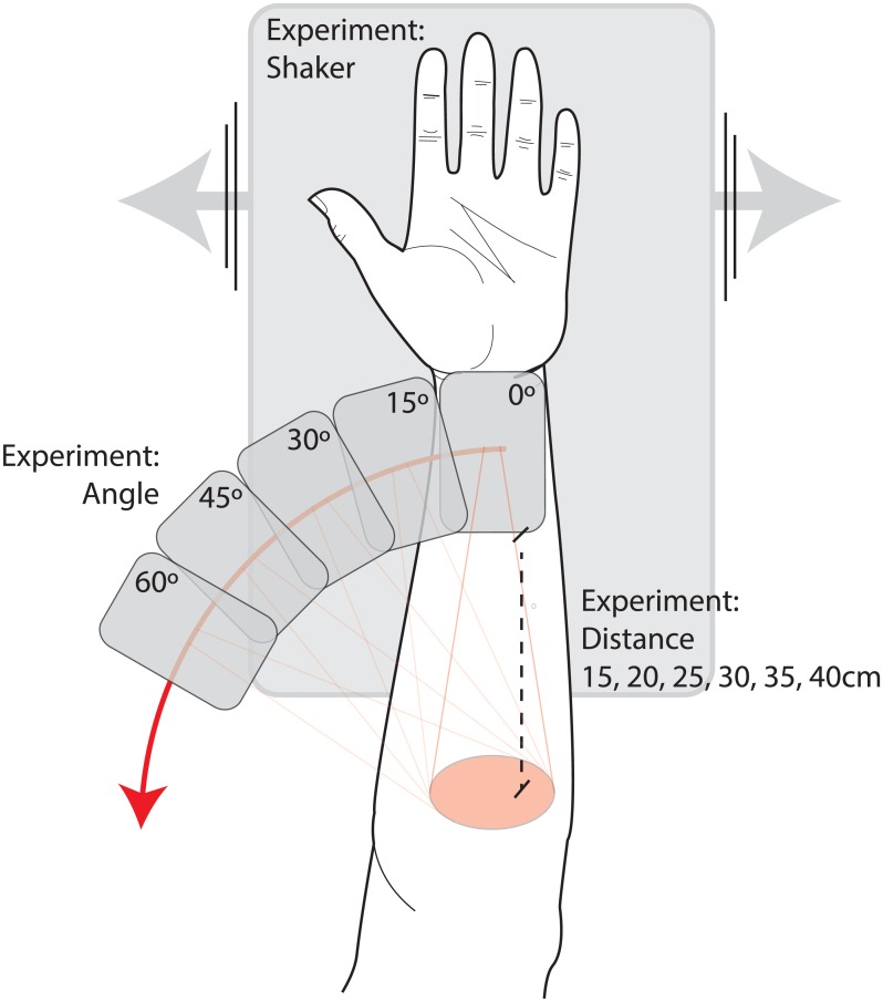
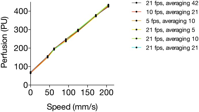
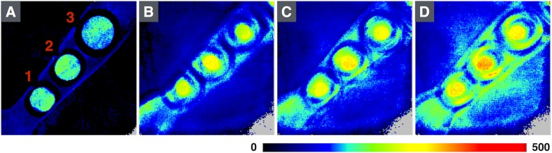
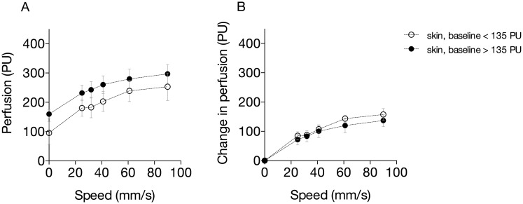
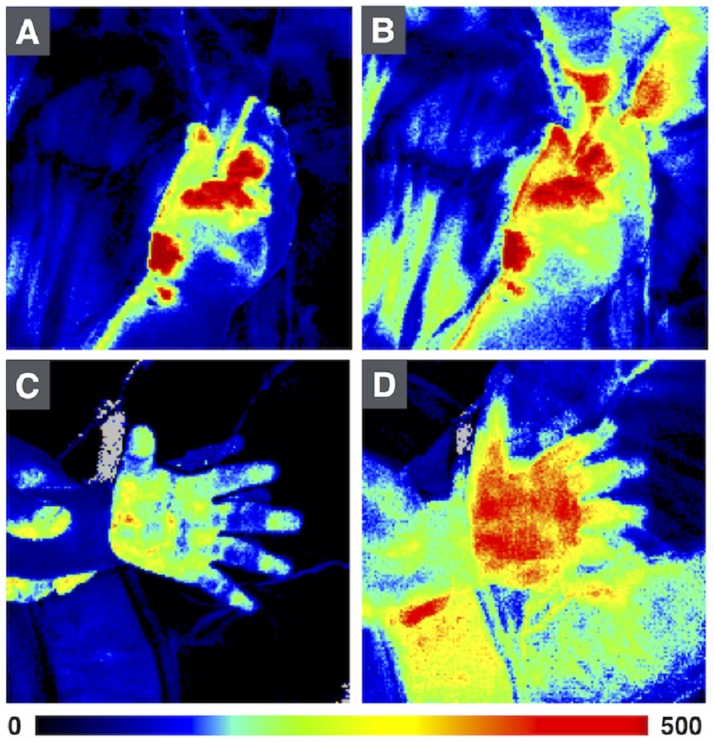
Methods: Methyl nicotinate-induced vasodilation in the forearm skin was measured using LSCI during controlled motion at different speeds, using different combinations of frame rate and number of frames/image, and at varying camera angles and distances. Experiments were made on healthy volunteers and on a cloth soaked in a colloidal suspension of polystyrene microspheres.
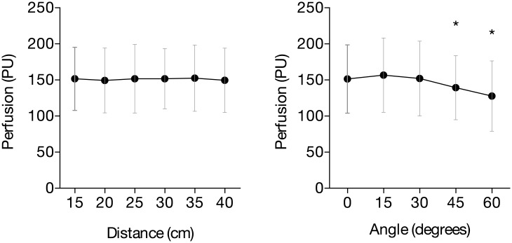
Results: Measured perfusion increased with tissue motion speed. The relation was independent of the absolute perfusion in the skin and of frame rate and number of frames/image. The measured perfusion decreased with increasing angles (16% at 60°, p = 0.01). Measured perfusion did not vary significantly between measurement distances from 15 to 40 cm (p = 0.77, %CV 0.9%).
Conclusion: Tissue motion increases and measurement angles beyond 45° decrease the measured perfusion in LSCI. These findings have to be taken into account when LSCI is used to assess moving or curved tissue surfaces, which is common in clinical applications.
Conflict of interest statement
Figures

References
-
- Ambrozy E, Waczulikova I, Willfort-Ehringer A, Ehringer H, Koppensteiner R, Gschwandtner ME. Microcirculation in mixed arterial/venous ulcers and the surrounding skin: clinical study using a laser Doppler perfusion imager and capillary microscopy. Wound repair and regeneration: official publication of the Wound Healing Society [and] the European Tissue Repair Society. 2009;17(1):19–24. Epub 2009/01/21. - PubMed
-
- Lindahl F, Tesselaar E, Sjoberg F. Assessing paediatric scald injuries using Laser Speckle Contrast Imaging. Burns: journal of the International Society for Burn Injuries. 2013;39(4):662–6. Epub 2012/10/25. - PubMed
-
- Mirdell R, Iredahl F, Sjoberg F, Farnebo S, Tesselaar E. Microvascular blood flow in scalds in children and its relation to duration of wound healing: A study using laser speckle contrast imaging. Burns: journal of the International Society for Burn Injuries. 2016;42(3):648–54. Epub 2016/01/27. - PubMed
-
- Monstrey S, Hoeksema H, Verbelen J, Pirayesh A, Blondeel P. Assessment of burn depth and burn wound healing potential. Burns: journal of the International Society for Burn Injuries. 2008;34(6):761–9. Epub 2008/05/31. - PubMed
MeSH terms
Substances
LinkOut - more resources
Full Text Sources
Other Literature Sources
