. 2017 Mar 30;12(3):e0174779.
doi: 10.1371/journal.pone.0174779. eCollection 2017.

Vector-free intracellular delivery by reversible permeabilization

Affiliations

Vector-free intracellular delivery by reversible permeabilization

Shirley O'Dea et al. PLoS One. .

Abstract

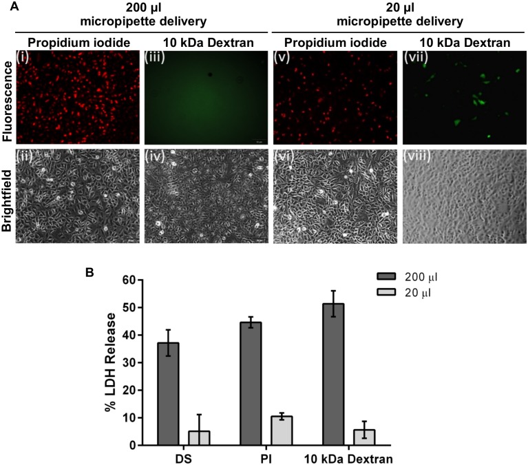
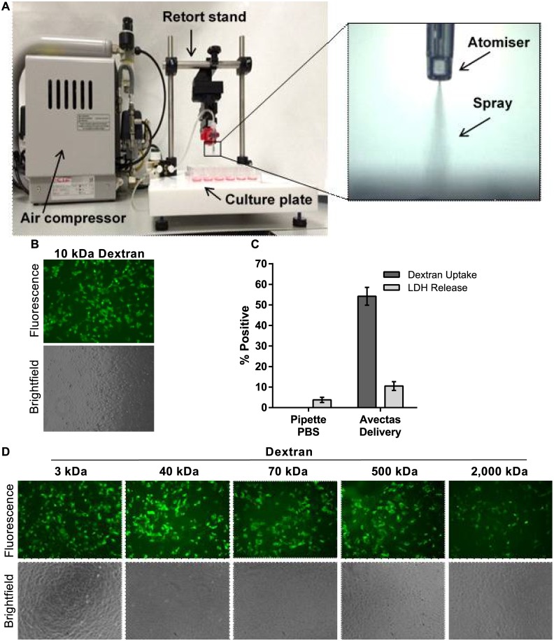
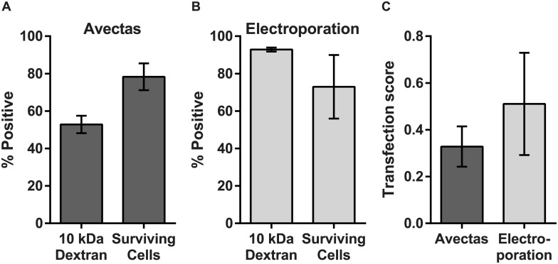
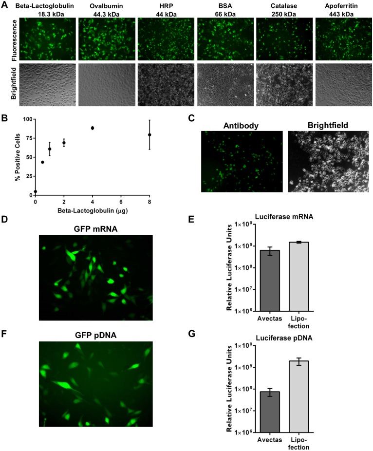
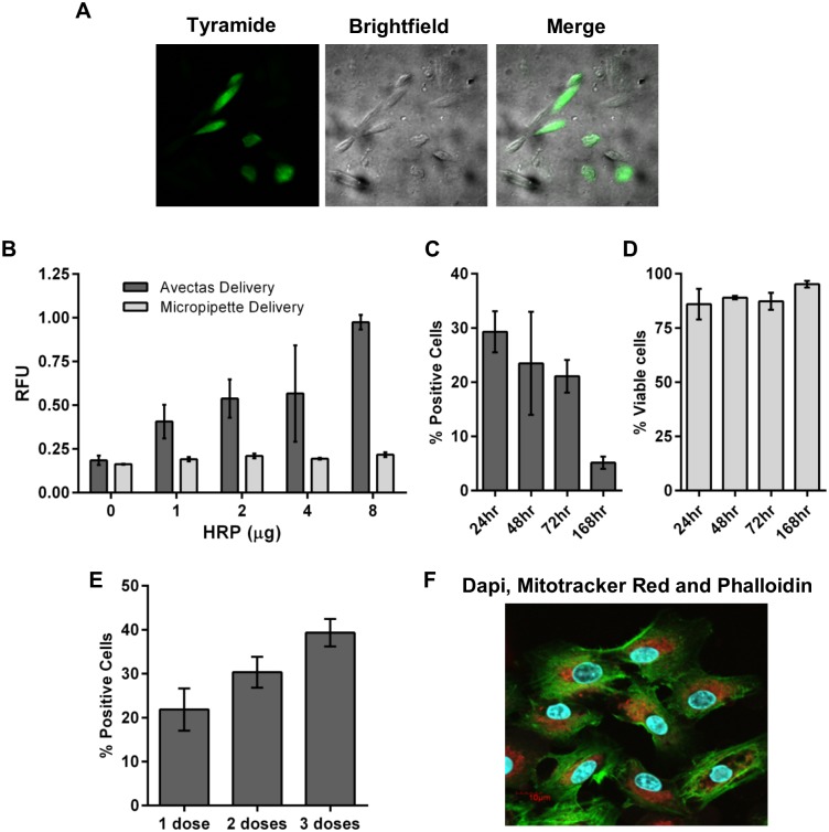
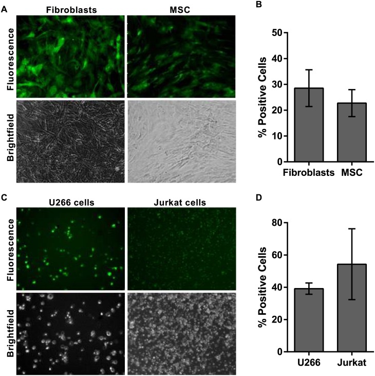
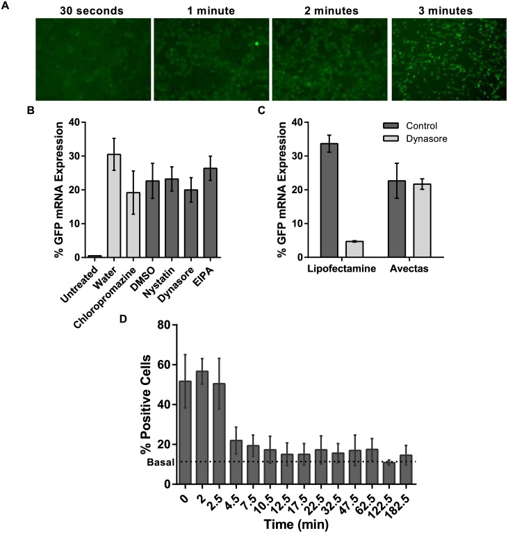
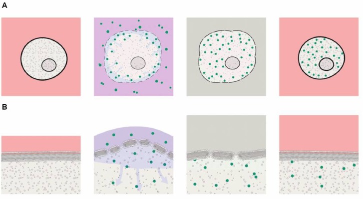
Despite advances in intracellular delivery technologies, efficient methods are still required that are vector-free, can address a wide range of cargo types and can be applied to cells that are difficult to transfect whilst maintaining cell viability. We have developed a novel vector-free method that uses reversible permeabilization to achieve rapid intracellular delivery of cargos with varying composition, properties and size. A permeabilizing delivery solution was developed that contains a low level of ethanol as the permeabilizing agent. Reversal of cell permeabilization is achieved by temporally and volumetrically controlling the contact of the target cells with this solution. Cells are seeded in conventional multi-well plates. Following removal of the supernatant, the cargo is mixed with the delivery solution and applied directly to the cells using an atomizer. After a short incubation period, permeabilization is halted by incubating the cells in a phosphate buffer saline solution that dilutes the ethanol and is non-toxic to the permeabilized cells. Normal culture medium is then added. The procedure lasts less than 5 min. With this method, proteins, mRNA, plasmid DNA and other molecules have been delivered to a variety of cell types, including primary cells, with low toxicity and cargo functionality has been confirmed in proof-of-principle studies. Co-delivery of different cargo types has also been demonstrated. Importantly, delivery occurs by diffusion directly into the cytoplasm in an endocytic-independent manner. Unlike some other vector-free methods, adherent cells are addressed in situ without the need for detachment from their substratum. The method has also been adapted to address suspension cells. This delivery method is gentle yet highly reproducible, compatible with high throughput and automated cell-based assays and has the potential to enable a broad range of research, drug discovery and clinical applications.


Conflict of interest statement

Competing Interests: The authors SOD, VA, LG, JM, EM, CB, JG, DM, MM are current or past employees of Avectas Ltd. FC was a paid Consultant for Avectas Ltd. SOD and MM are co-founders and Board members of Avectas Ltd. and have shares in Avectas Ltd. Avectas Ltd. is developing and intends to commercialise technology based in part on the work described in this manuscript. Avectas Ltd. has filed patents applications PCT/US2015/057247: ‘Delivery across cell plasma membranes’ and 62/273,284: ‘Vector-Free Delivery of Gene Editing Proteins to Cells and Tissues’. This declaration does not alter the authors' adherence to all PLOS ONE policies regarding sharing data and materials.

Figures

Fig 1
Fig 1. Delivery solution.
(A) 150 μM PI or 3 μM 10 kDa Dextran-Alexa488 in 200 μl delivery solution was delivered to A549 cells using a micropipette. Immediately after delivery, uptake of PI was visible throughout the cell population but no uptake of dextran was apparent. With 20 μl delivery solution, PI uptake was apparent where the solution first landed in the well (drop zone) but not in other areas. Low level uptake of 10 kDa Dextran-Alexa488 was also observed in the drop zone. (B) LDH release measured at 24 hr post-delivery indicated that 37.2±4.8%, 44.6±1.9% and 51.4±4.7% cells were damaged when 200 μl delivery solution alone, delivery solution containing PI or delivery solution containing 10 kDa Dextran-Alexa488 respectively was applied. In contrast, LDH release was 5.1±6.0, 10.5±1.3% and 5.6±3.1% respectively for these solutions when a 20 μl volume was applied. All photomicrographs are 10x magnification. n = 3, data are depicted as the mean ± standard deviation. (DS = delivery solution only; PI = propidium iodide; LDH = lactate dehydrogenase).
Fig 2
Fig 2. Spray method.
(A) The spray instrument comprised of an air compressor that delivered compressed air to an atomizer which was held in position on a retort stand. The culture plate was positioned on a stage below the atomizer. The atomizer could be moved vertically to adjust the distance between the atomizer and the cells. The air pressure levels could also be adjusted. Insert shows the atomizer and spray. (B) 10 kDa dextran-Alexa488 uptake was apparent in A549 cells and uptake was evenly distributed across the cell monolayer. (C) Delivery efficiency levels in A549 cells. Also LDH release was similar to PBS pipette controls. No delivery was observed in controls where 10 kDa dextran-Alexa488 was delivered in PBS using a micropipette. (D) A range of low and high molecular weight dextrans were successfully delivered to A549 cells using the method, 10x magnification. n = 3, data are depicted as the mean ± standard deviation. (LDH = lactate dehydrogenase).
Fig 3
Fig 3. Delivery and viability compared with electroporation.
Comparison of delivery efficiency (using 10kDa dextran-Alexa488) and cell viability and survival (using propidium iodide exclusion) for (A) the reversible permeabilization method and (B) electroporation. (C) The transfection score defined as (transfected cells/ total cells)x(viable cells/ total cells) for the two methods). n = 3, data are depicted as the mean ± standard deviation.
Fig 4
Fig 4. Examples of delivery of diverse cargoes to CHO cells.
(A) Proteins of varying sizes were labelled with FITC and 4 μg protein per well was delivered to CHO cells and analyzed by fluorescence microscopy at 2 hr post-delivery, 10x magnification. (B) Increased efficiency of delivery of beta-lactoglobulin was demonstrated with increasing concentration of protein delivered. (C) Full length anti-rabbit-Alexa488 secondary antibody was successfully delivered. (D) GFP mRNA (5 μg) was delivered twice (10 μg/well in total) into cells using the permeabilization method and expression of GFP protein was observed by fluorescence microscopy at 24 hr post-delivery. (E) Luciferase mRNA (5 μg) was delivered twice (10 μg/well total) into cells using the permeabilization method and expression of luciferase was quantified by luminometry at 24 hr post-delivery. For lipofection, luciferase mRNA (5 μg) was delivered per well. (F) pGFP (5 μg) was delivered twice (10 μg/well total) into cells using the permeabilization method and expression of GFP protein was observed by fluorescence microscopy at 24 hr post-delivery. (G) pGLuc (10 μg) was delivered into cells using the permeabilization method and expression of luciferase was quantified by luminometry at 24 hr post-delivery. For lipofection, 0.5 μg pGLuc was delivered per well. n = 3, data are depicted as the mean ± standard deviation. (GFP = green fluorescent protein; pGFP = plasmid encoding GFP; pGLuc = plasmid encoding Guassia luciferase).
Fig 5
Fig 5. Cell functionality and intracellular targeting.
(A) Alexa Fluor® 488-labelled tyramide substrate demonstrated activity and localization of HRP in CHO cells following delivery of HRP. (B) Increasing production of fluorescent DCF product with increasing dose of HRP delivered into CHO cells compared with cells where HRP was delivered by pipette. (C) GFP expression following delivery of GFP mRNA. (D) Cell viability remained above 75% up to 168hr post-delivery. (E) Up to 3 doses of GFP mRNA (4 μg) were delivered. GFP expression was analyzed 24 hr after the final dose. (F) Confocal microscopy image illustrates co-delivery to A549 cells: DAPI (300 nM), Mitotracker Red (50 μM) and Phalloidin-Alexa488 (0.33 μM) correspond to blue nuclei, red mitochondria and green actin filaments, respectively. n = 3, data are depicted as the mean ± the standard deviation. (HRP = horseradish peroxidase; DCF = dichlorofluorescein; GFP = green fluorescent protein; DAPI = 4',6-diamidino-2-phenylindole).
Fig 6
Fig 6. Cell-independent delivery.
(A) Delivery of 3 μM 10 kDa dextran-Alexa488 to primary human fibroblasts and primary human MSC. (B) Efficiency of delivery was quantified by flow cytometry at 2 hr post-delivery. (C) Delivery of BSA-FITC to U266 and Jurkat suspension cells. (D) Efficiency of delivery was quantified by flow cytometry at 2 hr post-delivery. All photomicrographs are 10x magnification. n = 3, data are depicted as the mean ± the standard deviation. (MSC = mesenchymal stem cells; BSA-FITC = bovine serum albumin-fluorescein isothiocyanate).
Fig 7
Fig 7. Testing mechanisms of cargo uptake and subsequent membrane resealing.
(A) Time course of uptake of 10 kDa dextran-Alexa488 into A549 cells analyzed by fluorescence microscopy consistent with simple diffusion post-delivery (10x mag.). (B) In A549 cells the uptake of EGFP mRNA was not inhibited either by pretreatment with Dynasore or chloropromazine to inhibit clathrin-mediated endocytosis or Nystatin or EIPA to inhibit caveolar-mediated endocytosis and micropinocytosis. (C) Lipofectamine 2000 was used as a positive control for endocytosis-mediated delivery. EGFP expression was reduced in lipofected cells treated with Dynasore. (D) PI uptake was analyzed by flow cytometry and the data indicate that the cells remain permeable to PI for up to 6 min post-treatment but then reseal and prevent uptake thereafter. n = 3, data are depicted as the mean ± standard deviation. (EIPA = 5-(N-Ethyl-N-isopropyl)amiloride; EGFP = enhanced green fluorescent protein; PI = propidium iodide; PBS = phosphate buffered saline).
Fig 8
Fig 8. Mechanism of action.
(A) Graphic representation of mechanism of action. Cells are initially in culture medium (pink). The cartoon illustrates a sequence lasting about 6 minutes. Ethanol the perturbs cell membrane and makes it susceptible to transient permeablization. Cell begins to swell as extracellular water moves into the cell due to the oncotic effects of the large molecules in the cytoplasm. Cargos now move across the membrane: For smaller molecules, the predominant mechanism would be diffusion. For larger molecules the osmotically-driven water influx (a process known as ‘solvent drag’) augments diffusion by carrying cargo into the cells and concentrating cargo at the cell membrane. Solvent drag may be particularly important for even larger molecules, where an additional tendency for molecules to be carried toward the membrane is a consequence of the spray mode of applying pressure-dependent mechanical force to the cells. We propose that the velocity of the spray droplets leads to a concentration of the larger molecules close to the cell membrane where they enter the cell by diffusion. The next critical step is resealing the membrane and restoration of cell viability. The standard histology fixation protocols use higher ethanol concentrations (>30%) and longer incubation times which result in loss of cell viability due to irreversible membrane permeabilization. Diluting the ethanol more than 50-fold with a PBS solution which is non-toxic to permeabilized cells enables the membrane to reseal (grey). After the cells are returned to culture medium water leaves the cell by the cell’s own regulatory processes as normal electrolyte osmotic gradients are restored. (B) A higher magnification view of the processes at the cell membrane. Exposure to ethanol (25%) thins the membrane so that the tension caused by cell swelling induces reversible permeabilization sufficient to allow entry of cargos as large as proteins and DNA plasmids. Subsequent washout of ethanol restores membrane thickness, reseals the cell membrane and enables recovery in culture medium.

References

    1. Stewart MP, Sharei A, Ding X, Sahay G, Langer R, Jensen KF. In vitro and ex vivo strategies for intracellular delivery. Nature. 2016;538(7624):183–92. 10.1038/nature19764 - DOI - PubMed
    1. Gurtovenko AA, Anwar J. Modulating the structure and properties of cell membranes: the molecular mechanism of action of dimethyl sulfoxide. J Phys Chem B. 2007;111(35):10453–60. 10.1021/jp073113e - DOI - PubMed
    1. Koynova R, Tenchov B. Enhancing nucleic acid delivery, insights from the cationic phospholipid carriers. Curr Pharm Biotechnol. 2014;15(9):806–13. - PubMed
    1. Waehler R, Russell SJ, Curiel DT. Engineering targeted viral vectors for gene therapy. Nat Rev Genet. 2007;8(8):573–87. 10.1038/nrg2141 - DOI - PMC - PubMed
    1. Li L, Wei Y, Gong C. Polymeric Nanocarriers for Non-Viral Gene Delivery. J Biomed Nanotechnol. 2015;11(5):739–70. - PubMed

LinkOut - more resources