. 2017 Mar 1;158(3):664-677.
doi: 10.1210/en.2016-1779.

Glucocorticoids Induce Bone and Muscle Atrophy by Tissue-Specific Mechanisms Upstream of E3 Ubiquitin Ligases

Affiliations

Glucocorticoids Induce Bone and Muscle Atrophy by Tissue-Specific Mechanisms Upstream of E3 Ubiquitin Ligases

Amy Y Sato et al. Endocrinology. .

Abstract

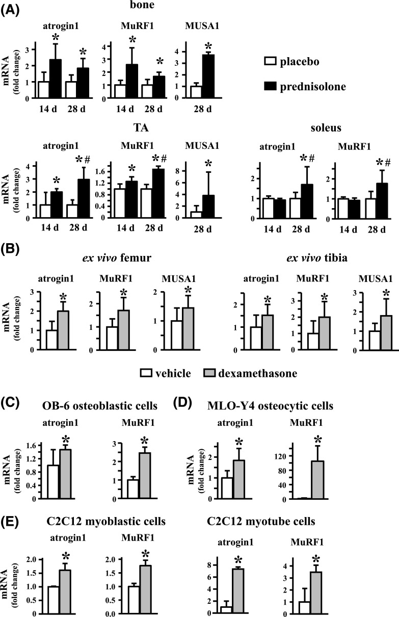
Glucocorticoid excess, either endogenous with diseases of the adrenal gland, stress, or aging or when administered for immunosuppression, induces bone and muscle loss, leading to osteopenia and sarcopenia. Muscle weakness increases the propensity for falling, which, combined with the lower bone mass, increases the fracture risk. The mechanisms underlying glucocorticoid-induced bone and muscle atrophy are not completely understood. We have demonstrated that the loss of bone and muscle mass, decreased bone formation, and reduced muscle strength, hallmarks of glucocorticoid excess, are accompanied by upregulation in both tissues in vivo of the atrophy-related genes atrogin1, MuRF1, and MUSA1. These are E3 ubiquitin ligases traditionally considered muscle-specific. Glucocorticoids also upregulated atrophy genes in cultured osteoblastic/osteocytic cells, in ex vivo bone organ cultures, and in muscle organ cultures and C2C12 myoblasts/myotubes. Furthermore, glucocorticoids markedly increased the expression of components of the Notch signaling pathway in muscle in vivo, ex vivo, and in vitro. In contrast, glucocorticoids did not increase Notch signaling in bone or bone cells. Moreover, the increased expression of atrophy-related genes in muscle, but not in bone, and the decreased myotube diameter induced by glucocorticoids were prevented by inhibiting Notch signaling. Thus, glucocorticoids activate different mechanisms in bone and muscle that upregulate atrophy-related genes. However, the role of these genes in the effects of glucocorticoids in bone is unknown. Nevertheless, these findings advance our knowledge of the mechanism of action of glucocorticoids in the musculoskeletal system and provide the basis for novel therapies to prevent glucocorticoid-induced atrophy of bone and muscle.


Figures

Figure 1.
Figure 1.
Glucocorticoids induce atrophy of bone and suppression of bone formation. Female C57BL/6 mice were implanted with pellets releasing 2.1 mg/kg/d of prednisolone or placebo, and BMD was assessed 14 or 28 days later. (A) Percentage of change in BMD was measured using dual energy x-ray absorptiometry and calculated from initial scans performed before pellet implantation and final scans performed at the indicated points. (B) Trabecular thickness was measured by micro-computed tomography analysis at the distal femur and proximal tibia metaphysis. Dynamic histomorphometric parameters on (C) the cancellous bone of proximal tibia and (D) periosteal and endocortical surfaces of the tibia mid-diaphysis were measured in mice treated for 14 days with placebo or prednisolone. Bars represent mean ± standard deviation; n = 7 to 10 mice. *P < 0.05 vs placebo-treated mice by Student’s t test. MAR, mineral apposition rate; MS/BS, mineralizing surface per bone surface.
Figure 2.
Figure 2.
Glucocorticoids induce muscle loss and weakness. (A) Percentage of change in whole body lean mass determined using dual energy x-ray absorptiometry. (B) Scheme depicting the position of the muscles tibialis anterior (TA), EDL, and soleus (SOL) relative to the tibia. (C) Wet weight of tibialis anterior and soleus muscles recorded 14 or 28 days after pellet implantation. (D) Wet weight of EDL and soleus muscles and ex vivo contractility muscle function testing recorded 28 days after pellet implantation. (E) Muscle function testing performed in vivo 14 and 28 days after pellet implantation. Bars represent mean ± standard deviation; n = 7 to 10 for (A and C) and n = 8 to 10 for (D and E). *P < 0.05 vs placebo by Student’s t test for (A to C) and muscle weights in (D). *P < 0.05 vs placebo by two-factor, mixed-model repeated measures ANOVA, followed by Tukey post hoc method for muscle functionality tests in (D and E). For the fatigue tests in (D), regression line equations for EDL muscles were as follows: for the first 10% of stimulations, y = −0.5387x + 22.784 (placebo) and y = −0.3549x + 17.033 (glucocorticoids) (P < 0.05 by t test); for the last 90% of stimulations, y = −0.1601x + 20.474 (placebo) and y = −0.102x + 15.181 (glucocorticoids) (P < 0.05 by t test). Regression line equations for soleus muscles were as follows: for the first 10% of stimulations, y = −0.0658x + 11.72 (placebo) and y = −0.0641x + 9.2375 (glucocorticoids) (P = NS by t test); for the last 90% of the stimulations, y = −0.0102x + 10.778 (placebo) and y = −0.0097x + 8.2384 (glucocorticoids) (P = NS by t test).
Figure 3.
Figure 3.
Glucocorticoids promote the transcription of atrophy genes in vivo, ex vivo, and in vitro. (A) mRNA levels were quantified in vertebral bone L4 (bone) and tibialis anterior (TA) and soleus muscle preparations. The transcripts were normalized to the levels of the housekeeping gene GAPDH. Bars represent the mean ± standard deviation; n = 5 to 7 for L4; n = 8 to 10 for tibialis anterior and soleus. *P < 0.05 vs placebo-treated mice; #P < 0.05 vs 14-day glucocorticoid-treated mice, by two-way ANOVA followed by Tukey post hoc method. (B) Gene expression in bones from C57BL/6 mice cultured with vehicle or 1 μm dexamethasone for 6 hours. Bars represent mean ± standard deviation; n = 9 to 12. *P < 0.05 vs vehicle-treated cells by Student’s t test. (C) OB-6 osteoblastic cells and (D) MLO-Y4 osteocytic cells were treated with vehicle or 1 μm dexamethasone for 24 hours. RNA was extracted, and atrogin1 and MuRF1 levels were quantified using quantitative polymerase chain reaction. Bars represent mean ± standard deviation; n = 5 to 6. *P < 0.05 vs vehicle-treated cells by Student’s t test. (E) C2C12 myoblasts were kept in growing media or induced to differentiate into myotubes for 6 days. Cells were treated with vehicle or 1 μm dexamethasone for 24 hours, RNA was extracted, and atrogin1 and MuRF1 levels were quantified by quantitative polymerase chain reaction. The transcripts were normalized to the levels of the housekeeping gene GAPDH. Bars represent mean ± standard deviation; n = 3 for myoblasts; n = 6 for myotubes. *P < 0.05 vs vehicle-treated cells by Student’s t test.
Figure 4.
Figure 4.
Glucocorticoids (GC) did not induce Notch activation in bone in vivo, ex vivo, or in vitro. The expression of the indicated genes normalized to GAPDH was measured by quantitative polymerase chain reaction in (A) bone preparations obtained from mice treated with placebo or prednisolone for 14 or 28 days and (B) bones from C57BL/6 mice cultured with vehicle or 1 μm dexamethasone for 6 hours. Bars represent mean ± standard deviation; n = 9 to 10 for A and n = 10 to 12 for B. *P < 0.05 vs placebo-treated mice or vehicle-treated bones by Student’s t test. Gene expression was measured in (C) OB-6 osteoblastic or (D) MLO-Y4 osteocytic cells treated with vehicle or 1 μm dexamethasone for 24 hours. Bars represent mean ± standard deviation; n = 5 to 6 for (C) and n = 6 for (D). *P < 0.05 vs vehicle-treated cells by Student’s t test.
Figure 5.
Figure 5.
Glucocorticoids increased the expression of Notch signaling pathway components in muscle in vivo. The expression of the indicated genes normalized to GAPDH was measured by quantitative polymerase chain reaction in tibialis anterior (TA) muscles from mice treated with placebo or prednisolone for 14 or 28 days. Bars represent mean ± standard deviation; n = 6. *P < 0.05 vs placebo-treated mice; #P < 0.05 vs 14-day glucocorticoid-treated mice by two-way ANOVA followed by Tukey post hoc method.
Figure 6.
Figure 6.
Glucocorticoids (GC) increased the expression of Notch signaling pathway components in muscle ex vivo, and Notch inhibition with GSI XX prevented glucocorticoid-induced atrophy-related gene expression in muscle and the reduction in myotube diameter. (A–D) Tibialis anterior muscles or tibial bones were isolated from C57BL/6 mice and cultured with vehicle or 1 μm dexamethasone in the presence and absence of the Notch inhibitor GSI XX for (A and C) 12 or (B) 24 hours for tibialis anterior and (D) 6 hours for tibia. RNA was isolated, and the levels of the indicated genes were measured by quantitative polymerase chain reaction and normalized to Rplp2. Bars are mean ± standard deviation; n = 5 to 7. *P < 0.05 vs corresponding vehicle-treated muscles; #P < 0.05 vs vehicle-treated muscles in the absence of GSI XX by two-way ANOVA, followed by Tukey post hoc test. (E) C2C12 cells were differentiated into myotubes and treated with vehicle or 1 μm dexamethasone in the absence or presence of GSI XX. Myotube diameter was measured as indicated in the Materials and Methods section. Scale bars = 200 μm. Bars represent mean ± standard deviation; n = 3. *P < 0.05 vs vehicle-treated cells by two-way ANOVA, followed by Tukey post hoc test. Data shown from a representative experiment of 4 experiments performed in which the decrease in myotube diameter was 17%, 24%, 24%, and 20%.
Figure 7.
Figure 7.
Glucocorticoids induced bone and muscle atrophy by distinct tissue-specific mechanisms upstream of atrogin1, MuRF1, and MUSA1. Glucocorticoids induced atrophy of both bone and muscle, although by different upstream mechanisms that converge in upregulation of atrophy-related E3 ubiquitin ligase genes atrogin1, MuRF1, and MUSA1.

References

    1. Weinstein RS. Clinical practice: glucocorticoid-induced bone disease. N Engl J Med. 2011;365(1):62–70. - PubMed
    1. Rizzoli R, Adachi JD, Cooper C, Dere W, Devogelaer JP, Diez-Perez A, Kanis JA, Laslop A, Mitlak B, Papapoulos S, Ralston S, Reiter S, Werhya G, Reginster JY. Management of glucocorticoid-induced osteoporosis. Calcif Tissue Int. 2012;91(4):225–243. - PubMed
    1. Reid IR. Glucocorticoid osteoporosis—mechanisms and management. Eur J Endocrinol. 1997;137(3):209–217. - PubMed
    1. Suzuki Y, Nawata H, Soen S, Fujiwara S, Nakayama H, Tanaka I, Ozono K, Sagawa A, Takayanagi R, Tanaka H, Miki T, Masunari N, Tanaka Y. Guidelines on the management and treatment of glucocorticoid-induced osteoporosis of the Japanese Society for Bone and Mineral Research: 2014 update. J Bone Miner Metab. 2014;32(4):337–350. - PubMed
    1. van Staa TP, Leufkens HG, Cooper C. The epidemiology of corticosteroid-induced osteoporosis: a meta-analysis. Osteoporos Int. 2002;13(10):777–787. - PubMed