. 2017 Jun 1;174(6):586-594.
doi: 10.1176/appi.ajp.2017.16070814. Epub 2017 Mar 31.

Selective Loss of Smaller Spines in Schizophrenia

Affiliations

Selective Loss of Smaller Spines in Schizophrenia

Matthew L MacDonald et al. Am J Psychiatry. .

Abstract

Objective: Decreased density of dendritic spines in adult schizophrenia subjects has been hypothesized to result from increased pruning of excess synapses in adolescence. In vivo imaging studies have confirmed that synaptic pruning is largely driven by the loss of large or mature synapses. Thus, increased pruning throughout adolescence would likely result in a deficit of large spines in adulthood. Here, the authors examined the density and volume of dendritic spines in deep layer 3 of the auditory cortex of 20 schizophrenia and 20 matched comparison subjects as well as aberrant voltage-gated calcium channel subunit protein expression linked to spine loss.

Method: Primary auditory cortex deep layer 3 spine density and volume was assessed in 20 pairs of schizophrenia and matched comparison subjects in an initial and replication cohort (12 and eight pairs) by immunohistochemistry-confocal microscopy. Targeted mass spectrometry was used to quantify postsynaptic density and voltage-gated calcium channel protein expression. The effect of increased voltage-gated calcium channel subunit protein expression on spine density and volume was assessed in primary rat neuronal culture.

Results: Only the smallest spines are lost in deep layer 3 of the primary auditory cortex in subjects with schizophrenia, while larger spines are retained. Levels of the tryptic peptide ALFDFLK, found in the schizophrenia risk gene CACNB4, are inversely correlated with the density of smaller, but not larger, spines in schizophrenia subjects. Consistent with this observation, CACNB4 overexpression resulted in a lower density of smaller spines in primary neuronal cultures.

Conclusions: These findings require a rethinking of the overpruning hypothesis, demonstrate a link between small spine loss and a schizophrenia risk gene, and should spur more in-depth investigations of the mechanisms that govern new or small spine generation and stabilization under normal conditions as well as how this process is impaired in schizophrenia.

Keywords: Calcium Channel; Dendritic Spines; Neuropathology; Proteomics; Schizophrenia.


Conflict of interest statement

Dr. MacDonald reports no competing interests

Dr. Alhassan reports no competing interests

Dr. Newman reports no competing interests

Dr. Richards reports no competing interests

Dr. Gu reports no competing interests

Dr. Sampson reports no competing interests

Dr. Fish reports no competing interests

Dr. Penzes reports no competing interests

Dr. Wills reports no competing interests

Dr. Sweet reports no competing interests

Figures

Figure 1
Figure 1. Spine attributes by volume
A. Phalloidin-labeled and spinophilin-immunoreactive puncta in deep layer 3 of primary auditory cortex. (Top) Phalloidin-labeled puncta (red) and spinophilin-immunoreactive puncta (green) colocalize throughout deep layer 3 of Brodmann area 41 and are found along microtubule-associated protein 2 (MAP2) immunoreactive processes (blue) suggesting spine structures along dendrites. (Bottom) Images are a magnification of the inset in the top panel highlighting the relationship between phalloidin-labeled (1) and spinophilin-immunoreactive objects (2) and their colocalization in presumptive spine structures (3). The volume of phalloidin objects, co-localized with spinophilin, was used to estimate spine volume. B. and C. show representative phalloidin-labeled presumptive spines (60×) from schizophrenia and a matched control, respectively. D. 20 pairs of subjects were assayed as an initial and an independent replication cohort: Cohort 1 (n = 12 pairs) and Cohort 2 (n = 8 pairs) (Supplemental Table 1). The results were equally significant between the two groups and are presented here as a single cohort for simplicity. The density of spines of different volumes (0.15 µM3 increments, with the final bin including all objects with volume > 1.35 µM3) for control and schizophrenia subjects from both cohorts are shown. The density of spines in the bins of smaller volumes differed significantly between control and schizophrenia. P-values are Bonferroni corrected. E. The mean phalloidin intensity (calculated as the total phalloidin intensity of the spine object divided by spine object volume) of spines of different volumes from control and schizophrenia subjects are shown. F. The sum phalloidin intensity of spines of different volumes from control and schizophrenia subjects are shown. Mean and sum phalloidin intensity was consistent between control and schizophrenia across all of the spine volume categories.
Figure 2
Figure 2. Correlation of ALFDFLK with spine density by bin
The CACNB peptide ALFDFLK was quantified by liquid chromatograph – selected reaction monitoring mass spectrometry. Expression of this peptide was plotted against the density of spine objects from each size bin, e.g. panel A (0–0.15 µM3) reports the correlation of ALFDFLK with the density of spine objects 0–0.15 µM3 in both control (black open circle) and schizophrenia (grey filled circle) subjects. ALFDFLK expression was significantly inversely correlated with the density of small, but not medium or large, spines across all subjects (solid black line). This effect was similar for the control (black dashed line) and schizophrenia (grey dashed line) cohorts when separated.
Figure 3
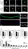
Figure 3. CACNB4 overexpression decreases small spine density in primary neuronal culture
E17 rat cortex primary neuronal cultures were transfected on day 12 with either GFP or GFP/CACNB4-Myc. A. shows representative 10x confocal images of transfected neurons. Endogenous CACNB4 expression was not observed in these cultures, in line with previous reports that CACNB4 is not expressed at this stage of development in the cortical cultures. CACNB4 transfection induced robust somatodendritic expression (A.) as well as expression in spines (B.). C. Shows processing for determination of dendritic spine volumes in another representative dendritic segment. Note that the filopodia is not masked. Spine and filopodia density was evaluated in three independent experiments. In each experiment spine density was significantly decreased in CACNB4 expressing neurons (D.). N (number of dendrites assayed in each experiment) is reported in each bar. In addition to reaching significance in each independent experiment, spine density was also significantly decreased across all three experiments (p = 6.5E−5). E. compares filopodia densities between GFP+ and GFP+/CACNB4-Myc+ neurons from all experiments, which were equivalent. Spine density was then assessed for spines of different sizes (F.). A significant reduction in density selective for small spines was observed (two-way ANOVA). The reported P-value is Bonferroni corrected.

Comment in

References

    1. Feinberg I. Schizophrenia: caused by a fault in programmed synaptic elimination during adolescence? J Psychiatr Res. 1982;17(4):319–334. - PubMed
    1. Vita A, et al. Progressive loss of cortical gray matter in schizophrenia: a meta-analysis and meta-regression of longitudinal MRI studies. Transl Psychiatry. 2012;2:e190. - PMC - PubMed
    1. Moyer CE, Shelton MA, Sweet RA. Dendritic spine alterations in schizophrenia. Neurosci Lett. 2014 - PMC - PubMed
    1. Chen X, et al. Functional mapping of single spines in cortical neurons in vivo. Nature. 2011;475(7357):501–505. - PubMed
    1. Midtgaard J. The integrative properties of spiny distal dendrites. Neuroscience. 1992;50(2):501–502. - PubMed

Publication types

MeSH terms

LinkOut - more resources