Review
. 2017 Oct:84:36-47.
doi: 10.1016/j.mcn.2017.03.007. Epub 2017 Mar 27.

It takes a village to raise a branch: Cellular mechanisms of the initiation of axon collateral branches

Affiliations
Review

It takes a village to raise a branch: Cellular mechanisms of the initiation of axon collateral branches

Lorena Armijo-Weingart et al. Mol Cell Neurosci. 2017 Oct.

Abstract

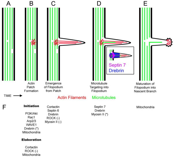
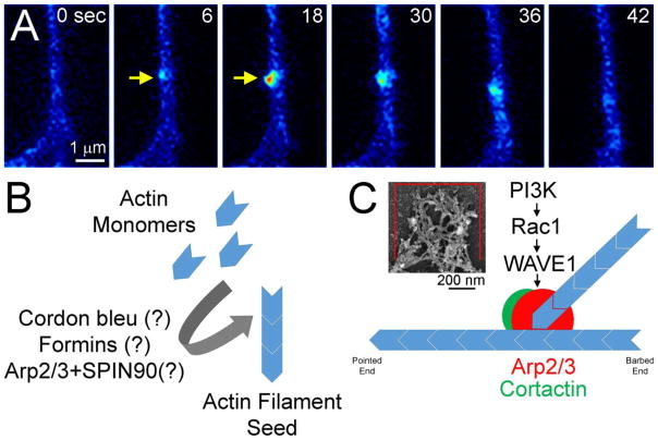
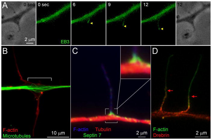
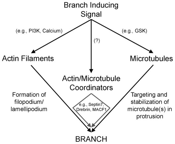
The formation of axon collateral branches from the pre-existing shafts of axons is an important aspect of neurodevelopment and the response of the nervous system to injury. This article provides an overview of the role of the cytoskeleton and signaling mechanisms in the formation of axon collateral branches. Both the actin filament and microtubule components of the cytoskeleton are required for the formation of axon branches. Recent work has begun to shed light on how these two elements of the cytoskeleton are integrated by proteins that functionally or physically link the cytoskeleton. While a number of signaling pathways have been determined as having a role in the formation of axon branches, the complexity of the downstream mechanisms and links to specific signaling pathways remain to be fully determined. The regulation of intra-axonal protein synthesis and organelle function are also emerging as components of signal-induced axon branching. Although much has been learned in the last couple of decades about the mechanistic basis of axon branching we can look forward to continue elucidating this complex biological phenomenon with the aim of understanding how multiple signaling pathways, cytoskeletal regulators and organelles are coordinated locally along the axon to give rise to a branch.

Keywords: Axon sprouting; Filopodia; Interstitial branch; Neurite; Neuronal morphogenesis.


Conflict of interest statement

Declaration of interest: The authors have no conflicts of interest to disclose.

Figures

Figure 1
Figure 1
Sequence of cytoskeletal events leading to the formation of axon collateral branches. The schematic shows the formation of a collateral branch, in time (left to right), along an axon (A). The first event in the formation of a branch is the emergence of an axonal filopodium (B–C). The emergence of an axonal filopodium is preceded by the formation of an axonal actin filament patch (B). The actin patch is a meshwork of actin filaments that gives rise to the bundle of actin filaments that defines the core of the filopodium (C). The targeting of an axonal microtubule into the filopodium is the next necessary step in the formation of a branch from a filopodium (D). The inset shows the general distributions of Drebrin and Septin 7 at axonal filopodia. Septin 7 localizes specifically to the base of filopodia and Drebrin localizes to actin patches and the proximal half of filopodia. Note that although not specifically shown, actin patches have shorter lifespans than filopodia and often are not present at the base of existing filopodia, although remnants may persist. Filopodia containing microtubules that remain in place have the potential to mature into nascent branches. The process of maturation involves the disassembly of the filopodial actin filament bundle, and the establishment of a distally polarized actin filament cytoskeleton giving rise a small growth cone-like structure at the tip of the nascent branch (E). Once the branch if established it then has the potential to continue elongating or undergo retraction back into the main axon. Each of the steps B–E has a given probability of occurring. In other words, only subsets of actin patches give rise to filopodia, only subsets of filopodia are targeted by microtubules, and only a subset of filopodia containing microtubules undergo maturation. Ultimately a branch arises from a segment of the axon that has successfully met the criteria for each of these steps. Not shown in this schematic is the local splaying of the microtubule array at sites of potential branching that is however discussed in the main text. The splaying occurs early in the process between steps A–B. (F) Summary of molecules discussed in the main text with specific identified roles in steps A–E. Each set of regulators is shown below the relevant step. Other regulators of branching which have not been assigned specific loci of regulation in the cytoskeletal basis of branching are not shown, but discussed in the text. Under (B) regulators are shown depending on whether they control the initiation of patches or their subsequent development (i.e., increase in size and lifespan). Unless denoted by (−) the role of the regulator is positive. If denoted by (−) the role is inhibitory. For example, Myosin II acts to suppress the emergence of filopodia from actin patches (C). In (B) Drebrin is denoted with a (*) to note that while it is required for patch initiation it is not sufficient to drive initiation. In (E) myosin II is denoted with (*). In this case, Myosin II does not regulate the entry of microtubules into filopodia but it serves to decrease the distance the microtubules penetrate into the filopodium.
Figure 2
Figure 2
Axonal actin filament patch formation and dynamics. (A) Example of actin patch formation (yellow arrow at 6 sec), elaboration (6–18 sec) and dissipation (30–42 sec) as imaged along the axon of an embryonic sensory neuron expressing eYFP-β-actin. (B) Possible actin filament nucleators involved in the initiation of actin patches. The specific nucleators required for patch initiation remains to be determined, as reflected by the (?). (C) The Arp2/3 complex is required for the detection and elaboration of axonal actin filament patches. The inset shows an example of the complex network of actin filaments in axonal actin patches, as detected using platinum replica electron microscopy (from a collaboration with Dr. T. Svitkina, University of Pennsylvania). PI3K activity drives the Rac1 GTPase that in turn activates the Arp2/3 complex through WAVE1. Cortactin serves to stabilize Arp2/3 mediated filament branches and positively regulates the elaboration of patches.
Figure 3
Figure 3
Microtubule dynamics, organization and microtubule-actin interactors. (A) Example of a microtubule plus tip decorated with eGFP-EB3 (denoted by yellow arrowheads) entering an axonal filopodium. The phase contrast images show the filopodium at 0 and 12 secs. The microtubule tip enters the filopodium at 6 sec and by 12 sec has traveled almost to the tip of the filopodium. (B) Example of the splaying of the microtubule array at sites of axonal protrusive activity, reflected by filopodia. The splaying of microtubules is denoted by the white]. Note that the adjacent axon segments exhibit a uniformly bundled array. (C) Example of Septin 7 localization to the base of an axonal filopodium (from a collaboration with Dr. E. Spiliotis, Drexel University). The inset shows a 2x empty magnification view of the base of the filopodium. (D) Example of the distribution of Drebrin restricted to the proximal 4–5 microns of axonal filopodia (denoted by red arrows) (from a collaboration with Dr. J. Chilton, University of Exeter). All examples are from embryonic chicken sensory neurons.
Figure 4
Figure 4
General summary model of the regulation of the axonal cytoskeleton during branching. Branch inducing signals activate multiple pathways that independently regulate the actin filament and microtubule cytoskeleton. The ensuing cytoskeletal dynamics promote branching by increasing actin filament dependent protrusive activity and microtubule tip polymerization or transport into protrusions. Molecules that coordinate the actin filament and microtubule cytoskeleton, in turn serve to promote interactions between the cytoskeletal elements. It remains to be determined if and by which signaling pathways cytoskeletal coordinators may be under regulation by branch inducing signals. The formation of a branch requires the formation of protrusive structures along the axon through the regulation of actin filaments, and the targeting and retention of microtubules in these protrusions. The means through which these ends are accomplished, at the molecular level, are likely diverse relying on different signaling pathways and effectors, as discussed in the concluding statement.

References

    1. Ageta-Ishihara N, Miyata T, Ohshima C, Watanabe M, Sato Y, Hamamura Y, Higashiyama T, Mazitschek R, Bito H, Kinoshita M. Septins promote dendrite and axon development by negatively regulating microtubule stability via HDAC6-mediated deacetylation. Nat Commun. 2013;4:2532. - PMC - PubMed
    1. Ahuja R, Pinyol R, Reichenbach N, Custer L, Klingensmith J, Kessels MM, Qualmann B. Cordon-bleu is an actin nucleation factor and controls neuronal morphology. Cell. 2007;131:337–350. - PMC - PubMed
    1. Akbik F, Cafferty WB, Strittmatter SM. Myelin associated inhibitors: a link between injury-induced and experience-dependent plasticity. Exp Neurol. 2012;235:43–52. - PMC - PubMed
    1. Alvarez J, Giuditta A, Koenig E. Protein synthesis in axons and terminals: significance for maintenance, plasticity and regulation of phenotype. With a critique of slow transport theory. Prog Neurobiol. 2000;62:1–62. - PubMed
    1. Andersen EF, Asuri NS, Halloran MC. In vivo imaging of cell behaviors and F-actin reveals LIM-HD transcription factor regulation of peripheral versus central sensory axon development. Neural Dev. 2011;6:27. - PMC - PubMed

Publication types