. 2017 Jul:103:600-610.
doi: 10.1016/j.wneu.2017.03.088. Epub 2017 Mar 27.

First Application of 7-T Magnetic Resonance Imaging in Endoscopic Endonasal Surgery of Skull Base Tumors

Affiliations

First Application of 7-T Magnetic Resonance Imaging in Endoscopic Endonasal Surgery of Skull Base Tumors

Thomas F Barrett et al. World Neurosurg. 2017 Jul.

Abstract

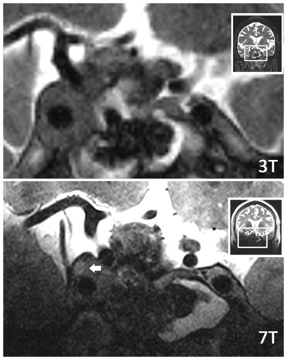
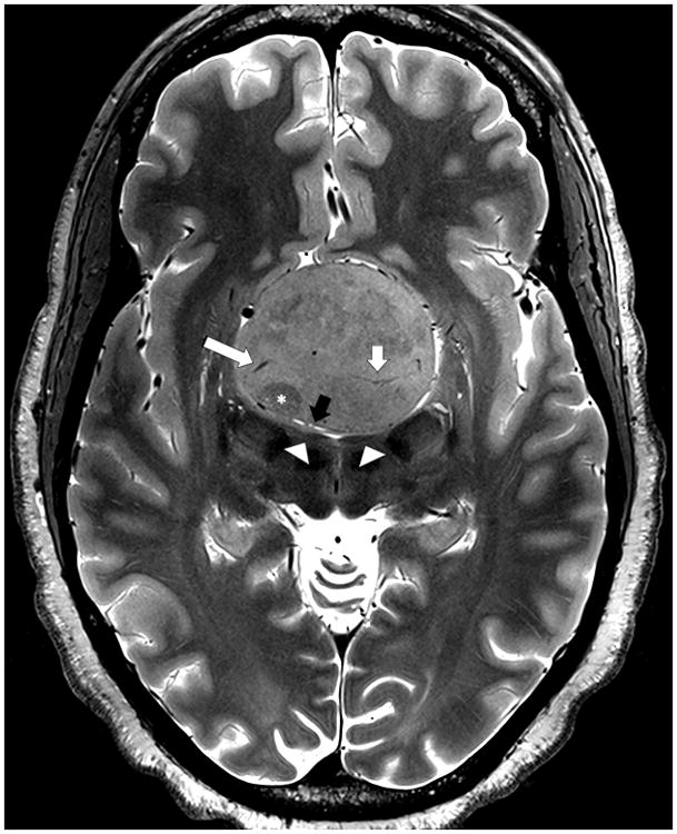
Background: Successful endoscopic endonasal surgery for the resection of skull base tumors is reliant on preoperative imaging to delineate pathology from the surrounding anatomy. The increased signal-to-noise ratio afforded by 7-T MRI can be used to increase spatial and contrast resolution, which may lend itself to improved imaging of the skull base. In this study, we apply a 7-T imaging protocol to patients with skull base tumors and compare the images with clinical standard of care.

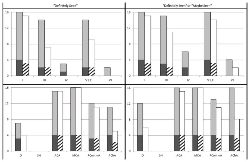
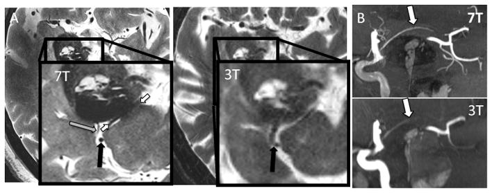
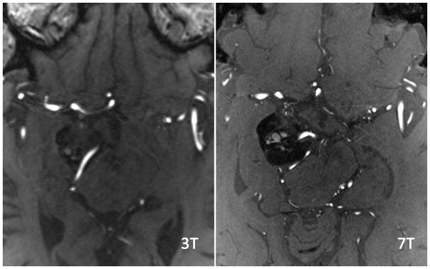
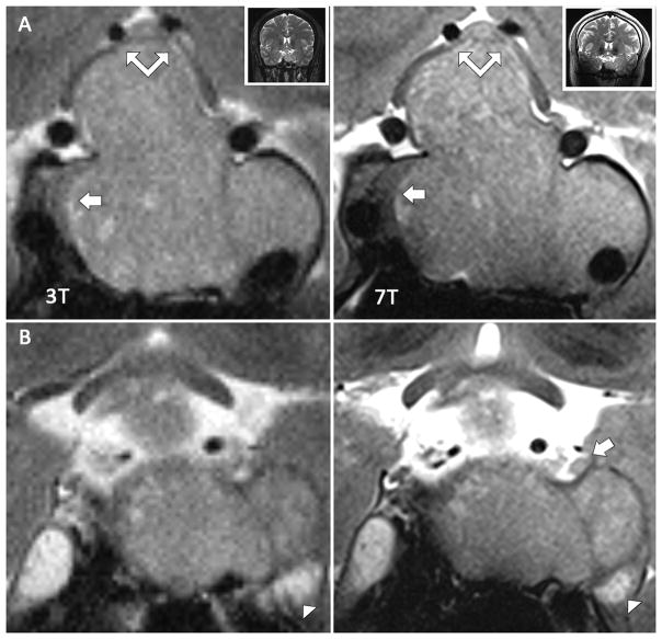
Methods: Images were acquired at 7 T on 11 patients with skull base lesions. Two neuroradiologists evaluated clinical 1.5-, 3-, and 7-T scans for detection of intracavernous cranial nerves and internal carotid artery (ICA) branches. Detection rates were compared. Images were used for surgical planning and uploaded to a neuronavigation platform and used to guide surgery.

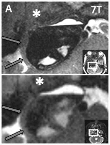
Results: Image analysis yielded improved detection rates of cranial nerves and ICA branches at 7 T. The 7-T images were successfully incorporated into preoperative planning and intraoperative neuronavigation.

Conclusions: Our study represents the first application of 7-T MRI to the full neurosurgical workflow for endoscopic endonasal surgery. We detected higher rates of cranial nerves and ICA branches at 7-T MRI compared with 3- and 1.5-T MRI, and found that integration of 7 T into surgical planning and guidance was feasible. These results suggest a potential for 7-T MRI to reduce surgical complications. Future studies comparing standardized 7-, 3-, and 1.5-T MRI protocols in a larger number of patients are warranted to determine the relative benefit of 7-T MRI for endonasal endoscopic surgical efficacy.

Keywords: 7-T MRI; Endoscopic endonasal surgery; Skull base tumors; Ultrahigh-field MRI.


Figures

Figure 1
Figure 1. Detection rates of cranial nerves and ICA branches
The top row displays detection rates of cranial nerves II–VI and the bottom row displays detection rates of ICA branches. Light gray bars represent 7T results in patients with 1.5T scans available for comparison. Dark gray bars represent 7T results in patients with 3T scans available for comparison. White bars represent 1.5T results and striped bars represent 3T results.
Figure 2
Figure 2. Tumor-trigeminal delineation at 3T and 7T
Axial T2W images of same patient at both 3T and 7T. The right trigeminal nerve (black arrow) is shown smoothly draped along the surface of the tumor, and distinction of nerve from tumor is easily made at 7T. At 3T, both the nerve and its border with the tumor are more difficult to resolve. The tumor margin is clearly delineated as it displaces the pons (P) with 7T, while the boundaries are blurred at 3T. A distinct fluid level (short white arrow) can be appreciated with 7T as well. Additionally, the fascicles of the left trigeminal nerve (long white arrow) are clearly defined as they approach Meckel’s cave with 7T and are less clearly resolved with 3T.
Figure 3
Figure 3. ICA branch detection at 3T and 7T
Sagittal Maximal Intensity Projection (MIP) of TOF MRA at both 3T and 7T. MRA acquisitions were fused to ensure identical tissue coverage. Imaging at 7T nicely shows the AChA (white) splayed over the tumor, while at 3T it is poorly seen over a shorter length. Additionally, other small vessels such as PCA tributaries projecting inferiorly are well visualized at 7T but not visualized at 3T.
Figure 4
Figure 4. Characterization of a macroadenoma within the right cavernous sinus and mass effect of tumor on adjacent cranial nerves
Two corresponding coronal T2W TSE images at 3T and 7T acquired per research protocol in same subject. A and B are in the same plane in the interpeduncular cistern at 3T and 7T, respectively, while C and D section more anteriorly through the cavernous sinuses. (A and B) At 7T, the left oculomotor (large black arrow), trochlear (small black arrow), and trigeminal nerve (small white arrow) can be seen approaching the cavernous sinus. At 3T, the oculomotor and trigeminal nerves are less clearly defined, and the trochlear nerve is not visible. The large macroadenoma clearly displaces the right oculomotor nerve superiorly (large white arrow) at 7T, while at 3T the nerve is not as well resolved. (C and D) At 7T, this nerve can be traced as it is courses around the large mass and eventually seen as a region of higher intensity in the cavernous sinus. At 3T, the course of the nerve is lost amid tumor signal.
Figure 5
Figure 5. Vessels feeding tumor
As in Figure 3, these images were acquired using TOF MRA per our research protocol at both 3T and 7T in the same patient. The slice angle is slightly different, and the slices were chosen based on the optimal slice at 3T to view the small feeding arteries. A comparable slice at 7T was then found. Small vessels feeding the tumor (white arrow) can be resolved at 7T that cannot be appreciated with 3T. Additionally, small perforating branches from the ACA and MCA territories can be appreciated throughout the 7T image.
Figure 6
Figure 6. Coronal Images of Pituitary Adenoma and Cavernous Structures
Coronal T2W TSE images from same patient acquired with both 3T and 7T research protocols. A and B are similar slices obtained through the cavernous sinus at 3T and 7T respectively. C and D are similar slices obtained slightly posterior to A and B. (A and B) At both 3T and 7T, a large isointense pituitary adenoma is appreciated with good delineation of the hypointense normal gland on the right. With 7T, however, the right medial wall of the cavernous sinus (short white arrow) is seen to be intact while with 3T it is unclear if the wall has been infiltrated. Additionally, the chiasm can be seen draped over the tumor at both field strengths (double arrow), but is better resolved at 7T. (C and D) At 7T, the left third nerve (white arrow) is appreciated being displaced superiorly by the invasive mass. At 3T, the nerve cannot be visualized. Additionally, the fascicles of the trigeminal nerve are better resolved in Meckel’s cave at 7T MRI (long white arrow).
Figure 7
Figure 7. 7T images of giant macroadenoma
Axial T2-weighted TSE image obtained at 7T in a patient with a giant pituitary adenoma. The image shows tumor heterogeneity including zone of lower signal suggesting a region of firmer tumor (asterisk), venous drainage (long white arrow), tumor arterial supply (short white arrow) and compressed adjacent anatomy including flattened mammillary bodies (arrowheads) and the lamina terminalis (black arrow).

References

    1. Alimohamadi M, Sanjari R, Mortazavi A, Shirani M, Moradi Tabriz H, Hadizadeh Kharazi H, et al. Predictive value of diffusion-weighted MRI for tumor consistency and resection rate of nonfunctional pituitary macroadenomas. Acta Neurochir (Wien) 2014;156:2245–2252. discussion 2252. - PubMed
    1. Amemiya S, Aoki S, Ohtomo K. Cranial nerve assessment in cavernous sinus tumors with contrast-enhanced 3D fast-imaging employing steady-state acquisition MR imaging. Neuroradiology. 2009;51:467–470. - PubMed
    1. Barrett TF, Sarkiss CA, Dyvorne HA, Lee J, Balchandani P, Shrivastava RK. Application of Ultrahigh Field Magnetic Resonance Imaging in the Treatment of Brain Tumors: A Meta-Analysis. World Neurosurg. 2016;86:450–465. - PubMed
    1. Blankena R, Kleinloog R, Verweij BH, van Ooij P, Ten Haken B, Luijten PR, et al. Thinner Regions of Intracranial Aneurysm Wall Correlate with Regions of Higher Wall Shear Stress: A 7T MRI Study. AJNR Am J Neuroradiol. 2016;37:1310–1317. - PMC - PubMed
    1. Blitz AM, Aygun N, Herzka DA, Ishii M, Gallia GL. High Resolution Three-Dimensional MR Imaging of the Skull Base: Compartments, Boundaries, and Critical Structures. Radiol Clin North Am. 2017;55:17–30. - PubMed

MeSH terms