. 2017 Mar 31;355(6332):1411-1415.
doi: 10.1126/science.aai7984. Epub 2017 Mar 30.

Breathing control center neurons that promote arousal in mice

Affiliations

Breathing control center neurons that promote arousal in mice

Kevin Yackle et al. Science. .

Abstract

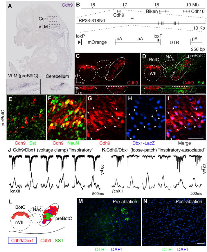
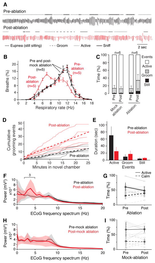
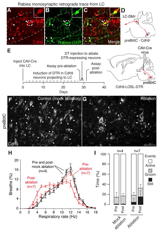
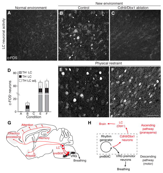
Slow, controlled breathing has been used for centuries to promote mental calming, and it is used clinically to suppress excessive arousal such as panic attacks. However, the physiological and neural basis of the relationship between breathing and higher-order brain activity is unknown. We found a neuronal subpopulation in the mouse preBötzinger complex (preBötC), the primary breathing rhythm generator, which regulates the balance between calm and arousal behaviors. Conditional, bilateral genetic ablation of the ~175 Cdh9/Dbx1 double-positive preBötC neurons in adult mice left breathing intact but increased calm behaviors and decreased time in aroused states. These neurons project to, synapse on, and positively regulate noradrenergic neurons in the locus coeruleus, a brain center implicated in attention, arousal, and panic that projects throughout the brain.


Figures

Fig. 1
Fig. 1. Identification and genetic ablation of Cdh9/Dbx1 double positive neurons in preBötC
A, Cdh9 mRNA expression (blue) in section of E14.5 mouse embryo (11). Insets, ventrolateral medulla, ventral cerebellum. B, (Top) Cdh9 locus on chromosome 15 (numbers, distance from centromere). (Middle) BAC RP23-318N6. Vertical lines, Cdh9 exons. (Bottom) Cdh9-LOSL-DTR BAC transgene: insertion at Cdh9 start codon of mOrange sequence and polyadenylation (pA) signals, flanked by loxP sites (triangles), followed by DTR sequence. C,D, Ventrolateral medulla sections of P0 Cdh9-LOSL-DTR mouse immunostained for mOrange to show Cdh9 expression (red) and P0 wild type mouse immunostained for Somatostatin (SST, green), shown aligned (registered by compact nucleus ambiguus (NAC), cranial nerve 7 (nVII), and ventral brainstem surface) in sagittal plane (upper) and transverse projection (lower panels). d, dorsal; c, caudal; m, medial. Bar, 200 μm. E–I, preBötC of P0 Cdh9 LOSL-DTR (E,F) or Cdh9-LOSL-DTR;Dbx1-lacZ (G–I) mouse immunostained for Cdh9-mOrange (E–G,I, red), SST (E, green), NeuN (F, green) or beta-galactosidase (Dbx1-LacZ, H,I, blue). Among Cdh9-neurons, none co-expressed SST (n=43 cells), all co-expressed NeuN (n=57), and 56% co-expressed Dbx1 reporter (n=292, arrowheads). Bar (for E–I), 50 μm. J,K, Whole cell voltage clamp recordings (top, pA, picoAmp) of Cdh9/Dbx1 preBötC neurons in slice preparations (top) and simultaneous integrated cnXII activity (bottom). Neuron in J (neuron 1, Table S1) shows bursts in all inspiratory events (“inspiratory pattern”). Neuron in K (neuron 3) shows more widespread activity but bursts only during some events (“inspiratory-associated”). Bars, 500 msec. L, Schematic of ventrolateral medulla. Cdh9/Dbx1 neurons (blue border) intermingle with SST (green) and other Cdh9 neurons (red) in preBötC. M,N, Intersectional genetic labeling of Cdh9/Dbx1 preBötC neurons with DTR (immunostain, green) in ~P35 Cdh9-LOSL-DTR;Dbx1-cre mice before (M) and after (N) intraperitoneal DT injection to ablate them. Bar, 50 μm.
Fig. 2
Fig. 2. Respiratory and behavior changes after Cdh9/Dbx1 neuron ablation
A, Plethysmography airflow traces of Cdh9-LOSL-DTR;Dbx1-Cre mice before (black) and 2 days after (red) Cdh9/Dbx1 ablation. Note more grooming (− −) and eupneic (−) breaths and less active breaths (− −) and sniffing (−−) after ablation. Bar, 2 seconds. B, Distribution of respiratory rates (Hz, bin size 1 Hz) in 40 minute assay of control (wild type, Cdh9-LOSL-DTR or Dbx1-Cre; black, n=5) and experimental (Cdh9-LOSL-DTR;Dbx1-Cre; red, n=5) animals before (dashed lines) and 2 days after (solid lines) Cdh9/Dbx1 ablation. C, Percent of time in plethysmography chamber spent still sitting (black), grooming (grey) or active (white) by control (n=6) or experimental (n=6) mice before (pre) or 2 days after (post) ablation or mock ablation. p-value comparing pre- and post-ablation behavior: active (0.02), grooming (0.02), still sitting (0.07). D, Grooming events in new chamber of Cdh9-LOSL-DTR;Dbx1-Cre mice before (black) or after (red) ablation. Solid lines, individual mice (n=6); dotted line, average. E, Duration of behaviors in C (mean ± S.D., n = 6). After ablation, active episodes shortened (p = 0.005), grooming and still sitting showed non-significant trend to lengthening (p=0.24 and 0.21, respectively). F,H, ECoG power spectral analysis (average (solid lines) ± S.E.M) of 20-minute recording (trial 1) of Cdh9-LOSL-DTR;Dbx1-Cre (F, n = 5) or control Cdh9-LOSL-DTR (H, n = 4) mice before (black) or 4–10 days after (red) ablation. δ, delta wave. V, voltage. Active behavior correlates with faster breathing (fig. S15C–E). G,I, Time spent in active (solid black line, mean ± S.E.M) and calm (dashed black line) behavioral states defined by EMG and ECoG (fig. S15) of individual animals in F,H (gray lines) during two 20-minute assays pre- and post-Cdh9/Dbx1 ablation. Note decreased active and increased calm periods following ablation in experimental animals (p = 0.001 and 0.02, respectively, paired t-test) and no change in controls (p = 0.86 and 0.81, respectively).
Fig. 3
Fig. 3. Effect on breathing and behavior of ablation of Cdh9 neurons that project to and synapse on LC neurons
A–D, Rabies virus monosynaptic retrograde trace from dopamine beta hydroxylase (Dbh)-expressing locus coeruleus (LC) neurons. Section through contralateral preBötC (A–C) of adult Cdh9-LOSL-DTR;Dbh-Cre mouse 5 days after unilateral LC injection of rabies-GFP and helper virus, immunostained to show Cdh9 neurons (mOrange, red). Arrowheads, colocalization of GFP and mOrange. Insets, boxed regions. Bar, 50 μm. D, Schematic of monosynaptic projection (red line) from Cdh9 preBötC neurons (red circle) to contralateral LC, which projects to higher brain structures (arrow). E, Scheme for ablating only Cdh9-expressing preBötC neurons that project to LC. CAV-Cre virus injected bilaterally into LC of adult Cdh9-LOSL-DTR mice (right) is taken up by Cdh9 preBötC neurons that project there (red). Cre induces DTR expression, and DT injection induces ablation. F–G, preBötC Cdh9-mOrange expression (white) in control uninjected (F, mock ablation) and CAV-Cre injected (G, ablation) Cdh9-LOSL-DTR mice 2 days after DT injection. Bar, 50 μm. Quantification showed 32% (mean) and 50% (maximal) reduction in mOrange neurons (n = 15 sections), close to the value expected if all Cdh9/Dbx1 preBötC neurons (50% of Cdh9 neurons) project to LC. H, Distribution of respiratory rates in 40 min assay (as in Fig. 2B) of CAV-Cre injected Cdh9-LOSL-DTR adult mice (red, n = 7) or wild type littermates (black, n = 4) before (dashed) and 2 days after (solid) DT injection. I, Behavioral analysis (as in Fig. 2C) of mice in H. Pre vs. post-ablation p-values: active (0.015), grooming (0.37), and still sitting (0.015). The increased calm events in pre-ablation experimental versus control mice was reproducible; it may be due to toxicity of DTR induced in adult neurons, which curiously is not observed in Cdh9-LOSL-DTR;Dbx1-Cre mice when DTR is expressed in early development, perhaps due to developmental compensation.
Fig. 4
Fig. 4. Effect of Cdh9/Dbx1 neuron ablation on LC neuronal activity
A–C, E–F, c-FOS immunostaining (arrowheads) in LC of adult wild type mouse in normal environment (home cage, A) and of control (wild type, Cdh9-LOSL-DTR, or Dbx1-Cre mice, B,E) and Cdh9/Dbx1-ablated mice (Cdh9-LOSL-DTR;Dbx1-cre mice 2 days after DT injection, C,F) after 1 hour in new chamber (B,C) or in a conical tube under physical restraint (E,F). Bar, 50 μm. D, Quantification of c-FOS+ neurons in A–C and E–F (mean ± SD) per 25 μm section of LC: A, 0.4 ± 0.8 neurons (n = 39 sections, 6 animals); B, 13.8 ± 6.5 neurons (14 sections, 4 animals); C, 0.1 ± 0.3 neurons (17 sections, 6 animals); E, 61.4 ± 31.4 neurons (5 sections, 3 animals); F, 63.8.8 ± 19.9 neurons (6 sections, 3 animals). c-FOS+/TH+ neurons, gray; c-FOS+/TH neurons embedded within TH+, black; c-FOS+/TH neurons directly surrounding TH+ LC region, white. G, Ascending neural circuit from preBötC. Cdh9/Dbx1 preBötC neurons (red) provide monosynaptic excitatory input to noradrenergic locus coeruleus (LC) neurons (red), which project throughout brain to promote arousal and active behaviors. Also shown is the classical circuit from preBötC rhythm-generating neurons (black) to premotoneurons in ventral respiratory group (VRG, black). H, Model of preBötC with Cdh9/Dbx1 neurons distinct from, but regulated by rhythm generating neurons. This provides an ascending respiratory corollary signal to LC on to rest of brain, separate from classical descending motor circuit. Hence, when breathing speeds up or is otherwise altered, Cdh9/Dbx1 neurons activate LC to induce or maintain an aroused state. (Less direct circuits or downstream events from Cdh9/Dbx1 neurons could also contribute to LC activation, and because LC also regulates sensory modalities (27, 28), sensory alterations could also contribute to LC-induced behaviors. Also, a direct contribution of Cdh9/Dbx1 neurons to preBötC breathing rhythm generation cannot be excluded, because compensatory mechanisms may obscure them.)

Comment in

  • Breathing to inspire and arouse.
    Sheikhbahaei S, Smith JC. Sheikhbahaei S, et al. Science. 2017 Mar 31;355(6332):1370-1371. doi: 10.1126/science.aan1466. Science. 2017. PMID: 28360280 Free PMC article. No abstract available.

References

    1. Brown RP, Gerbarg PL. Yoga breathing, meditation, and longevity. Ann N Y Acad Sci. 2009;1172:54–62. - PubMed
    1. Brown RP, Gerbarg PL. Sudarshan Kriya Yogic breathing in the treatment of stress, anxiety, and depression: part II-clinical applications and guidelines. J Altern Complement Med. 2005;11:711–717. - PubMed
    1. Nardi E, Freire RC, Zin WA. Panic disorder and control of breathing. Respir Physiol Neurobiol. 2009;167:133–143. - PubMed
    1. Parshall MB, et al. An official american thoracic society statement: update on the mechanisms, assessment, and management of dyspnea. Am J of Respir Crit Care Med. 2012;185:435–452. - PMC - PubMed
    1. Feldman JL, Del Negro CA, Gray PA. Understanding the rhythm of breathing: so near, yet so far. Annu Rev Physiol. 2013;75:423–452. - PMC - PubMed

Publication types